SHORT COMMUNICATION
Lawsonella clevelandensis as a Mimic of Mycobacterial Infection Presenting as Umbilical Angiomatosis-like Lesion
Shaoheng WANG1 , Jing ZHANG1, Yun JIN1* and Xiaohua TAO1*
1Dermatology Hospital of Jiangxi Province, Jiangxi Provincial Clinical Research Center For Skin Diseases, Candidate Branch of National Clinical Research Center for Skin Diseases, JXHC Key Laboratory of Skin Infection and Immunity, The Affiliated Dermatology Hospital of Nanchang University, Nanchang, China. *Emails: jinyun-8420@163.com; taoxiaohua@126.com
Citation: Acta Derm Venereol 2026; 106: adv-2026-0919. DOI: https://doi.org/10.2340/actadv.v106.adv-2026-0919.
Copyright: 2026 ©Author(s). Published by MJS Publishing, on behalf of the Society for Publication of Acta Dermato-Venereologica. This is an Open Access article distributed under the terms of the Creative Commons Attribution-NonCommercial 4.0 International License (https://creativecommons.org/licenses/by-nc/4.0/).
Submitted: Aug 5, 2026. Accepted after revision: Sept 15, 2026.
Published: Oct 6, 2026.
Competing interests and funding: The authors have no conflicts of interest to declare.
The data that support the findings of this study are available from the corresponding author upon reasonable request.
This case report was conducted in accordance with the Declaration of Helsinki. Written informed consent was obtained from the patient for publication of this case report and any accompanying images. Ethical approval was not required for this case report under our institutional policy.
Lawsonella clevelandensis belongs to the suborder Corynebacterineae and is a Gram-positive, anaerobi, non-spore-forming bacillus that exhibits marked pleomorphism, ranging from coccoid to variable-length rod forms. Its most notable microbiological feature is partial acid-fastness – it stains positive on Ziehl–Neelsen or other acid-fast methods, making it easily mistaken for a mycobacterial species in routine clinical microbiology, thereby potentially misleading the diagnostic work-up and therapeutic decisions. This novel species was first formally described in 2016 and, albeit rarely, has been implicated in human infections (1, 2). The clinical significance of this organism is increasingly recognized, particularly in the context of polymicrobial infections where its unique staining properties can confound diagnosis. We report the first case of an umbilical angiomatosis-like lesion caused by L. clevelandensis in a previously healthy adult female. Acid-fast staining was positive, initially suggesting a mycobacterial aetiology. Metagenomic next-generation sequencing (mNGS) identified a polymicrobial anaerobic community including L. clevelandensis. After oral amoxicillin-clavulanate plus metronidazole, the lesion resolved clinically.
CASE REPORT
A 35-year-old otherwise healthy woman visited our dermatology outpatient clinic with a 1-month history of a slowly enlarging, painless angiomatosis-like lesion protruding from the umbilicus, accompanied by persistent purulent discharge (Fig. 1A). She reported no relevant travel history, unusual dietary habits or known sick contacts. Laboratory screening revealed no abnormalities in fasting blood glucose, complete blood count or HIV serology. She was treated at a local clinic with oral cefuroxime for 1 month, which did not improve the discharge. Dermoscopy showed a hypervascular lesion with red to whitish areas (Fig. 1B). Gram stain of pus revealed numerous gram-negative rods and a few gram-positive cocci. Unexpectedly, direct acid-fast staining was strongly positive for abundant acid-fast bacilli (Fig. 1C), which raised suspicion of a mycobacterial infection, such as a sinus tract caused by mycobacterial infection. However, mycobacterial culture was negative and ultrasonography did not reveal any sinus tract or urachal remnants.
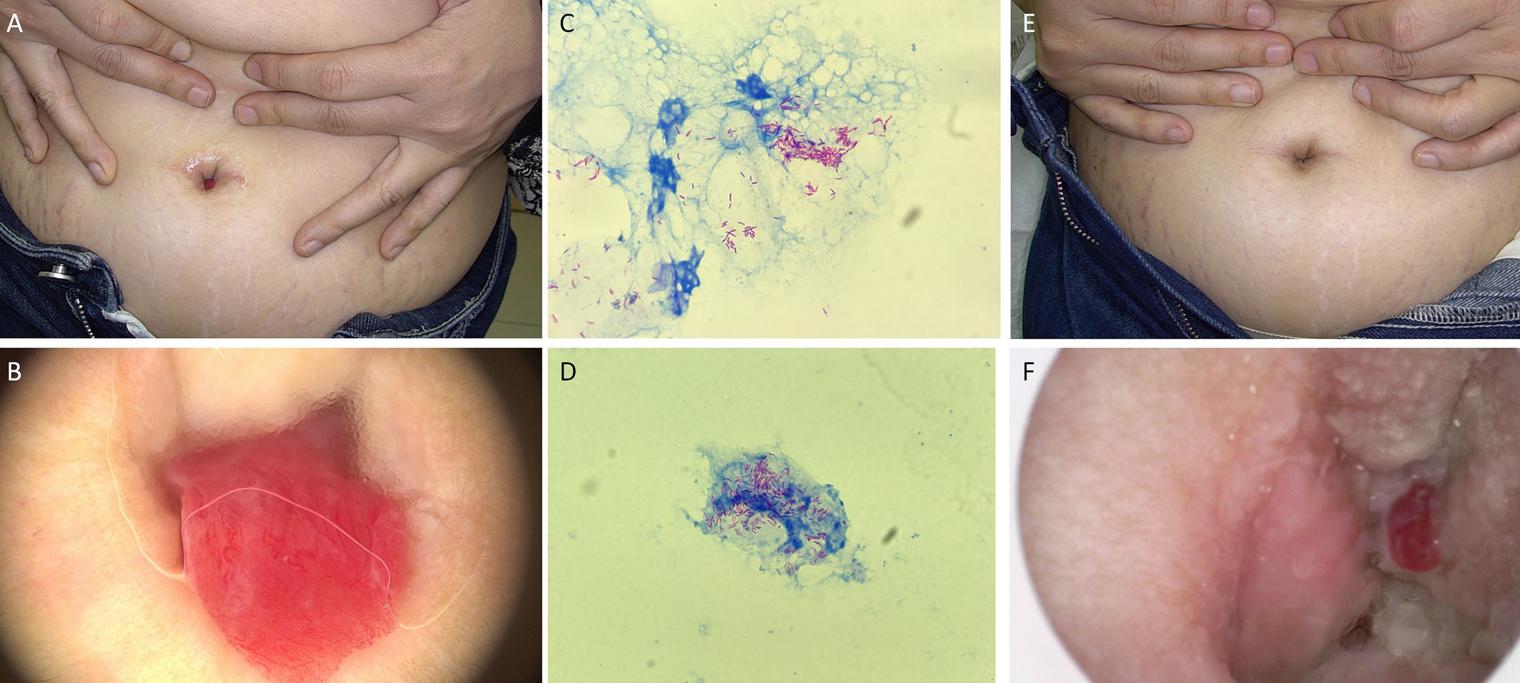
Fig. 1. (A) Clinical photograph at initial presentation showing an erythematous, exophytic, angiomatosis-like lesion with purulent discharge. (B) Dermoscopic image of the lesion revealing a hypervascular pattern with prominent tortuous vessels and red-to-whitish areas. The sinuous pale line across the lesion is the edge of the immersion fluid. (C) Direct acid-fast staining of the purulent exudate demonstrating abundant acid-fast bacilli. (D) Repeat acid–fast staining after 3 weeks of doxycycline plus clarithromycin therapy, still showing numerous persistent acid–fast bacilli. (E) Clinical photograph after switching to amoxicillin–clavulanate plus metronidazole, showing complete resolution of the umbilical mass and cessation of purulent discharge. (F) Fine-tip dermoscopic examination of the deep umbilical fossa after apparent clinical healing, disclosing a residual focus.
We performed both routine bacterial cultures on the pus sample. Bacterial cultures on blood agar and China Blue agar grew Helcococcus and non-fermenting Corynebacterium species, while anaerobic culture yielded 4 distinct colonies, which were subsequently identified by 16S rRNA sequencing as Mobiluncus, Winkia neuii, Finegoldia magna and Aedoeadaptatus urinae. Notably, L. clevelandensis was not isolated via culture. Metagenomic next-generation sequencing (mNGS) of the same pus sample detected Lawsonella clevelandensis, Mobiluncus massiliensis, Winkia neuii, Corynebacterium uberis and Peptoniphilus lacrimalis, each with >10 000 sequence reads. Additionally, Finegoldia magna had approximately 6,000 reads, and Anaerococcus vaginalis and Anaerococcus obesiensis each had fewer than 5,000 reads.
While awaiting mNGS results, the patient was started on empirical oral doxycycline 100 mg twice daily and clarithromycin 250 mg twice daily. After 3 weeks of this therapy, there was no visible improvement in the discharge or the angiomatosis-like lesion, and repeat acid-fast staining of the umbilical exudate still revealed numerous acid-fast bacilli (Fig. 1D). Based on the mNGS findings, therapy was switched to amoxicillin-clavulanate 875/125 mg twice daily plus metronidazole 400 mg 3 times daily. After 3 weeks of this regimen, the purulent discharge ceased completely and the angiomatosis-like lesion resolved to the naked eye, leaving a clinically normal umbilicus (Fig. 1E). However, examination of the deep umbilical fossa with a fine-tip dermoscope disclosed a residual focus (Fig. 1F). The combined antibiotic therapy was continued.
DISCUSSION
This case illustrates a diagnostically challenging umbilical lesion where the positive acid-fast staining strongly suggested a mycobacterial infection, but mNGS ultimately revealed a polymicrobial anaerobic infection with L. clevelandensis as a prominent member. Abdominal tuberculosis may rarely present as an umbilical sinus but typically with marked abdominal symptoms and constitutional signs (3). In a solitary umbilical lesion without nodules, swelling or other systemic features, even a positive acid-fast stain should raise suspicion of non-mycobacterial pathogens such as L. clevelandensis.
L. clevelandensis is a fastidious anaerobe with slow growth and high nutritional requirements. It rarely forms visible colonies on standard anaerobic media, and its isolation by culture is notoriously difficult (2, 4). Previous studies suggest that this organism may exist as a commensal component of the normal human skin and nasal microbiota, with a particular predilection for sebaceous-rich areas such as the nasal alar groove, glabella and occipital region (5). In recent years, accumulating case reports have linked L. clevelandensis to localized abscess formation, occurring both in immunocompromised individuals ((e.g.) those with diabetes mellitus, malignancies or immunosuppressant use) and, less frequently, in immunocompetent hosts (2, 6, 7, 8).
The persistence of numerous acid-fast bacilli on repeat staining after 3 weeks of doxycycline and clarithromycin indicates that the L. clevelandensis in this patient was resistant to macrolides or tetracyclines. Combined with inadequate anaerobic coverage for Mobiluncus and Peptoniphilus, this explains the lack of response to the initial regimen and supports the switch to amoxicillin-clavulanate plus metronidazole, a combination supported by previous reports of successful amoxicillin-clavulanate therapy for L. clevelandensis infections (9). The observed gram-negative rods likely correspond to Mobiluncus or Gram-stain-variable L. clevelandensis (1, 10). mNGS enabled pathogen identification and guided a regimen that covered both aerobic and anaerobic components, leading to rapid macroscopic resolution. The deep dermoscopic detection of subclinical residual focus tissue underscores the value of thorough inspection in apparently healed lesions. mNGS should be considered early for culture-negative, acid-fast-positive cutaneous infections, and treatment must target the full polymicrobial spectrum. To our knowledge, umbilical angiomatosis-like lesion caused by L. clevelandensis has not been reported previously.
REFERENCES
- Bell ME, Bernard KA, Harrington SM, Patel NB, Tucker TA, Metcalfe MG, et al. Lawsonella clevelandensis gen. nov., sp. nov., a new member of the suborder Corynebacterineae isolated from human abscesses. Int J Syst Evol Microbiol 2016; 66: 2929–2935. https://doi.org/10.1099/ijsem.0.001122
- Morton AB, Boyle E, Pettengill MA, Gancher E. The brief case: Strictly anaerobic and staining acid fast. J Clin Microbiol 2023; 61: e0015022. https://doi.org/10.1128/jcm.00150-22
- Vetri R, Kannichamy V, Jacob VA, Sainath S. Abdominal tuberculosis presenting as an umbilical sinus: A case report. Cureus 2022; 14: e31184. https://doi.org/10.7759/cureus.31184
- Goldenberger D, Naegele M, Steffens D, Eichenberger R, Egli A, Seth-Smith HMB. Emerging anaerobic and partially acid-fast Lawsonella clevelandensis: extended characterization by antimicrobial susceptibility testing and whole genome sequencing. Clin Microbiol Infect 2019; 25: 1447–1448. https://doi.org/10.1016/j.cmi.2019.07.008
- Escapa IF, Chen T, Huang Y, Gajare P, Dewhirst FE, Lemon KP. New insights into human nostril microbiome from the expanded human oral microbiome database (eHOMD): A resource for the microbiome of the human aerodigestive tract. mSystems 2018; 3: e00187-18. https://doi.org/10.1128/mSystems.00187-18
- Favila Menezes M, Sousa MJ, Paixão P, Atouguia J, Negreiros I, Simões MJ. Lawsonella clevelandensis as the causative agent of a breast abscess. IDCases 2018; 12: 95–96. https://doi.org/10.1016/j.idcr.2018.03.014
- Gonzales Zamora JA, Romero Alvarez M, Henry Z, Baracco GJ, Dickinson G, Lichtenberger P. Liver abscess caused by Lawsonella clevelandensis in a patient with rheumatoid arthritis: A case report and literature review. IDCases 2020; 20: e00734. https://doi.org/10.1016/j.idcr.2020.e00734
- Inthasorn P, Chirasuthat P. A false-positive bacillary index caused by lawsonella clevelandensis: A diagnostic pitfall. Dermatol Pract Concept 2026; 16: 7115. https://doi.org/10.5826/dpc.1601a7115
- Liu Y, Yue X, Zhou Y, Wang J, Yang F, Jin Y, et al. Recurrent breast abscess caused by Lawsonella clevelandensis: A case report and literature review. Medicine (Baltimore) 2025; 104: e44218. https://doi.org/10.1097/MD.0000000000044218
- Abou Chacra L, Bonnet M, Heredia M, Haddad G, Armstrong N, Alibar S, et al. Peptoniphilus genitalis sp. nov. and Mobiluncus massiliensis sp. nov.: Novel Bacteria Isolated from the Vaginal Microbiome. Curr Microbiol 2024; 81: 97. https://doi.org/10.1007/s00284-023-03584-7