SHORT COMMUNICATION
Haemophagocytic Lymphohistiocytosis Following Severe Erysipelas with EBV Reactivation
Thilo GAMBICHLER1,2,3* , Josefine BRUNE1 and Laura SUSOK1
1Department of Dermatology, Dortmund Hospital gGmbH, University Witten-Herdecke, Dortmund, Germany, 2Department of Dermatology, Christian Hospital Unna, Unna, Germany, and 3Department of Dermatology, Ruhr-University Bochum, Bochum, Germany. *Email: thilo.gambichler@klinikumdo.de
Citation: Acta Derm Venereol 2026; 106: adv-2026-0822. DOI: https://doi.org/10.2340/actadv.v106.adv-2026-0822.
Copyright: 2026 ©Author(s). Published by MJS Publishing, on behalf of the Society for Publication of Acta Dermato-Venereologica. This is an Open Access article distributed under the terms of the Creative Commons Attribution-NonCommercial 4.0 International License (https://creativecommons.org/licenses/by-nc/4.0/).
Submitted: Jun 26, 2026. Accepted after revision: Aug 24, 2026.
Published: Oct 7, 2026.
Competing interests and funding: The authors have no conflicts of interest to declare.
All crucial data generated or analysed during this case study are included in this published article.
Written informed consent for publication of the clinical details and clinical images was obtained from the patient.
Secondary haemophagocytic lymphohistiocytosis (HLH) is a potentially life-threatening hyperinflammatory syndrome that may mimic sepsis and should be considered in patients with persistent fever, cytopenias, organomegaly and marked hyperferritinaemia (1). Dermatological manifestations of HLH are heterogeneous, but erysipelas or cellulitis as a precipitating event is very rarely documented; cellulitis-like presentations may also reflect opportunistic infection or occult lymphoma (2, 3, 4, 5, 6, 7, 8, 9). We report a case of steroid-responsive HLH developing during treatment of severe streptococcal erysipelas, accompanied by transient EBV-DNAemia.
CASE REPORT
A 44-year-old patient was transferred to our dermatology department because of severe erysipelas of the left leg, accompanied by chills, fever, extensive erythematous swelling, blistering and erosions, with marked distal accentuation (Fig. 1). Initial laboratory evaluation showed marked leukocytosis and elevated C-reactive protein (CRP). A profoundly increased anti-streptolysin titre of 17,586 U/mL supported ongoing or recent streptococcal immune stimulation, whereas repeated blood cultures remained negative for bacterial and fungal growth.
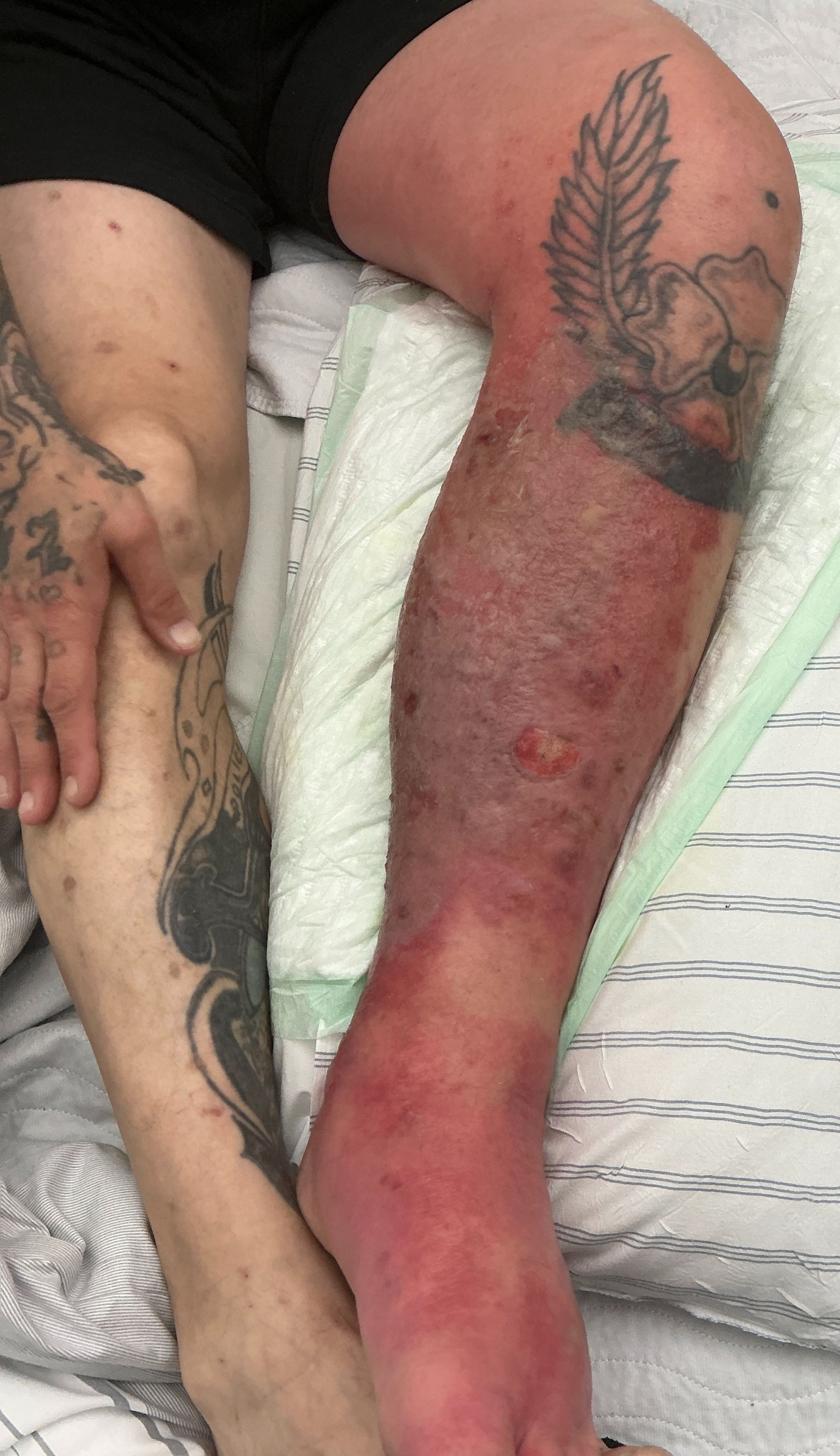
Fig. 1. Severe erythema, blistering, swelling and erosions involving the entire left leg, most pronounced on the lower part.
Despite gradual improvement of the cutaneous findings and initial defervescence during 3 weeks of broad-spectrum combination antibiotic therapy, the patient developed recurrent high fever of upto 39.7°C and marked pancytopenia, with leukocytes decreasing to 0.53×10⁹/L, platelets to 54×10⁹/L and erythrocytes to 2.82×10¹²/L. Peripheral blood smear showed pronounced erythrocyteanisocytosis, reduced platelets and almost complete absence of nucleated cells, with no circulating blasts or pathological cells. Laboratory findings revealed hyperferritinaemia up to 28,256 µg/L, hypertriglyceridaemia up to 413 mg/dL, hypofibrinogenaemia of 1.52g/L, elevated soluble interleukin-2 receptor levels of 176 RU/mL, markedly increased lactate dehydrogenase of 4,385 U/L and CRP peaking at 326.44 mg/L. Abdominal ultrasound demonstrated hepatosplenomegaly. Liver involvement was reflected by AST elevation up to 518U/L, whereas ALT was only moderately increased up to 57U/L. EBV-DNA was detectable at 8,520 copies/mL, while CMV-DNA was negative. Hepatitis screening revealed positive anti-HCV antibodies and a positive confirmatory line immunoassay, but HCV-RNA was negative; HBV-PCR was also negative.
Flow cytometry was limited by low cellularity but showed T-cell predominance, an inverted CD4/CD8 ratio of 0.48, low B-cell numbers and no evidence of a circulating malignant population. Bone marrow examination, performed after initiation of intravenous high-dose corticosteroid therapy, did not reveal haemophagocytosis. However, absence of haemophagocytosis, particularly after treatment initiation, did not exclude HLH. Hence, the HScore, a validated clinical and laboratory score estimating the probability of reactive HLH, was 284, corresponding to a >99% probability of HLH (1).
High-dose intravenous corticosteroid therapy resulted in rapid clinical and laboratory improvement. The patient became afebrile within the first days, ferritin and transaminases decreased markedly, and EBV-DNA became undetectable after 99 of treatment. After 5 weeks, the patient was discharged with complete normalization of the blood count and undetectable EBV-DNA in EDTA blood. Ferritin had decreased to 624, transaminases were nearly normal, and CRP had fallen to 15.51, while triglycerides remained moderately elevated. Corticosteroids were tapered gradually, and close short-term clinical and laboratory monitoring was scheduled.
DISCUSSION
This case illustrates the diagnostic difficulty of HLH emerging in the setting of an apparently improving bacterial skin and soft-tissue infection. The exceptionally high anti-streptolysin titre supported a strong streptococcal immune stimulus, but erysipelas alone is not an established HLH trigger. EBV-DNAemia was only moderate and became rapidly undetectable under corticosteroid therapy; therefore, EBV should be interpreted as a plausible contributor or amplifier of immune activation rather than as proven primary cause of HLH in this case (10). Moreover, LDH was strikingly elevated, supporting extensive systemic cell turnover and tissue injury, although not part of formal HLH criteria. Alternative infectious and malignant conditions were considered: repeated blood cultures remained negative, CMV-DNA, HBV-PCR and HCV-RNA were negative, and peripheral blood smear and flow cytometry did not provide evidence of leukaemia or a circulating lymphoid malignancy; bone marrow examination did not reveal haemophagocytosis. Nevertheless, cellulitis-like lesions in HLH should prompt careful exclusion of mimickers, particularly disseminated fungal infection and subcutaneous panniculitis-like T-cell lymphoma (7, 8, 9).
In this clinical context, recurrent fever, cytopenias and hyperferritinaemia despite improving skin findings should prompt immediate HLH work-up.
REFERENCES
- Khare N, Jinkala SR, Kanungo S. Performance of HScore in reactive hemophagocytic lymphohistiocytosis. Indian J Hematol Blood Transfus 2021; 37: 256–263. https://doi.org/10.1007/s12288-020-01342-4
- Chang CC, Hsiao PJ, Chiu CC, Chen YC, Lin SH, Wu CC, et al. Catastrophic hemophagocytic lymphohistiocytosis in a young man with nephrotic syndrome. Clin Chim Acta 2015; 439: 168–171. https://doi.org/10.1016/j.cca.2014.10.025
- Boggio F, Lora V, Cota C, Pereira A, Müllegger R, Prieto-Torres L, et al. Cutaneous hemophagocytosis: clinicopathologic features of 21 cases. J Am Acad Dermatol 2018; 78: 377–382. https://doi.org/10.1016/j.jaad.2017.08.041
- Kuntz N, Grovas LJ, Tang R, Armanyous Y, Hoerig C, Torno L, et al. Cutaneous manifestations in an infant with hemophagocytic lymphohistiocytosis: case report. J Pediatr Hematol Oncol Nurs 2025; 42: 202–209. https://doi.org/10.1177/27527530251356970
- Hall AD, Salibindla D, Dubocq-Cabrera L, Hunt M, Rotert LM, Johnson RS, et al. Progressive disseminated histoplasmosis with dermatomyositis and macrophage activation syndrome/hemophagocytic lymphohistiocytosis. Am J Trop Med Hyg 2025; 113: 773–780. https://doi.org/10.4269/ajtmh.24-0802
- Baba M, Fenech M, Saeed MU, McCormick A, Jawad M. A rare case of orbital inflammation complicated by hemophagocytic lymphohistiocytosis. Orbit 2024; 43: 751–754. https://doi.org/10.1080/01676830.2023.2274868
- Puing AG, Raghavan SS, Aleshin MA, Ho DY. Hemophagocytic lymphohistiocytosis secondary to progressive disseminated histoplasmosis presenting as cellulitis. Med Mycol Case Rep 2021; 33: 18–20. https://doi.org/10.1016/j.mmcr.2021.06.002
- Tzeng HE, Teng CL, Yang Y, Young JH, Chou G. Occult subcutaneous panniculitis-like T-cell lymphoma with initial presentations of cellulitis-like skin lesion and fulminant hemophagocytosis. J Formos Med Assoc 2007; 106: S55–9. https://doi.org/10.1016/s0929-6646(09)60354-5
- Liu D, Pei X, Zhang X, Xu L, Wang Y, Yan C, et al. Clinical manifestations and outcomes of EBV-related secondary hemophagocytic lymphohistiocytosis. Ann Hematol 2025; 104: 5605–5613. https://doi.org/10.1007/s00277-025-06677-4
- La Rosée P, Horne A, Hines M, von Bahr Greenwood T, Machowicz R, Berliner N, et al. Recommendations for the management of hemophagocytic lymphohistiocytosis in adults. Blood 2019; 133: 2465–2477. https://doi.org/10.1182/blood.2018894618