SHORT COMMUNICATION
Molluscum Contagiosum, Warts and Condylomas during JAK Inhibitor Therapy for Atopic Dermatitis: a Retrospective Series of 17 Cases
Giuseppe RUSSO1,2* , Emmanuel MAHÉ3,4, Florence TETART5, Audrey LORETAN1, Audrey NOSBAUM6,7, Marie TAUBER6,7, Nadia RAISON-PEYRON8, Estelle BURLE9, Angèle SORIA10, Emilie BRENAUT11, Anne SAUSSINE2, Thibault MAHÉVAS2, Jean-David BOUAZIZ2, Emmanuel LAFFITTE1, Marie JACHIET2, FRench Atopic DErmatitis Network from the GREAT Research group, and French Society of Dermatology (SFD)
1Department of Dermatology, Geneva university hospitals HUG, Genève, Suisse, Switzerland, 2Department of Dermatology, Saint-Louis Hospital, Assistance Publique-Hôpitaux de Paris, Paris, France, 3Department of Dermatology, Hôpital Victor Dupuy, Argenteuil, France, 4Versailles-Saint-Quentin-en-Yvelines Unviersity, Montigny-le-Bretonneux, France, 5Department of Dermatology and Allergology, CHU Rouen, Rouen, France, 6Hospices Civils de Lyon, Department of allergy and Clinical Immunology, Centre Hospitalier Lyon Sud, Pierre-Bénite, France, 7CIRI, Centre International de Recherche en Infectiologie (Team Epidermal Immunity and Allergy Team), INSERM U1111, Université Lyon1, Ecole Normale Supérieure de Lyon, CNRS UMR 5308, Lyon, France, 8Department of Dermatology, Centre Hospitalier Universitaire Montpellier, Montpellier, France, 9Department of Dermatology, CHU Toulouse-Larrey, Toulouse, France, 10Sorbonne Université, Department of Dermatology and Allergology, Tenon Hospital, Assistance Publique-Hôpitaux de Paris, Paris, INSERM 1155, France, and 11Department of Dermatology, CHU de Brest, Brest, France. *Email: giuseppe.russo@hug.ch
Citation: Acta Derm Venereol 2026; 106: adv-2026-0779. DOI: https://doi.org/10.2340/actadv.v106.adv-2026-0779.
Copyright: 2026 ©Author(s). Published by MJS Publishing, on behalf of the Society for Publication of Acta Dermato-Venereologica. This is an Open Access article distributed under the terms of the Creative Commons Attribution-NonCommercial 4.0 International License (https://creativecommons.org/licenses/by-nc/4.0/).
Submitted: Jun 10, 2026. Accepted after revision: Sept 9, 2026.
Published: Oct 12, 2026.
Competing interests and funding: The data that support the findings of this study are available on request from the corresponding author. The data are not publicly available due to privacy restrictions.
All procedures adhered to the ethical standard set by the Helsinki Declaration. All patients provided written informed consent for images. Identifying information has been removed.
MJ and EL have received personal fees for consulting and/or speaking activities or travel grants and congress registration support from AbbVie, Almirall, AstraZeneca, Galderma, Incyte, LEO Pharma, Lilly, Pfizer, Sanofi. These relationships are outside the submitted work. GR has received personal fees for speaking activities and/or travel grants and congress registration support from Sanofi, Johnson & Johnson, Pfizer and UCB. These relationships are outside the submitted work. EM has undertaken activities as a paid consultant, adviser or speaker for AbbVie, Almirall, Amgen, Biolane, Janssen-Cilag, Leo Pharma, Lilly, Novartis, Sanofi, and UCB. AN has served as an investigator, speaker, and/or advisor for AbbVie, Almirall, Sanofi Regeneron, Pierre Fabre, Janssen Cilag, Celgene, Lilly, Leo Pharma, L'Oréal, Galderma, Leo Pharma, Medac, Novartis. NRP has served as an investigator for AbbVie and Pierre Fabre and has received personal fees for speaking activities and/or for travel grants and congress registration support from Sanofi and ALK. AS has received support or personal fees for consulting and/or speaking activities from Novartis, Sanofi, AbbVie, Lilly, LEO Pharma, Pfizer, Bioprojet, Almirall and research grants from Sanofi. MT has served as an investigator, speaker, and/or advisor for AbbVie, Almirall, Sanofi Regeneron, Pierre Fabre, Janssen Cilag, Lilly, Leo Pharma, Galderma, Leo Pharma, Medac, Novartis.
Janus kinase inhibitors (JAK-i) represent a major therapeutic advancement for inflammatory dermatoses such as atopic dermatitis (AD), alopecia areata and vitiligo. By blocking intracellular signal transduction of multiple cytokines, including type I and II interferons, they exert potent anti-inflammatory effects but may also impair antiviral immune defence (1). Although herpes-zoster reactivation and other varicella-zoster virus (VZV) infections are well-recognized adverse events, data on nonherpetic cutaneous viral infections remain limited (2). We report a multicentre retrospective case series of 17 patients who developed cutaneous viral infections (molluscum contagiosum (MC), warts or condylomas) during JAK-i treatment for AD.
Our cohort comprised 17 patients (13 males, 4 females; mean age 35.6±11.4 years, median 35 years, range 13–63 years) treated for AD with JAK-i in 9 dermatology centres in France and Switzerland. Upadacitinib was the most prescribed agent (94.1%, n=16; 14 patients at 30 mg/day, 2 at 15 mg/day). Nine patients had received previous immunosuppressive therapy, most commonly ciclosporin and prior JAK-i. Patients were not systematically screened for underlying immunodeficiency; however, no clinical history suggestive of primary or secondary immunodeficiency was reported. Among the cohort, 6 patients (35.3%) had a documented history of MC, warts or condylomas prior to JAK-i initiation. Cutaneous viral infections developed at a median of 6 months (range: 1–24 months, mean 9.2±7.1 months) after JAK-i initiation. The predominant infection types were condylomas (47.1%, n=8), warts (47.1%, n=8) and molluscum contagiosum (41.2%, n=7), with 23.8% (n=5) of patients exhibiting multiple concurrent infections (Table I). Lesion burden varied considerably according to infection type, with most patients presenting either 10–50 lesions (n=5) or fewer than 10 lesions; however extensive disease (>50 lesions) occurred in 3 patients, predominantly in those with condylomas or molluscum contagiosum (Fig. 1).
Table I. Clinical characteristics and outcomes of 17 patients with cutaneous viral infections during Janus kinase inhibitor (JAKi) therapy for atopic dermatitis
| Patient | Age/Sex | Prior immunosuppression | Prior viral infection | JAKi (dose, mg/day) | Time to infection (months)a | Infection type | Lesion count | JAKi management |
|---|---|---|---|---|---|---|---|---|
| 1 | 32 /M | CsA, BARI, ABRO | No | UPA (30) | 6 | Molluscum contagiosum | Unknown | Switched JAKi |
| 2 | 63 /M | None | Molluscum contagiosum | UPA (30) | 3 | Molluscum contagiosum | Unknown | Continued |
| 3 | 30 /F | None | No | UPA (30) | 15 | Molluscum contagiosum | >50 | Stopped |
| 4 | 44 /M | None | Molluscum contagiosum, condyloma, warts | UPA (30) | 6 | Condyloma, warts | Unknown | Stopped |
| 5 | 36 /M | None | Herpes zoster, warts | UPA (30) | 6 | Molluscum contagiosum, warts | 10–50 | Reduced |
| 6 | 39 /M | None | No | UPA (30) | 9 | Condyloma | 10–50 | Reduced |
| 7 | 28 /M | None | No | UPA (15) | 2 | Molluscum contagiosum | >50 | Continued |
| 8 | 44 /M | BARI, ABRO | No | UPA (30) | 11 | Condyloma | >50 | Stopped |
| 9 | 35 /M | CsA, BARI, ABRO | No | ABRO (200) | 24 | Condyloma, warts | <10 | Stopped |
| 10 | 31 /M | CsA | Condyloma | UPA (30) | 1 | Condyloma | <10 | Continued |
| 11 | 53 /M | CsA, MTX | Herpes zoster, Condyloma | UPA (30) | 1 | Condyloma, warts | 10–50 | Switched JAKi |
| 12 | 22 /F | CsA | No | UPA (30) | 15 | Condyloma | 10–50 | Stopped |
| 13 | 13 /F | MTX, OCS | Warts | UPA (15) | 12 | Warts | <10 | Continued |
| 14 | 31 /M | BARI | No | UPA (30) | 9 | Molluscum contagiosum | 10–50 | Switched JAKi |
| 15 | 31 /F | BARI | No | UPA (30) | 6 | Warts | <10 | Reduced |
| 16 | 35 /M | None | No | UPA (30) | 6 | Warts | <10 | Continued |
| 17 | 38 /M | None | No | UPA (30) | 24 | Molluscum contagiosum, condyloma, warts | 10–50 | Continued |
|
aTime to infection represents the interval from JAK-i initiation to viral infection onset. Reduced: Upadacitinib (UPA) switched to 15 mg/day. ABRO: abrocitinib; ADA: adalimumab; BARI: baricitinib; CsA: ciclosporine; HSV: herpes simplex virus; MTX: methotrexate; OCS: oral corticosteroids. |
||||||||
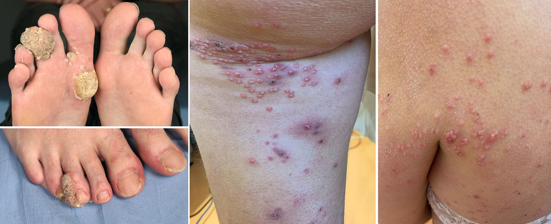
Fig. 1. Cutaneous viral infections arising during treatment with upadacitinib in patients with atopic dermatitis. Giant plantar warts (left); extensive molluscum contagiosum eruption (right)
JAK-i therapy was modified in 11 patients (64.7%), including discontinuation (n=5, 29.4%), dose reduction (n=3, 17.6%) or switching to another JAK-i (n=3, 17.6%) in response to viral infections. Therapeutic interventions included cryotherapy, topical imiquimod, topical keratolytic agents and surgical excision. Among the 5 patients in whom JAK-i were discontinued, cutaneous viral lesions completely regressed in 4 cases, including 3 patients with disseminated MC and 1 with condylomas.
JAK-i may increase susceptibility to viral infections through suppression of type I interferon signalling in epidermal and mucosal tissues, a pathway critical for keratinocyte antiviral responses and cytotoxic lymphocyte activation (3).
A cohort study of 1793 AD patients reported higher overall infection rates with JAK-i treatment compared to biologic treatment, particularly for herpes infections (4). Beyond herpesviruses, isolated case reports have described MC and human papillomavirus (HPV)-related warts arising during JAK-i therapy. Notably, a case of disseminated MC was reported in a patient receiving ruxolitinib, a JAK1/2 inhibitor, highlighting the role of interferon pathway blockade in impaired viral clearance (5).
Most recently, several reports documented the occurrence of HPV-related warts following JAK-i initiation (6, 7, 8). Collectively, these observations support a mechanistic link between JAK inhibition and reduced cutaneous antiviral immunity beyond herpesvirus reactivation.
Our retrospective series suggests that JAK-i may predispose patients to opportunistic cutaneous viral infections particularly HPV-related condylomas, warts, and MC. Patients with AD and/or a prior history of viral skin infections may represent a population at increased risk. Clinicians should therefore remain alert to the development of new or progressive viral skin lesions during JAK-i therapy.
The main limitations of this study are the relatively young cohort and the predominance of upadacitinib exposure, limiting the generalizability of our findings to older patients and to other JAK inhibitors.
We recommend patient education, baseline assessment of viral infection history and periodic skin examination during JAK-i therapy. Reporting such cases is essential to better define their incidence, risk factors, and outcomes across dermatologic indications.
REFERENCES
- Jarneborn A, Kopparapu PK, Jin T. The Dual-Edged Sword: Risks and benefits of JAK inhibitors in infections. Pathogens 2025; 14: 324. https://doi.org/10.3390/pathogens14040324
- Ireland PA, Verheyden M, Jansson N, Sebaratnam D, Sullivan J. Infection risk with JAK inhibitors in dermatoses: a meta-analysis. Int J Dermatol 2025; 64: 24–36. https://doi.org/10.1111/ijd.17501
- Ravlo E, Ianevski A, Skipperstøen MN, Lysvand H, Schjølberg JO, Solheim O, et al. JAK inhibitors remove innate immune barriers facilitating viral propagation. NAR Mol Med 2025; 2: ugaf017. https://doi.org/10.1093/narmme/ugaf017
- van der Gang LF, Atash K, Zuithoff NPA, Haeck I, Boesjes CM, Bacoş-Cosma OI, et al. Infection risk in atopic dermatitis patients treated with biologics and JAK inhibitors: BioDay results. J Eur Acad Dermatol Venereol 2025; 39: 2056–2068. https://doi.org/10.1111/jdv.20674
- Kinoshita M, Ogawa Y, Kawamura T, Kirito K, Shimada S. Case of disseminated molluscum contagiosum caused by ruxolitinib, a Janus kinase 1 and 2 inhibitor. J Dermatol 2016; 43: 1387–1388. https://doi.org/10.1111/1346-8138.13404
- Seale E, Gavigan G. Exacerbation of human papillomavirus infection with initiation of upadacitinib for atopic dermatitis. JAAD Case Rep 2023; 36: 60–62. https://doi.org/10.1016/j.jdcr.2023.04.019
- Kennoki N, Nohata K, Nakamori Y. Impaired Human Papillomavirus (HPV) clearance in a patient on upadacitinib for atopic dermatitis: A case of refractory genital warts resolved after drug withdrawal. Cureus 2025; 17: e97974. https://doi.org/10.7759/cureus.97974
- Takahashi N, Shimizu T, Kondoh A, Anzawa K, Shimizu A, Mabuchi T. Human Papilloma Virus Type 94-associated flat warts developing repeatedly during tofacitinib treatment: A case report and review. J Dermatol 2025. https://doi.org/10.1111/1346-8138.17814