ORIGINAL ARTICLE
Serum Mediators in Patients with Both Type 2 Diabetes Mellitus and Pruritus
Guan-Yi HE1–3, Tai-Yi HSU4–6,, Ching-Wen CHEN7, Feng-Jung NIEN8, Huan-Yuan CHEN9, Chia-Yu CHU3 and Li-Fang WANG3
1Department of Dermatology, National Taiwan University Hospital Yunlin Branch, Yunlin, 2Graduate Institute of Clinical Medicine, College of Medicine, National Taiwan University, 3Department of Dermatology, National Taiwan University Hospital and National Taiwan University College of Medicine, Taipei City, 4School of Medicine, College of Medicine, China Medical University, 5Department of Emergency Medicine, China Medical University Hospital, 6Department of Public Health, China Medical University, Taichung City, 7Subdivision of Endocrinology and Metabolism, Department of Internal Medicine, National Taiwan University Hospital Yunlin Branch, Yunlin, 8Department of General Medicine, National Taiwan University Cancer Center and 9Institute of Biomedical Sciences, Academia Sinica, Taipei City, Taiwan
Chronic pruritus is an unpleasant sensory perception that negatively affects quality of life and is common among patients with type 2 diabetes mellitus. Current antipruritic therapies are insufficiently effective. Thus, the mediation of diabetic pruritus by histamine-independent pathways is likely. The aim of this study was to identify possible mediators responsible for diabetic pruritus. A total of 87 patients with type 2 diabetes mellitus were analysed, of whom 59 had pruritus and 28 did not. The 2 groups were assessed for baseline demographics, serum biochemistry parameters, cytokines, and chemokines. This study also investigated the associations of these factors with the severity of itching. Neither haemoglobin A1c nor serum creatinine levels were correlated with severity of itching. Significantly higher levels of interleukin-4 (p = 0.004), interleukin-13 (p = 0.006), granulocyte-macrophage colony-stimulating factor (p < 0.001) and C-X-C motif chemokine ligand 10 (p = 0.028) were observed in the patients with pruritus than in those without pruritus. Moreover, the levels of these mediators were positively correlated with the severity of itching. Thus, novel antipruritic drugs can be developed to target these molecules. This is the first study to compare inflammatory mediators comprehensively in patients with diabetes mellitus with pruritus vs those without pruritus.
Key words: chemokine CXCL10; diabetes mellitus; granulocytemacrophage colony-stimulating factor; interleukin-4; interleukin-13; pruritus.
SIGNIFICANCE
Chronic pruritus is common in patients with type 2 diabetes, severely affects quality of life, and is notoriously difficult to treat. This study identified possible mediators involved in the pathophysiology of diabetic pruritus. It was observed that, among patients with type 2 diabetes, those with pruritus had significantly elevated levels of specific molecules. Further exploration of the roles of these molecules may help treat diabetic pruritus.
Citation: Acta Derm Venereol 2023; 103: adv00875. DOI: https://doi.org/10.2340/actadv.v103.4863.
Copyright: © Published by Medical Journals Sweden, on behalf of the Society for Publication of Acta Dermato-Venereologica. This is an Open Access article distributed under the terms of the Creative Commons Attribution-NonCommercial 4.0 International License (https://creativecommons.org/licenses/by-nc/4.0/)
Accepted: Jan 31, 2023; Published: Feb 28, 2023
Corr: Li-Fang Wang, Department of Dermatology, National Taiwan University Hospital and National Taiwan University College of Medicine, No. 8, Zhongshan S. Road, Zhongzheng District, Taipei City 100226, Taiwan. E-mail: lifangwa@ntu.edu.tw
Competing interests and funding: The authors have no conflicts of interest to declare.
INTRODUCTION
The prevalence of diabetes mellitus (DM) and its associated complications is increasing worldwide (1). One-third of patients with DM have skin disorders (2). Among these skin manifestations, chronic pruritus is common and considerably impairs individuals’ quality of life. Patients may have generalized or localized pruritus (3). Chronic generalized pruritus prompts a DM screening, whereas localized pruritus necessitates a survey for a dermatological disease (4). Studies have revealed that DM is a statistically significant predictor of chronic pruritus in older adults (5). The reported prevalence of pruritus in patients with DM varies from 18.4% to 27.5%, and may be underestimated (4). Current therapies for chronic pruritus include emollients, topical antipruritic agents, systemic antipruritic agents (H1 antihistamine, doxepin, or gabapentinoids), and phototherapy (6). However, the effects of these antipruritic therapies are often insufficient, because the pathophysiology of chronic pruritus in patients with DM is poorly understood. Studies have reported that postprandial blood glucose (7), fasting plasma glucose (3, 8), age, duration of DM, diabetic neuropathy, diabetic retinopathy, and diabetic kidney disease (8) are associated with diabetic pruritus. Nevertheless, results from different studies are inconsistent and occasionally contradictory.
A proinflammatory state has been observed in patients with DM, but few studies have investigated the profile of cytokines and chemokines in diabetic pruritus (9, 10). This study therefore investigated whether clinical characteristics, biochemistry parameters, cytokines, and chemokines are associated with diabetic pruritus.
MATERIALS AND METHODS
Study design and setting
This cross-sectional study was conducted in a single university-affiliated hospital. All participants provided written informed consent. The study was approved by the Institutional Review Board of National Taiwan University Hospital (IRB number: 201810098RINA).
Patient selection
Patients with type 2 DM undergoing treatment at National Taiwan University Hospital Yunlin Branch between 2019 and 2020 were evaluated for enrollment. Exclusion criteria were: patients with known pruritic skin diseases, such as psoriasis, atopic dermatitis (AD), chronic urticaria, autoimmune bullous disease, contact dermatitis, and scabies, on the basis of skin examinations performed by a dermatologist. Patients with invasive cancers or end-stage renal disease were also excluded. A total of 90 patients were enrolled. Among the included patients, 60 (31 men and 29 women) had concurrent pruritus and 30 (15 men and 15 women) did not. Baseline characteristics, including age, sex, duration of diabetes, concurrent antipruritic medication, comorbidities, and DM complications (diabetic neuropathy, diabetic nephropathy, and proteinuria) were recorded. A diagnosis of diabetic neuropathy was made if a patient’s medical charts contained the disease codes or records of diabetic neuropathy.
Methods and measurements
Itching assessment. The intensity of pruritus was assessed using a numerical rating scale (NRS; 0 = ”no pruritus,” 1–2 = ”mild pruritus,” 3–6 = ”moderate pruritus,” 7–8 = ”severe pruritus”, 9–10 = ”very severe pruritus”) (11) and the 12-item Pruritus Severity Score (12–PSS, 3–6 points = ”mild pruritus,” 7–11 points = ”moderate pruritus,” and 12–22 points = ”severe pruritus”) (12). The NRS score reflects mean pruritus within the past 24 h. The treatment modalities for itching, including systemic and topical drugs, were also documented.
Multiplex immunoassay. The mediator levels in 87 sera samples were analysed by the Inflammation Core Facility, Institute of Biomedical Sciences, Academia Sinica, Taiwan. The core facility is funded by the Academia Sinica Core Facility and Innovative Instrument Project (AS-CFII-108-118; support number: 206f-1083818). Blood samples were collected using an antiblocked reagent and then diluted 4- and 7-fold for the quantification of mediator levels. The levels of 21 mediators were measured in duplicate, and these mediators are outlined as follows: interleukin (IL)-1α, IL-1β, IL-2, IL-4, IL-6, IL-8, IL-9, IL-12, IL-13, IL-15, IL-17A, IL-23, tumour necrosis factor (TNF)-α, interferon-γ (IFN-γ), IL-31, granulocyte colony-stimulating factor (G-CSF), granulocyte–macrophage colony-stimulating factor (GM-CSF), C-X-C motif chemokine ligand 10 (CXCL10), monocyte chemoattractant protein-1 (MCP-1), high-sensitivity C-reactive protein, and transforming growth factor beta (TGF-β). In brief, antibody-coupled magnetic beads were incubated with standard or sera samples for 2 h. After being washed, the beads were incubated with detection antibody for 1 h, washed, and subsequently incubated with 1 μg/mL streptavidin–phycoerythrin (SA–PE) for 30 min. The beads were rewashed, suspended in 100 μL assay buffer, and analysed through a Bio-Plex 200 system (Bio-Rad, Hercules, CA, USA). All assays were protected from light and performed at room temperature. The detection limits of the 21 mediators are listed in Table SI.
Biochemistry. Venous blood samples were collected to measure fasting plasma glucose (fasting period of at least 8 h), postprandial (2 h after a meal) plasma glucose, glycated haemoglobin A1c (HbA1c), creatinine, estimated glomerular filtration rate (eGFR), and alanine aminotransferase (ALT) levels by using central laboratory systems.
Statistical analysis
All statistical analyses were performed using SAS software (version 9.4; SAS Institute, Cary, NC, USA). A 2-sided p-value < 0.05 was considered statistically significant. Descriptive statistics were used to compare the distribution of factors, including baseline demographics and laboratory test results, in the 2 groups. Categorical variables are presented as proportions, and non-normally distributed continuous variables are presented as medians (interquartile range; IQR). Univariate analysis was performed using the χ2 or Fisher’s exact test for categorical variables and the Mann–Whitney U test for continuous variables to identify predictors of pruritus. Spearman’s rank correlation coefficient was applied to assess the relationship between 2 variables. Moreover, multivariable linear regression was conducted to determine the significance between variables and the severity of itching (NRS score and 12-PSS). The levels of cytokines and chemokines were logarithmically transformed before the application of the Spearman’s rank correlation coefficient and multivariable regression analysis.
RESULTS
Three patients were excluded from the study due to incorrect inclusion: 1 had type 1 DM, 1 had cervical cancer and underwent radiotherapy, and 1 had hepatitis C virus infection and underwent antiviral therapy. Thus, a total of 87 patients were included in the final analysis, of whom 59 (31 men and 28 women) were in the pruritus group and 28 (15 men and 13 women) were in the non-pruritus group. In the pruritus group, 7 patients who were undergoing treatments for itching (3 patients received levocetirizine, 3 received levocetirizine plus topical steroids, and 1 received levocetirizine, oral doxepin, and topical steroids). All enrolled patients had chronic pruritus after the diagnosis of DM. None of the patients in the non-pruritus group underwent anti-itching therapy. The distributions of NRS and 12-PSS scores are shown in Fig. S1. The median (IQR) of the NRS score is 5 (3–7), and of the 12-PSS score is 6 (4–8). The 2 groups did not differ significantly in age, sex, or comorbidities (Table I), and they did not differ significantly in biochemistry parameters for peripheral blood, including fasting glucose, postprandial glucose, HbA1c, serum creatinine, and eGFR levels. The serum levels of IL-4, IL-13, GM-CSF, and CXCL-10, were significantly higher in the pruritus group (p = 0.004, 0.006, < 0.001, and 0.028, respectively; Table II and Fig. 1). The other 17 mediators that were not significantly different between the 2 groups are listed in Table SII.
| Variables | Total patients n=87 | Diabetic patients with pruritus n=59 | Diabetic patients without pruritus n=28 | p-value | |||
| Biochemistry data, median (IQR) | |||||||
| Glucose, mg/dL, fasting | 129 (106–152) | 128 (107–157) | 130.5 (105.25–151.25) | 0.784 | |||
| Glucose, mg/dL, postprandial 2 h | 187.5 (138.25–250.25) | 195 (150.5–242) | 162 (113–252) | 0.328 | |||
| Haemoglobin A1c (%) | 7.2 (6.6–8.0) | 6.7 (7.4–8.0) | 7.05 (6.6–7.75) | 0.416 | |||
| Creatinine, mg/dL | 0.9 (0.7–1.1) | 0.9 (0.7–1.1) | 0.86 (0.73–1.18) | 0.21 | |||
| eGFR, mL/min/1.73 m2 | 79.7 (61.4–102.5) | 81.7 (65.2–104.8) | 74.25 (58.55–88.85) | 0.195 | |||
| Alanine aminotransferase, U/L | 18.5 (14–25) | 20 (13–24.25) | 17.5 (14–19.25) | 0.955 | |||
| Cytokines and chemokines, pg/mL | Median (IQR) | Mean (SD) | Median (IQR) | Mean (SD) | Median (IQR) | Mean (SD) | |
| Interleukin-4 | 0 (0–0) | 0.10 (0.44) | 0 (0–0.04) | 0.15 (0.53) | 0 (0–0) | 0 (0) | 0.004* |
| Interleukin-13 | 0 (0–11.89) | 19.86 (89.36) | 0.84 (0–14.55) | 27.69 (107.69) | 0 (0–0) | 3.35 (10.34) | 0.006* |
| GM-CSF | 0.06 (0–0.37) | 0.38 (1.32) | 0.25 (0.06–0.47) | 0.55 (1.57) | 0 (0–0) | 0.04 (0.15) | < 0.001** |
| CXCL10 | 0 (0–4.83) | 10.67 (35.51) | 0 (0–16.21) | 15.20 (39.95) | 0 (0–0) | 1.12 (2.87) | 0.028* |
| IQR: interquartile range; eGFR: estimated glomerular filtration rate; SD: standard deviation; GM-CSF: granulocyte-macrophage colony-stimulating factor; CXCL10: C-X-C motif chemokine ligand 10; *p < 0.05; **p < 0.001. | |||||||
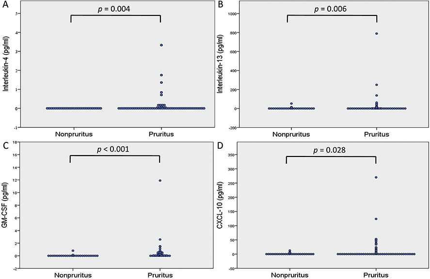
Fig. 1. Comparison between patients with type 2 diabetes mellitus with pruritus and those without pruritus. (A) Interleukin-4. (B) Interleukin-13. (C) Granulocyte-macrophage colony-stimulating factor (GM-CSF). (D) C-X-C motif chemokine ligand 10 (CXCL10).
Spearman’s rank correlation coefficients for the 21 mediators were examined, and the results revealed that IL-2, IL-4, IL-13, IFN-γ, CXCL-10, and GM-CSF were correlated with the severity of itching. Moreover, IL-2, GM-CSF, and CXCL-10 levels were positively correlated with both NRS and 12-PSS scores (p < 0.05). IL-4, IL-13, and IFN-γ levels were positively correlated with NRS only. IL-31 levels were not correlated with NRS or 12-PSS (Table III).
| Spearman coefficient rho | NRS | 12-PSS | |
| Interleukin-2 | ρ | 0.211 | 0.238 |
| p-value | 0.049* | 0.027* | |
| Interleukin-4 | ρ | 0.315 | 0.153 |
| p-value | 0.003* | 0.157 | |
| Interleukin-13 | ρ | 0.328 | 0.144 |
| p-value | 0.002* | 0.184 | |
| Interleukin-31 | ρ | 0.141 | 0.062 |
| p-value | 0.194 | 0.568 | |
| Interferon-γ | ρ | 0.277 | 0.112 |
| p-value | 0.009* | 0.302 | |
| GM-CSF | ρ | 0.576 | 0.496 |
| p-value | < 0.001** | < 0.001** | |
| CXCL10 | ρ | 0.266 | 0.27 |
| p-value | 0.013* | 0.009* | |
| NRS: numerical rating scale; 12-PSS: 12-Item Pruritus Severity Score; GM-CSF: granulocyte-macrophage colony-stimulating factor; CXCL10: C-X-C motif chemokine ligand 10. *p < 0.05; **p < 0.001. | |||
The 7 mediators were used to perform a multivariable linear regression analysis. The results showed that only logarithmically transformed GM-CSF and CXCL-10 were significant predictors of pruritus severity, as determined using the NRS or 12-PSS after adjustment for other variables (GM-CSF: p = 0.001 for NRS and p = 0.001 for 12-PSS; CXCL-10: p = 0.003 for NRS and p = 0.002 for 12-PSS; Table IV).
| Covariate | Numerical rating scale | 12-Item Pruritus Severity Scale | ||||
| Parameter estimate | Standard error | p-value | Parameter estimate | Standard error | p-value | |
| Intercept | 1.835 | 0.419 | < 0.001** | 2.818 | 0.586 | < 0.001** |
| Log interleukin-2 | –1.953 | 1.043 | 0.065 | –1.438 | 1.461 | 0.328 |
| Log interleukin-4 | –5.842 | 4.096 | 0.158 | –6.999 | 5.737 | 0.226 |
| Log interleukin-13 | 0.292 | 0.545 | 0.594 | –0.546 | 0.764 | 0.477 |
| Log interleukin-31 | –0.014 | 0.685 | 0.984 | 0.02 | 0.959 | 0.983 |
| Log interferon-γ | 2 | 1.034 | 0.057 | 1.443 | 1.448 | 0.322 |
| Log GM-CSF | 11.616 | 3.238 | 0.001* | 16.052 | 4.535 | 0.001* |
| Log CXCL10 | 1.363 | 0.45 | 0.003* | 2.056 | 0.63 | 0.002* |
| GM-CSF: granulocyte-macrophage colony-stimulating factor; CXCL10: C-X-C motif chemokine ligand 10. *p < 0.05; **p < 0.001. | ||||||
DISCUSSION
Baseline demographic characteristics and biochemistry parameters
Previous studies have noted different risk factors for diabetic pruritus (8, 13). Stefaniak et al. (3) reported a significantly higher fasting plasma glucose level in patients with type 2 DM with pruritus than in those without pruritus. Poor DM control might subsequently cause skin dryness and diabetic neuropathy. Skin xerosis and diabetic neuropathy are the most common assumptions for diabetic pruritus (3, 4). Ko et al. (7) reported that postprandial glucose, not fasting plasma glucose or HbA1c, levels were associated with generalized pruritus in patients with type 2 DM. The inconsistency between these results might arise from the use of non-standardized tests for blood glucose in different studies, including various fasting periods and food intake. Thus, the association between glycaemic control and diabetic pruritus is still poorly defined and controversial (4). In the current study, the 2 groups did not differ significantly in terms of fasting plasma glucose, postprandial glucose, HbA1c, serum creatinine, or eGFR levels. Most studies have demonstrated that HbA1c was not strongly correlated with the presence of diabetic pruritus (7).
Regarding comorbidities, diabetic neuropathy is commonly considered to cause diabetic pruritus (3, 8). The current results reveal that underlying comorbidities, such as hypertension, hyperlipidaemia, diabetic neuropathy, or cardio-cerebrovascular diseases, were not significantly correlated with diabetic pruritus. However, most studies, including the present study, have identified diabetic neuropathy through medical records; thus, a diagnosis bias may exist.
Cytokines and chemokines
Cytokines and chemokines are crucial messenger proteins that modulate various immune cell functions (14). Many cytokines and chemokines play a role in chronic pruritus (15). Among these cytokines, Th1 cytokines, including IFN-γ and IL-2, and Th2 cytokines, including IL-4, IL-5, IL-13, and IL-31, are the most commonly mentioned (9, 15, 16).
In the current study, the 2 groups did not differ significantly in terms of serum IFN-γ or IL-31 levels. IFN-γ levels were relatively high in chronic AD skin lesions and patients with DM (10, 17). However, in a study with 224 patients with type 2 DM, IFN-γ levels were not significantly higher among patients with pruritus vs those without pruritus (8), similar to our results. IL-31 is a crucial cytokine that contributes to pruritus of AD and has been reported to be elevated in many pruritic skin diseases (18–21). Ko et al. and Oweis et al. have reported that IL-31 might play a major role in the pathophysiology of uraemic pruritus (19, 21). Regarding DM as a common cause of uraemia, the current study also investigated the role of IL-31. It was shown that the 2 groups did not differ significantly in terms of IL-31 levels. Thus, IL-31 might not be a crucial mediator of diabetic pruritus. The current study is the first to explore the role of IL-31 in diabetic pruritus.
Dupilumab, an IL-4Rα antibody targeting IL-4 and IL-13, has been shown to have a significant effect on itching relief in AD, prurigo nodularis, and many pruritic skin disorders (22, 23). This indicates that IL-4 and IL-13 are vital mediators in type 2 inflammation-related pruritus, especially in AD (22, 24). Previous studies have reported increased IL-4 and IL-13 levels in patients with type 2 DM (10). In the current study, the pruritic group exhibited higher IL-4 and IL-13 levels than did the other group (Table II). Moreover, serum levels of IL-4 and IL-13 were positively correlated with itching extent, as measured using NRS scores (Table III).
The 2 groups in the current study did not differ significantly in terms of the levels of other cytokines associated with pruritus, such as IL-2, IL-12, IL-17A, and IL-23. Serum IL-2 levels were elevated in patients on haemodialysis with pruritus vs those without pruritus (25). IL-12 and IL-17A antagonists might have antipruritic effects in some conditions. IL-23 was positively associated with pruritus in patients with AD (26). However, the current data suggest that these cytokines might not be key cytokines in diabetic pruritus.
Surendar et al. (27) reported increased serum GM-CSF levels in patients with type 2 DM compared with healthy controls, and the increase was associated with the activation of dendritic cells. The expression of GM-CSF was also noted to be elevated in activated eosinophils, keratinocytes from patients with AD, and in chronic spontaneous urticaria (28, 29). These results might indicate a relationship between GM-CSF with type 2 inflammation and itching. The current study revealed significantly higher serum GM-CSF levels in the pruritic group vs the non-pruritic group (p < 0.001). Moreover, multivariable linear regression analysis showed that GM-CSF levels were positively correlated with the severity of itching after adjustment for other variables (p = 0.001 for NRS and p = 0.001 for 12-PSS; Table IV).
Notably, it was observed that CXCL10 levels were significantly higher in the pruritic group than in the non-pruritic group. CXCL10, also known as IFN-γ inducible protein 10 (IP-10), is a Th1 proinflammatory chemokine that belongs to the C-X-C chemokine subfamily (30). CXCL10 is secreted by cells of various types, including monocytes, neutrophils, keratinocytes, endothelial cells, and fibroblasts (31). CXCL10 attracts activated T cells through its receptor, CXCR3 (32, 33). Recent studies have reported the expression of CXCR3 in neuronal cells, such as the dorsal root ganglion (34, 35). Qu et al. (35) conducted a study on a murine model and indicated that CXCL10 directly activated a subset of cutaneous dorsal root ganglion neurones through neuronal CXCR3, and the upregulation of CXCL10–CXCR3 signalling might contribute to itching in allergic contact dermatitis; they reported that the addition of a CXCR3 antagonist reduced itch behaviour. In addition, CXCR3 was noted to be involved in the pathophysiology of pruritus of AD (36). IFN-γ-induced CXCL10 in keratinocytes was also observed in the lesional skin of patients with AD through immunohistochemical staining (37). The findings of these studies suggest that CXCL10 might contribute to chronic pruritus in several skin disorders.
Elevated plasma CXCL10 levels were noted in patients with type 2 DM (38). Schulthess et al. (39) provided evidence that CXCL10 might impair pancreatic β cell function and viability in patients with DM. Therefore, CXCL10 might be a critical mediator in the pathogenesis of DM (40). In the current study, serum CXCL10 levels were significantly higher in patients with DM with pruritus (p = 0.028). Multivariable linear regression analysis conducted after adjustment for other variables showed that CXCL10 levels were positively correlated with pruritus (p = 0.003 for NRS and p = 0.002 for 12-PSS; Table IV), as observed for GM-CSF. Diabetic pruritus is usually resistant to conventional antihistamines; thus, the mediation of diabetic pruritus by histamine-independent pathways is likely. Combining the role of CXCL10 in chronic pruritus and in DM development, it is possible that the CXCL10–CXCR3 signalling pathway might play a role in the pathophysiology of diabetic pruritus. Further research to investigate the roles of IL-4, IL-13, GM-CSF, and CXCL10–CXCR3 signalling pathways in diabetic pruritus can help the development of new drugs.
Study limitations
To our knowledge, this is the first study to demonstrate the associations of GM-CSF and CXCL10 with diabetic pruritus. However, this study has some limitations. First, the study design was cross-sectional. Secondly, because it was planned to perform a subgroup analysis of the pruritus group, more patients were enrolled into the pruritus group than into the non-pruritus group. Although the decision to not enrol the same number of patients for the 2 groups was a practical one, it was, nevertheless, a limitation. Thirdly, cutaneous levels of cytokines and chemokines were not assessed because a skin biopsy would have been relatively invasive. Finally, the levels of cytokines and chemokines were low in most patients, and detection limitations unavoidably exist.
Conclusion
IL-4, IL-13, CXCL-10 and GM-CSF levels were significantly higher in patients with both type 2 DM and pruritus than in those with only type 2 DM. Moreover, CXCL-10 and GM-CSF levels were positively correlated with severity of itching.
ACKNOWLEDGEMENTS
This study was supported by a grant from National Taiwan University Hospital Yunlin Branch (NTUHYL S013).
IRB approval status. The study was conducted in accordance with the guidelines of the Declaration of Helsinki and was approved by the National Taiwan University Institutional Review Board in conjunction with the National Taiwan University Ethics Committee (201810098RINA).
REFERENCES
- Khan MAB, Hashim MJ, King JK, Govender RD, Mustafa H, Al Kaabi J. Epidemiology of type 2 diabetes – global burden of disease and forecasted trends. J Epidemiol Glob Health 2020; 10: 107–111.
- Lima AL, Illing T, Schliemann S, Elsner P. Cutaneous manifestations of diabetes mellitus: a review. Am J Clin Dermatol 2017; 18: 541–553.
- Stefaniak AA, Krajewski PK, Bednarska-Chabowska D, Bolanowski M, Mazur G, Szepietowski JC. Itch in adult population with type 2 diabetes mellitus: clinical profile, pathogenesis and disease-related burden in a cross-sectional study. Biology (Basel) 2021; 10: 1332.
- Stefaniak A, Chlebicka I, Szepietowski J. Itch in diabetes: a common underestimated problem. Adv Dermatol Allergol/Postępy Dermatologii i Alergologii 2019; 38: 177–183.
- Teoh Y, Yeo B, Koh M, Teo R. Pruritus in the elderly and its impact on quality of life. J Am Acad Dermatol 2012; 66: AB11.
- Weisshaar E, Szepietowski JC, Dalgard FJ, Garcovich S, Gieler U, Giménez-Arnau AM, et al. European S2k guideline on chronic pruritus. Acta Derm Venereol 2019; 99: 469–506.
- Ko MJ, Chiu HC, Jee SH, Hu FC, Tseng CH. Postprandial blood glucose is associated with generalized pruritus in patients with type 2 diabetes. Eur J Dermatol 2013; 23: 688–693.
- Yang QP, Chen YY, Li Z, Xu M. Major risk factors analysis of pruritus complicated by type 2 diabetes mellitus and the effect of comprehensive nursing intervention. Front Surg 2022; 9: 842884.
- Wang F, Kim BS. Itch: a paradigm of neuroimmune crosstalk. Immunity 2020; 52: 753–766.
- Mahlangu T, Dludla PV, Nyambuya TM, Mxinwa V, Mazibuko-Mbeje SE, Cirilli I, et al. A systematic review on the functional role of Th1/Th2 cytokines in type 2 diabetes and related metabolic complications. Cytokine 2020; 126: 154892.
- Reich A, Chatzigeorkidis E, Zeidler C, Osada N, Furue M, Takamori K, et al. Tailoring the cut-off values of the visual analogue scale and numeric rating scale in itch assessment. Acta Derm Venereol 2017; 97: 759–760.
- Reich A, Bożek A, Janiszewska K, Szepietowski JC. 12-item pruritus severity scale: development and validation of new itch severity questionnaire. Biomed Res Int 2017; 2017: 3896423.
- Chen YY, Yang QP, Peng SP, Li ZZ, Yang HY, Fan XG, et al. Risk factors and psychological condition of pruritus in type 2 diabetes mellitus: a retrospective, propensity score-matched study. Eur Rev Med Pharmacol Sci 2022; 26: 5353–5361.
- Turner MD, Nedjai B, Hurst T, Pennington DJ. Cytokines and chemokines: at the crossroads of cell signalling and inflammatory disease. Biochim Biophys Acta 2014; 1843: 2563–2582.
- Storan ER, O’Gorman SM, McDonald ID, Steinhoff M. Role of cytokines and chemokines in itch. Handb Exp Pharmacol 2015; 226: 163–176.
- Shibuya R, Takimoto-Ito R, Kambe N, Kabashima K. A new era with the development of cytokine-based therapy for pruritus. J Invest Dermatol 2022; 142: 47–52.
- Al-Shukaili A, Al-Ghafri S, Al-Marhoobi S, Al-Abri S, Al-Lawati J, Al-Maskari M. Analysis of inflammatory mediators in type 2 diabetes patients. Int J Endocrinol 2013; 2013: 976810.
- Furue M, Yamamura K, Kido-Nakahara M, Nakahara T, Fukui Y. Emerging role of interleukin-31 and interleukin-31 receptor in pruritus in atopic dermatitis. Allergy 2018; 73: 29–36.
- Ko MJ, Peng YS, Chen HY, Hsu SP, Pai MF, Yang JY, et al. Interleukin-31 is associated with uremic pruritus in patients receiving hemodialysis. J Am Acad Dermatol 2014; 71: 1151–1159.e1151.
- Hashimoto T, Nattkemper LA, Kim HS, Kursewicz CD, Fowler E, Shah SM, et al. Itch intensity in prurigo nodularis is closely related to dermal interleukin-31, oncostatin M, IL-31 receptor alpha and oncostatin M receptor beta. Exp Dermatol 2021; 30: 804–810.
- Oweis AO, Al-Qarqaz F, Bodoor K, Heis L, Alfaqih MA, Almomani R, et al. Elevated interleukin 31 serum levels in hemodialysis patients are associated with uremic pruritus. Cytokine 2021; 138: 155369.
- Gooderham MJ, Hong HC, Eshtiaghi P, Papp KA. Dupilumab: a review of its use in the treatment of atopic dermatitis. J Am Acad Dermatol 2018; 78: S28–s36.
- Husein-ElAhmed H, Steinhoff M. Dupilumab in prurigo nodularis: a systematic review of current evidence and analysis of predictive factors to response. J Dermatolog Treat 2022; 33: 1547–1553.
- Le Floc’h A, Allinne J, Nagashima K, Scott G, Birchard D, Asrat S, et al. Dual blockade of IL-4 and IL-13 with dupilumab, an IL-4Rα antibody, is required to broadly inhibit type 2 inflammation. Allergy 2020; 75: 1188–1204.
- Fallahzadeh MK, Roozbeh J, Geramizadeh B, Namazi MR. Interleukin-2 serum levels are elevated in patients with uremic pruritus: a novel finding with practical implications. Nephrol Dial Transplant 2011; 26: 3338–3344.
- Hon KL, Tsang KY, Kung JS, Leung TF, Lam CW, Wong CK. Clinical signs, staphylococcus and atopic eczema-related seromarkers. Molecules 2017; 22: 291.
- Surendar J, Mohan V, Pavankumar N, Babu S, Aravindhan V. Increased levels of serum granulocyte-macrophage colony-stimulating factor is associated with activated peripheral dendritic cells in type 2 diabetes subjects (CURES-99). Diabetes Technol Ther 2012; 14: 344–349.
- Pastore S, Fanales-Belasio E, Albanesi C, Chinni LM, Giannetti A, Girolomoni G. Granulocyte macrophage colony-stimulating factor is overproduced by keratinocytes in atopic dermatitis. Implications for sustained dendritic cell activation in the skin. J Clin Invest 1997; 99: 3009–3017.
- Chen Q, Zhong H, Chen WC, Zhai Z, Zhou Z, Song Z, et al. Different expression patterns of plasma Th1-, Th2-, Th17- and Th22-related cytokines correlate with serum autoreactivity and allergen sensitivity in chronic spontaneous urticaria. J Eur Acad Dermatol Venereol 2018; 32: 441–448.
- Luster AD. Chemokines – chemotactic cytokines that mediate inflammation. N Engl J Med 1998; 338: 436–445.
- Ahmadi Z, Arababadi MK, Hassanshahi G. CXCL10 activities, biological structure, and source along with its significant role played in pathophysiology of type I diabetes mellitus. Inflammation 2013; 36: 364–371.
- Tokuriki A, Seo N, Ito T, Kumakiri M, Takigawa M, Tokura Y. Dominant expression of CXCR3 is associated with induced expression of IP-10 at hapten-challenged sites of murine contact hypersensitivity: a possible role for interferon-gamma-producing CD8(+) T cells in IP-10 expression. J Dermatol Sci 2002; 28: 234–241.
- Jinquan T, Jing C, Jacobi HH, Reimert CM, Millner A, Quan S, et al. CXCR3 expression and activation of eosinophils: role of IFN-gamma-inducible protein-10 and monokine induced by IFN-gamma. J Immunol 2000; 165: 1548–1556.
- Nelson TE, Gruol DL. The chemokine CXCL10 modulates excitatory activity and intracellular calcium signaling in cultured hippocampal neurons. J Neuroimmunol 2004; 156: 74–87.
- Qu L, Fu K, Yang J, Shimada SG, LaMotte RH. CXCR3 chemokine receptor signaling mediates itch in experimental allergic contact dermatitis. Pain 2015; 156: 1737–1746.
- Walsh CM, Hill RZ, Schwendinger-Schreck J, Deguine J, Brock EC, Kucirek N, et al. Neutrophils promote CXCR3-dependent itch in the development of atopic dermatitis. Elife 2019; 8: e48448.
- Klunker S, Trautmann A, Akdis M, Verhagen J, Schmid-Grendelmeier P, Blaser K, et al. A second step of chemotaxis after transendothelial migration: keratinocytes undergoing apoptosis release IFN-gamma-inducible protein 10, monokine induced by IFN-gamma, and IFN-gamma-inducible alpha-chemoattractant for T cell chemotaxis toward epidermis in atopic dermatitis. J Immunol 2003; 171: 1078–1084.
- Xu H, Nakayama K, Ogawa S, Sugiura A, Kato T, Sato T, et al. [Elevated plasma concentration of IP-10 in patients with type 2 diabetes mellitus]. Nihon Jinzo Gakkai Shi 2005; 47: 524–530 (in Japanese).
- Schulthess FT, Paroni F, Sauter NS, Shu L, Ribaux P, Haataja L, et al. CXCL10 impairs beta cell function and viability in diabetes through TLR4 signaling. Cell Metab 2009; 9: 125–139.
- Chang CC, Wu CL, Su WW, Shih KL, Tarng DC, Chou CT, et al. Interferon gamma-induced protein 10 is associated with insulin resistance and incident diabetes in patients with nonalcoholic fatty liver disease. Sci Rep 2015; 5: 10096.