REVIEW ARTICLE
Skeletal maturation evaluation: which is the reliability of dental calcification Demirjian method versus hand-wrist X-ray in growing subjects? A systematic review
Martina Ferrilloa , Mario Migliariob, Claudio Curcic, Filippo Renòd, Amerigo Giudicea and Alessandro de Siree,f
aDepartment of Health Sciences, University of Catanzaro “Magna Graecia”, Catanzaro, Italy; bDepartment of Translational Medicine, University of Eastern Piedmont, Novara, Italy; cDepartment of Neurosciences, ASST Carlo Poma, Mantova, Italy; dDepartment of Health Sciences, University of Milan, Milan, Italy; eDepartment of Medical and Surgical Sciences, University of Catanzaro “Magna Graecia”, Catanzaro, Italy; fResearch Center on Musculoskeletal Health, MusculoSkeletalHealth@UMG, University of Catanzaro “Magna Graecia”, Catanzaro, Italy
ABSTRACT
Objectives: This systematic review aimed at evaluating the reliability of dental maturation (DM) according to Demirjian method compared to hand and wrist maturation (HWM) to assess skeletal maturity (SM) in growing subjects, to identify the teeth and the corresponding mineralisation stages related to the pubertal growth spurt (PGS).
Materials and Methods: PubMed, Scopus, and Web of Science were systematically searched until January 5th, 2024, to identify observational cross-sectional studies that assessed the reliability of Demirjian method compared to the HWM methods (i.e., Grave and Brown and Fishman) in growing subjects. The quality assessment was evaluated using the Joanna Briggs Institute (JBI) Critical Appraisal Checklist.
Results: Out of 136 papers suitable for title/abstract screening, 19 included studies. Of them, 17 papers showed the reliability of Demirjian DM method compared to HWM Fishman and Grave and Brown methods to assess SM in growing subjects. According to JBI Critical Appraisal Checklist, 12 papers were high-quality studies and 7 papers were medium-quality studies.
Conclusions: The mandibular second molar might be considered as the best indicator compared to other teeth and that the peak of growth occurs no earlier than stage F in females and stage G in males according to Demirjian method. Also, the mandibular canine might be analysed as indicator of SM in males, and results suggest that the peak of growth occurs no earlier than maturation stage F according to Demirjian method, only in male subjects. Further studies are needed to confirm these findings.
KEYWORDS: Growth and development; dental maturity; skeletal maturity; Demirjian’s method; tooth calcification
Citation: ACTA ODONTOLOGICA SCANDINAVICA 2024; VOL. 83: 230–237. DOI: https://doi.org/10.2340/aos.v83.40485.
Copyright: © 2024 The Author(s). Published by MJS Publishing on behalf of Acta Odontologica Scandinavica Society. This is an Open Access article distributed under the terms of the Creative Commons Attribution 4.0 International License (http://creativecommons.org/licenses/by/4.0/), allowing third parties to copy and redistribute the material in any medium or format and to remix, transform, and build upon the material, with the condition of proper attribution to the original work.
Received: 10 February 2024; Accepted: 28 February 2024; Published: 3 May 2024.
CONTACT Martina Ferrillo martina.ferrillo@unicz.it Dentistry Unit, Department of Health Sciences, University of Catanzaro “Magna Graecia”, Viale Europa, 88100 Catanzaro, Italy
Competing interests and funding: The authors report there are no competing interests to declare.
This research received no external funding.
Introduction
Skeletal discrepancies might have an impact on dentofacial features in growing patients, and the treatment timing plays a key role in achieving optimal results [1]. Indeed, the degree of skeletal maturity (SM) is a crucial aspect of orthodontic planning and the accelerated growth that occurs during the pubertal growth spurt (PGS) can significantly contribute to correct the dentoskeletal disharmonies [1–3].
Chronological age is not considered as an adequate indicator of skeletal age, because of variations in the timing of the PGS [4]. Thus, biologic age should be assessed evaluating the degree of maturation of different systems, and SM has most commonly been determined using hand-wrist radiographs evaluating the morphological changes and the ossification degree of hand and wrist bones [5–12]. However, to avoid additional X-ray exams, dental maturation (DM) assessment has been proposed to identify the PGS on panoramic radiographs, routinely used for diagnosis in orthodontics [7, 13, 14].
Dental maturation has been reported to be a potential predictor of SM and high correlation coefficients have been reported between dental calcification development and SM [13, 15–18]. However, it should be taken into account that high correlations with SM could represent a natural tendency because dental and skeletal structures are still considered as processes in progress. Moreover, these correlations could not provide information about which teeth and which mineralisation stage is satisfactory for the identification of the PGS.
In 2018, a meta-analysis by Bittencourt et al. [14] evaluated the correlations between DM and SM and concluded that studies suggested a strong correlation, but the authors pointed out a not clear association between teeth developmental stages and PGS, and also a high heterogeneity and methodology errors in the included studies. Other research has been conducted on this topic and a recent study concluded that the beginning of the PGS occurs when the roots of the canines or second premolars are almost totally mineralised [18], albeit other studies showed that the development of the second molar roots could be considered related to the PGS [13, 19].
However, to date, although the correlation between DM and SM in growing subjects has been proved, there is still a controversy in the scientific literature regarding the teeth and the corresponding stages of maturation that could identify the PGS.
Therefore, this systematic review aimed at evaluating the reliability of DM measurement according to Demirjian method compared to hand and wrist maturation (HWM) to assess SM in growing subjects to identify the teeth and the corresponding mineralisation stages related to the PGS.
Methods
Protocol and registration
This systematic review has been conducted according to Preferred Reporting Items for Systematic Reviews and Meta-analyses (PRISMA) guidelines [20], the protocol of this systematic review has been registered on the International Prospective Register of Systematic Reviews (PROSPERO) with number CRD42024499694.
Search strategy
On January 5th, 2024, two authors systematically searched three different databases (PubMed, Scopus, and Web of Science) adopting the search strategy reported in Table 1. Furthermore, a manual search of the references of previous systematic reviews on similar topic was conducted as well.
Eligibility criteria
We evaluated for inclusion observational cross-sectional studies answering the question: ‘Which is the reliability of dental calcification Demirjian method versus hand-wrist X-ray in growing subjects?’.
Specifically, all studies were screened and considered as eligible according to the following PICO model:
P) Participants consisted of growing subjects;
I) Intervention consisted of Demirjian method for the SM assessment;
C) Comparator consisted of HWM methods commonly used in the clinical practice for the SM assessment (i.e., Grave and Brown and Fishman);
O) Outcome measure consisted of the reliability of Demirjian method compared to HWM for the SM assessment.
Exclusion criteria were: 1) studies with cleft lip/palate patients; 2) papers written in a language different from English; 3) other study designs (e.g., case reports, case series, and reviews); 4) full text unavailable (e.g., posters, conference abstracts, etc.); 5) book chapters; 6) animal studies.
Study selection and data extraction
After removing the duplicates, two reviewers independently screened all the documents for title and abstract and then, for full text. A third author was asked to solve any disagreement by collegial discussion.
Then, two reviewers independently extracted data from eligible full-text papers utilising a customised data extraction form in Microsoft Excel. Key data were presented from each study relevant to the specific research questions. Any disagreement was solved by discussion between the two reviewers or by consulting a third reviewer.
The following data were extracted: 1) authors; 2) scientific journal; 3) publication year; 4) nationality of study participants; 5) population and number of patients; 6) age of subjects; 7) SM assessment methods; and 8) main findings.
Quality assessment
To estimate the potential most relevant bias for the study, we used the Joanna Briggs Institute (JBI) Critical Appraisal Checklist for analytical cross-sectional studies [21]. Any disagreement was discussed until a consensus was reached with a third reviewer.
Results
Out of 136 search results, 94 duplicates were removed, and 42 studies were considered as eligible for inclusion and screened for title and abstract. Out of these, we included 25 papers for full-text screening [22–40]. Six articles not respecting eligibility criteria were excluded by the systematic review.
Thus, 19 papers were included in the synthesis, as depicted by PRISMA Flow Diagram in Figure 1. The main characteristics of the included papers are depicted in detail in Table 2.
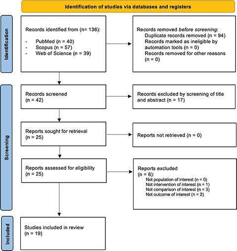
Figure 1. PRISMA 2020 Flow Diagram.
Correlation between Grave and Brown and Demirjian methods
Six papers [23, 26, 27, 31, 33, 39] compared Demirjian method with HWM Grave and Brown method. Only Camacho-Basallo et al. [23] showed no significant correlations (P > 0.05) between the two methods, whereas five studies [26, 27, 31, 33, 39] demonstrated significant correlations for all teeth (P < 0.01), and Lopes et al. [31] showed significant correlations for second molar and first premolar (P < 0.05) in females, and for all teeth except first premolars (P > 0.05) in males. More in detail, the second molar showed the strongest coefficient of correlation in these studies, with values ranging from 0.760 to 0.829 in male and from 0.790 to 0.880 in females.
Correlation between Fishman and Demirjian methods
A total of 14 papers [22–25, 28–30, 32, 34–38, 40] compared Demirjian method with HWM Fishman method.
Of them, 12 studies [22–25, 28, 30, 32, 34, 35, 37, 38, 40] reported statistically significant correlations (P < 0.05) between Demirjian method and HWM Fishman method, and only two studies [29, 36] showed that correlations were not significant.
The studies showed differences in terms of highest correlation (expressed as CC) with teeth involved: Krailassiri et al. [28] reported second premolars in both sexes (0.69 in female; 0.66 in male); Lecca-Morales et al. [30] reported second molars in both sexes (0.792 in female; 0.800 in male); both Ojha et al. [35] and Yadav et al. [40] reported second molars in female (0.882 and 0.845, respectively) and canines in male (0.835 and 0.755, respectively); Jeong. et al. [25] reported strongest correlations for the first premolars (0.780 among boys; 0.812 among girls); Sahin Sağlam et al. [38] reported second molars in both female (0.647) and male (0.550).
Reboucas et al. [37] stated that highest correlations were reported for mandibular canine (0.712) and second molar (0.735). Bagherpour et al. [22] showed a significant correlation only for left (P = 0.006) and right mandibular canine (P = 0.014) only in males, although they evaluated only correlations for mandibular canines and second molars. Camacho-Basallo et al. [23] reported significant correlations (P < 0.001) between the two methods with the exception of the first molars that showed a low significant correlation in female (CC = 0.263; P < 0.05) and not significant correlation in male (CC = 0.187; P > 0.05). Magat et al. [32] evaluated only third molars and found only a moderate correlation between the dental developmental stages of third mandibular molars and the start of PGS (P < 0.05) in both males and females.
Evidence of quality and risk of bias
In the present systematic review, the risk of bias of the included studies was analysed using the JBI Critical Appraisal Checklist for analytical cross-sectional studies [21].
Twelve papers were high-quality studies and seven papers medium-quality studies. Further details about the quality assessment of each study included in this systematic review are shown in Table 3.
Discussion
The main finding of this systematic review was that dental calcification according to Demirjian method might be considered reliable to assess SM in growing subjects.
Specifically, among the 19 studies included in the present systematic review, 12 studies [22–25, 28, 30, 32, 34, 35, 37, 38, 40] reported statistically significant correlations between Demirjian method and HWM Fishman method and 5 studies [26, 27, 31, 33, 39] demonstrated significant correlations between Demirjian method and HWM Grave and Brown method. Only 2 studies [29, 36] showed that correlations were not significant.
Mandibular canine and skeletal maturation
In terms of correlation between the dental developmental stages of mandibular canines and skeletal maturation, the results could suggest that the maturation of the mandibular canine may be related to the onset of the pubertal growth peak in male subjects.
Indeed, Lopes et al. and Bagherpour et al. [22] showed that the mandibular canine was not statistically significant as predictors of the maturation stages for girls (P > 0.05), whereas they showed a significant correlation between the start of PGS and the developmental stages of the mandibular canine in male subjects (P < 0.05). Accordingly, Ojha et al. [35] and Yadav et al. [40] showed that all correlations between Demirjian method and HWM Fishman method were statistically significant (P < 0.05), with a CC ranging from 0.676 to 0.845. Moreover, Ojha et al. [35] concluded that canines reported the highest correlation in male subjects (CC = 0.835) and Yadav et al. [40] concluded the same, reporting that the canines showed the highest correlation in in males (CC = 0.755).
Uysal et al. [39] found that 80% of male subjects in MP3cap stage according to Grave and Brown methos presented the mandibular canine in calcification stage H, and the 20% in stage G. No canines in stages D, E, and F were found in MP3cap stage.
Using the same HWM SM method, Motghare et al. [33] reported that 100% of male subjects had mandibular canine in stage H and no canines in stages D, E, F, and G were found in MP3cap stage.
Accordingly, Krailassiri et al. [28] found that 77.8% of subjects in MP3cap stage according to Fishman presented the mandibular canine in calcification stage H, and the 14.8% in stage G. No canines in stages D and E were found in MP3cap stage.
Rebouças et al. [37] reported that during the onset of the growth spurt, 80.9% of the subjects had the canine in stage F and G and during the peak, 92.9% had the canines in stage G and H.
The same findings were previously reported by Chertkow [41], Chertkow and Fatti [42] and Coutinho et al. [43]. The authors showed a high correlation between mandibular canine calcification and SM indicators, and stated that the skeletal maturation phase can be determined by the mandibular canine mineralisation phase.
Thus, the results of this systematic review suggest that peak growth occurs no earlier than the mandibular canine maturation stage F in males.
Mandibular second molar and skeletal maturation
Analysing the correlation between mandibular second molar developmental stages and skeletal maturation, Lopes et al. [31] showed that the second molar was statistically significant as predictors of the maturation stages (P < 0.05). Specifically, the second molar mineralisation stage was reported to have 4.34 times (in females) and 6.8 times (in males) more chance to predict the skeletal maturation than the other teeth. Also, Koçak et al. [27], Lecca-Morales et al. [30], Mothgare et al. [33], Uysal et al. [39], and Yadav et al. [40] showed that the second molar reached the highest correlation coefficient with HWM evaluated using Grave and Brown method (0.826 to 0.877 in female; 0.706 to 0.790 in male).
Ojha et al. [35] showed that all correlations between Demirjian method and HWM Fishman method were statistically significant (P < 0.05), with a CC ranging from 0.676 to 0.835 for males and from 0.812 to 0.882 for females. Furthermore, the authors concluded that second molars reported the highest correlation in female (CC = 0.882).
Analysing the DM stages, Lopes et al. [31] demonstrated that the second molars mineralisation stages F and G appeared mostly in females and the mineralisation stage G in males at the peak. Accordingly, Rebouças et al. [37] reported that the majority of second molars were identified in stages F or G in females and stages G or H among the boys.
Furthermore, Koçak et al. [27] showed that the second molar stage G in the boys and stages G–H in the girls showed the highest percentage distribution at the peak. Camacho-Basallo et al. [23] showed that Fishman’s method was significantly associated with all stages of maturation in boys and girls, and reported that mandibular second molar stages F-G appeared in 66.3% of both males and females.
Thus, the results of this systematic review suggest that peak growth occurs no earlier than the mandibular second molar maturation stage F in females and stage G in males.
These results are in line with an interesting study by Oyonarte et al. [13] The authors conducted a longitudinal study analysing one hand and wrist radiograph and one 45° oblique radiograph from each side, in 60 subjects at age 6, 9, 12, 14, 16, 18, and 20 years. Moreover, they collected annual records of height and weight, and identified the onset of the pubertal growth peak. The authors showed high levels of correlation between Fishman and Demirjian methods and pubertal growth peak. Specifically, the stage F for females and stage G for males were identified as cutoff points between pre-pubertal and post-pubertal ages.
Mandibular premolars and skeletal maturation
In terms of correlation between the dental developmental stages of mandibular premolars and skeletal maturation, Jeong et al. [25] found that the strongest correlations were found for the first premolars (r = 0.780 among males; 0.812 among females). On the other hand, Jourieh et al. [26] reported that the first premolars showed the lowest correlation (P < 0.001) in both genders.
Günen Yılmaz et al. [24] stated that the PGS was best assessed through the dental evaluation of second premolar, and accordingly, Krailassiri et al. [28] indicated that the second premolar showed the highest correlation (0.69 in female; 0.66 in male).
Mandibular third molar and skeletal maturation
Analysing the correlation between third mandibular molar developmental stages and skeletal maturation, Magat et al. [32] found a moderate correlation (P < 0.05) in both males and females. Krailassiri et al. [28] showed that all the correlations between Demirjian method and HWM were statistically significant (P < 0.01), but the third molars showed the lowest correlation (0.31 in female; 0.47 in male). Similarly, Bagherpour et al. [22] and Uysal et al. [39] showed a significant correlation between the start of PGS and the developmental stages mandibular third molar, but this tooth showed the lowest correlation compared to the other teeth.
Thus, the results showed a poor association between the third molar calcification and the SM, as previously reported [41, 44]. This great variation in tooth development may adversely affect the identification of the relationship between dental and skeletal development.
This systematic review had some limitations that should be taken into consideration. The inclusion of only cross-sectional studies hinders the assessment of growth on longitudinal analysis. Moreover, the heterogeneity of the teeth assessed in the different included studies could have affected the results.
Conclusions
Taken together, the findings of this systematic review showed the reliability of Demirjian dental calcification method compared to HWM Fishman and Grave and Brown in growing subjects to assess SM.
Moreover, the data analysis of the included studies could suggest that the mandibular second molar might be considered as the best indicator compared to other teeth and that the peak of growth occurs no earlier than stage F in females and stage G in males. Also, the mandibular canine might be analysed as indicator of SM in males, and the results suggest that the peak of growth occurs no earlier than maturation stage F in male subjects.
Further studies are needed to confirm these findings in order to improve the knowledge on the correlation between dental and skeletal development also using different methods that could be useful of dentists and physicians involved in the management of growing subjects.
Acknowledgements
None.
Informed consent
Not applicable.
ORCID
Martina Ferrillo https://orcid.org/0000-0003-1878-4694
References
[1] Baccetti T, Franchi L, McNamara JA. The cervical vertebral maturation (CVM) method for the assessment of optimal treatment timing in dentofacial orthopedics. Semin Orthod. 2005;11:119–29.
[2] Lewis AB, Roche AF, Wagner B. Pubertal spurts in cranial base and mandible. Comparisons within individuals. Angle Orthod. 1985;55(1):17–30.
[3] Nanda RS: The rates of growth of several facial components measured from serial cephalometric roentgenograms. Am J Orthod. 1955;41:658–73.
[4] Mappes MS, Harris EF, Behrents RG. An example of regional variation in the tempos of tooth mineralization and hand-wrist ossification. Am J Orthod Dentofacial Orthop. 1992;101:145–51.
[5] Grave KC, Brown T. Skeletal ossification and the adolescent growth spurt. Am J Orthod. 1976;69:611–9.
[6] Fishman LS. Radiographic evaluation of skeletal maturation; a clinically oriented method based on hand wrist films. Angle Orthod. 1982;52:88–112.
[7] Demirjian A, Goldstein H, Tanner JM. A new system of dental age assessment. Hum Biol. 1973;45:211–27.
[8] Lewis AB, Garn SM: The relationship between tooth formation and other maturation factors. Angle Orthod. 1960;30:70–7.
[9] Hägg U, Taranger J. Menarche and voice change as indicators of the pubertal growth spurt. Acta Odontol Scand. 1980;38(3):179–86.
[10] Franchi L, Baccetti T, McNamara Jr, JA. Mandibular growth as related to cervical vertebral maturation and body height. Am J Orthod Dentofacial Orthop. 2000;118(3):335–40.
[11] Jeon JY, Kim CS, Kim JS, Choi SH. Correlation and correspondence between skeletal maturation indicators in hand-wrist and cervical vertebra analyses and skeletal maturity score in korean adolescents. Children (Basel). 2021;8(10):910.
[12] Ferrillo M, Curci C, Roccuzzo A, Migliario M, Invernizzi M, de Sire A. Reliability of cervical vertebral maturation compared to hand-wrist for skeletal maturation assessment in growing subjects: A systematic review. J Back Musculoskelet Rehabil. 2021;34(6):925–36.
[13] Oyonarte R, Sánchez-Ugarte F, Montt J, et al. Diagnostic assessment of tooth maturation of the mandibular second molars as a skeletal maturation indicator: A retrospective longitudinal study. Am J Orthod Dentofacial Orthop. 2020;158:383–90.
[14] Bittencourt MV, Cericato G, Franco A, et al. Accuracy of dental development for estimating the pubertal growth spurt in comparison to skeletal development: a systematic review and meta-analysis. Dentomaxillofac Radiol. 2018;47:20170362.
[15] Kumar S, Singla A, Sharma R, Virdi MS, Anupam A, Mittal B. Skeletal maturation evaluation using mandibular second molar calcification stages. Angle Orthod. 2012;82(3):501–6.
[16] Chen J, Hu H, Guo J, et al. Correlation between dental maturation and cervical vertebral maturation. Oral Surg Oral Med Oral Pathol Oral Radiol Endod. 2010;110:777–83.
[17] Barreto BCT, Marañón-Vásquez GA, da Costa Barreto LS, Masterson D, de Souza MMG, Maia LC. Is there a correlation between dental and cervical vertebrae maturation stages in growing subjects? A systematic review with meta-analysis. Clin Oral Investig. 2022;26(5):3823–42.
[18] Poulsen AR, Sonnesen L. Association between dental and skeletal maturation in Scandinavian children born between 2005 and 2010. Acta Odontol Scand. 2023;81(6):464–72.
[19] Ercan DE, Yüksel S. Skeletal, dental, and sexual maturation as an indicator of pubertal growth spurt. Am J Hum Biol. 2023;35(12):e23957.
[20] Moher D, Altman DG, Liberati A, Tetzlaff J. PRISMA statement. Epidemiology. 2011;22(1):128.
[21] Institute, TJB. JBI Critical Appraisal Tools. [cited 2024 Jan 5]. Available from: https://jbi.global/critical-appraisal-tools
[22] Bagherpour A, Pousti M, Adelianfar E. Hand skeletal maturity and its correlation with mandibular dental development. J Clin Exp Dent. 2014;6(3):e275–9.
[23] Camacho-Basallo P, Yáñez-Vico RM, Solano-Reina E, Iglesias-Linares A. Five radiographic methods for assessing skeletal maturity in a Spanish population: is there a correlation? Acta Odontol Scand. 2017;75:106–12.
[24] Günen Yılmaz S, Harorlı A, Kılıç M, Bayrakdar İŞ. Evaluation of the relationship between the Demirjian and Nolla methods and the pubertal growth spurt stage predicted by skeletal maturation indicators in Turkish children aged 10–15: investigation study. Acta Odontol Scand. 2019;77:107–13.
[25] Jeong MJ, Lee KE, Chae YK, Nam OH, Lee HS, Choi SC. Correlations between skeletal maturity and dental calcification stages in Korean children. Eur J Paediatr Dent. 2022;23(2):101–5.
[26] Jourieh A, Khan H, Mheissen S, Assali M, Alam MK. The Correlation between Dental Stages and Skeletal Maturity Stages. Biomed Res Int. 2021;2021:9986498.
[27] Koçak T, Akan B. Assessment of maturation indicators in individuals with different skeletal malocclusion. J Orofac Orthop. 2021;82(3):187–97.
[28] Krailassiri S, Anuwongnukroh N, Dechkunakorn S. Relationships between dental calcification stages and skeletal maturity indicators in Thai individuals. Angle Orthod. 2002;72:155–66.
[29] Kumari S, Sahu AK, Rajguru J, Bishnoi P, Garg AJ, Thakur R. Age Estimation by Dental Calcification Stages and Hand-Wrist Radiograph. Cureus. 2022;14(9):e29045.
[30] Lecca-Morales RM, Carruitero MJ. Relationship between dental calcification and skeletal maturation in a Peruvian sample. Dental Press J Orthod. 2017;22:89–96.
[31] Lopes LJ, de Oliveira Gamba T, Visconti MA, Ambrosano GM, Haiter-Neto F, Freitas DQ. Utility of panoramic radiography for identification of the pubertal growth period. Am J Orthod Dentofacial Orthop. 2016;149(4):509–15.
[32] Magat G, Ozcan S. Assessment of maturation stages and the accuracy of age estimation methods in a Turkish population: A comparative study. Imaging Sci Dent. 2022;52(1):83–91.
[33] Motghare PC, Bedia AS, Degwekar SS, Indurkar AD, Bedia S. Correlation of calcification of permanent mandibular canine, mandibular premolars, and permanent mandibular first and second molars with skeletal maturity in Indian population. J Forensic Dent Sci. 2016;8:67–73.
[34] Mustafa S, Raj AC, Anekar J, Divakar DD, Al Kheraif AA, Ramakrishnaiah R, Khan AA, Alshahrani OA, Rai NP. Evaluation of dental and skeletal maturity using digital panoramic radiographs and digital cephalograms. Asian Biomed. 2015;9:335–42.
[35] Ojha A, Prasanth MA, Singh V, Sihag T, Bhati V, Tomar H. Assessment of correlation between dental calcification stages and skeletal maturity indicators. J Forensic Dent Sci. 2018;10(3):132–6.
[36] Ojha A, Chawla R, Sihag T, Ahmed A, Qurishi AA, Rajkumari L. Relationship between skeletal maturity indicators and dental calcification stages in a sample pediatric population. Indian J Dent Res. 2023;34(2):150–4.
[37] Rebouças PRM, Alencar CRB, Arruda MJALLA, Lacerda RHW, Melo DP, Bernardino ÍM, Bento PM. Identification of dental calcification stages as a predictor of skeletal development phase. Dental Press J Orthod. 2021;26(4):e2119292.
[38] Sahin Sağlam AM, Gazilerli U. The relationship between dental and skeletal maturity. J Orofac Orthop. 2002 Nov;63(6):454–62.
[39] Uysal T, Sari Z, Ramoglu SI, Basciftci FA. Relationships between dental and skeletal maturity in Turkish subjects. Angle Orthod. 2004;74:657–64.
[40] Yadav V, Loomba A, Autar R. A comparative evaluation of dental calcification stages and skeletal maturity indicators in North-Indian children. Natl J Maxillofac Surg. 2017;8:26–33.
[41] Chertkow S. Tooth mineralization as an indicator of the pubertal growth spurt. Am J Orthod. 1980;77:79–91.
[42] Chertkow S, Fatti P. The relationship between tooth mineralization and early evidence of the ulnar sesamoid. Angle Orthod. 1979;49:282–8.
[43] Coutinho S, Buschang PH, Miranda F. Relationship between mandibular canine calcification stages and skeletal maturity. Am J Orthod. 1993;104:262–8.
[44] Garn SM, Lewis AB, Bonne B. Third molar formation and its developmental course. Angle Orthod. 1962;44:270–6.