RESEARCH ARTICLE
Assessing position changes of impacted third molars in treatment of class II malocclusion with premolars extraction
Hatice Gokalpa and Murat Kaan Erdemb
aDepartment of Orthodontics, Faculty of Dentistry, Ankara University, Ankara, Turkey; bDepartment of Oral and Maxillofacial Surgery, Faculty of Dentistry, Lokman Hekim University, Ankara, Turkey
ABSTRACT
Introduction: Third molars (M3) remaining impacted in Class II malocclusion characterised with sagittal mandibular deficiency is a high probability. The null hypothesis of this study is that mesioangular M3s changes position through the eruption way in Class II malocclusion treatment requiring moderate anchorage with four first premolars extraction. The aim of this study is to reconsider the decision to surgically extract impacted third molars in four premolars extraction treatment of Class II malocclusion.
Material and Methods: The materials consisted of the pre-treatment and post-treatment lateral cephalograms and orthopantomographs of 30 individuals with skeletal and dental Class II malocclusion with a mean chronological age of 13.48 years, who were treated by the same clinician (H.G.) with four first premolar extractions via the straight wire technique at the Ankara University Faculty of Dentistry Department of Orthodontics, Ankara, Turkey. The sagittal position of the upper and lower incisors and molars, M3 position and M3 space were evaluated with the paired-t test; the relationship between the sagittal position of the upper and lower incisors and molars and the change in M3 position were evaluated with correlation analysis.
Results: The study found the retroclination and mesial movement of the upper incisors and molars, and an increase in the M3 space by the fixed orthodontic treatment. An insignificant steepening of both the upper right M3 position and the lower right M3 position was found. A statistically significant increase in the lower right and left side M3 spaces was found. Positive correlations between lower right M3 angulation and the sagittal position of the lower incisors and first molars were found.
Conclusion: Improvement in the mesioangulation of the M3s and an increase in the M3 space were achieved in this study. Based on the findings, it is useful to review the decision for prophylactic surgical extraction of the M3s before orthodontic treatment in such cases, taking into account the risks of postoperative complications.
KEYWORDS: Class II malocclusion; moderate anchorage; orthodontic therapy with extraction; third molar impaction
Citation: ACTA ODONTOLOGICA SCANDINAVICA 2024; VOL. 83: 582–587. DOI: https://doi.org/10.2340/aos.v83.42076.
Copyright: © 2024 The Author(s). Published by MJS Publishing on behalf of Acta Odontologica Scandinavica Society. This is an Open Access article distributed under the terms of the Creative Commons Attribution 4.0 International License (http://creativecommons.org/licenses/by/4.0/), allowing third parties to copy and redistribute the material in any medium or format and to remix, transform, and build upon the material, with the condition of proper attribution to the original work.
Received: 17 August 2024; Accepted: 23 September 2024; Published: 9 October 2024.
CONTACT Murat Kaan Erdem dtmuratkaanerdem@gmail.com Dentistry, Department of Oral and Maxillofacial Surgery, Lokman Hekim University, Sogutozu Mahallesi, Cankaya, Ankara, Turkey
Competing interests and funding: The authors declare no conflict of interest or financial interest.
No funding was obtained for this study.
Introduction
Tooth impaction is a common occurrence, affecting 0.8% – 3.6% of the general population, mainly because of space constraints in the dental arches or other conditions that hinder eruption. These are influenced by systemic, local, genetic, and racial factors [1, 2]. The teeth most frequently impacted include maxillary and mandibular third molars, maxillary canines, mandibular premolars, and maxillary central incisors [3, 4].
Mandibular third molar (M3) impaction was initially attributed to inadequate space between the second molars (M2) and the ascending ramus. Later studies identified additional contributing factors, such as mandibular growth deficiency, vertical condylar growth, and the backward eruption path of the dentition [5, 6].
In recent times, changes in eating habits that affect chewing patterns, coupled with insufficient jaw lengthening, have resulted in an increased prevalence of M3 impaction, ranging from 16.7% to 68.6% [7]. Fossil records indicate a reduction in both the number and size of individual teeth and jaw size throughout evolution. Initially, primates had more teeth, but over time, the third premolars and fourth molars disappeared in mammals. Presently, it is not uncommon for M3s, second premolars, and lateral incisors to sometimes fail to form [8].
Orthodontic treatment often requires the extraction of first premolars to achieve desired treatment outcomes. However, in treatments characterised by a forward mandibular growth pattern, the length of the dental arch may decrease, leading to the impaction of M3s because of factors such as ramus anterior remodelling, late mandibular growth spurt, and retrusion of the lower incisors [1, 6, 8–12]. In orthodontic treatment plans, regardless of whether extractions are involved, the common approach is to prophylactically extract asymptomatic M3s, independent of craniofacial growth characteristics. However, the margin of error in estimating the risk of impaction is 40% [13, 14]. This substantial uncertainty is particularly concerning given that surgically extracting asymptomatic M3s in adolescents can lead to long-lasting neurological and psychological complications [15, 16]. Therefore, it is crucial to evaluate the prognosis of M3 eruption alongside anchorage requirements, craniofacial growth patterns, M3 angulation, and M3 spacing to minimise surgical complications.
In cases of Class II malocclusion requiring maximum anchorage, distal molar movement may increase the risk of M3 impaction because of the influence of craniofacial growth direction on the remodelling of the ascending ramus [6].
Numerous studies on M3 impaction in the treatment of Class I, II, and III malocclusions, with or without extraction and during or after the growth period, are retrospective. These studies often fail to prioritise the relationship between anchorage requirements and craniofacial growth characteristics in M3 impaction. However, the craniofacial growth pattern significantly influences tooth alignment within the alveolus.
The null hypothesis of this study is that mesioangular M3s undergo positional changes during the eruption process in the course of treating Class II malocclusion, particularly in cases that require moderate anchorage and involve the extraction of all four first premolars. The primary aim of this study is to critically reevaluate the decision-making process regarding the surgical extraction of impacted third molars within the framework of Class II malocclusion treatments that include the extraction of four premolars.
Materials and methods
Sample design
This study was conducted on the lateral cephalograms and orthopantomograms (OPG) of 30 patients with skeletal and dental Class II malocclusion requiring moderate anchorage. These patients were treated by the same orthodontist (H.G.) using the straight wire technique with four premolar extractions at the Ankara University Faculty of Dentistry, Department of Orthodontics. All premolars were extracted by the same surgeon (M.K.E.). A power analysis determined that a sample size of 30 was adequate. All radiographs were taken by the same technician using a Planmeca ProMax Device, set at 66 kV and 9 mA, with the patients’ mouths closed. The mean treatment duration was 2.7 years (range: 1.90–4.50 years).
The average chronological age of the individuals at the beginning of treatment was 13.48 years (range: 11.80–19.30 years). Inclusion criteria were:
- Maxillary and mandibular arch length discrepancy with moderate anchorage requirements: −7.45 mm and −5.19 mm, respectively; Class II molar and canine relationship; overjet: 4.90 mm (range: 1.00–15.00 mm); overbite: 2.15 mm (range: −4.0 to 5.0 mm).
- SNA, 85º; SNB, 79º; ANB, 6º.
- GoGn-SN, 35.4º.
- All third molars present and in a mesioangular position on OPG, according to Archer’s and Winter’s classifications ([17, 18]; Figures 1 and 2).
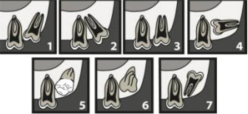
Figure 1. Archer’s classification of upper third molars according to their inclination to the long axis of the upper second molar. (1) mesioangular, (2) distoangular, (3) vertical, (4) horizontal, (5) buccoangular, (6) linguoangular, (7) inverted.
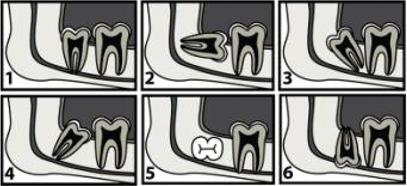
Figure 2. Winter’s classification (19): Third molars are classified according to their inclination to the long axis of the second molar. (1) vertical angulation, (2) horizontal angulation, (3) distoangular angulation, (4) mesioangular angulation, (5) transversal angulation, (6) inverse angulation.
At the end of orthodontic treatment, occlusion was achieved in accordance with Andrews’ normal occlusion criteria [19]. On OPG, all M3s were present, and at least one-third of root formation was completed. Temporary intraoral anchorage systems or extraoral anchorage applications were not used during fixed orthodontic treatment. Changes in M3 space and angulation were evaluated on lateral cephalograms and OPGs at the end of the treatment (Figures 3 and 4).
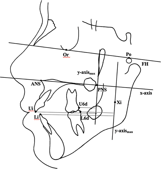
Figure 3. Points and reference lines for measurement of Lateral cephalograms. Points. 1. ANS, anterior nasal spine; 2. PNS, posterior nasal spine; 3. Ricketts Xi point 4. Ui, upper incisor edge, 5. U6d, upper first molar distal edge, 6. Li, lower incisor edge, 7. L6d, lower first molar distal edge. Reference Lines: 1. FH, Frankfort horizontal line; 2. X-axis is made between the ANS and PNS points. 3. Y-axis for maxilla is perpendicular line from PNS point to x-axis. 4. Y-axis for mandible is perpendicular line from Xi point to x-axis. Measurements: 1. Ui- y-axismax,2. U6d- y-axismax, 3. L6d- y-axismax 4. L6d- y-axisman. 5. M3 space for maxilla: distance between U6d and y-axismax. 6. Distance between L6d and y-axisman.
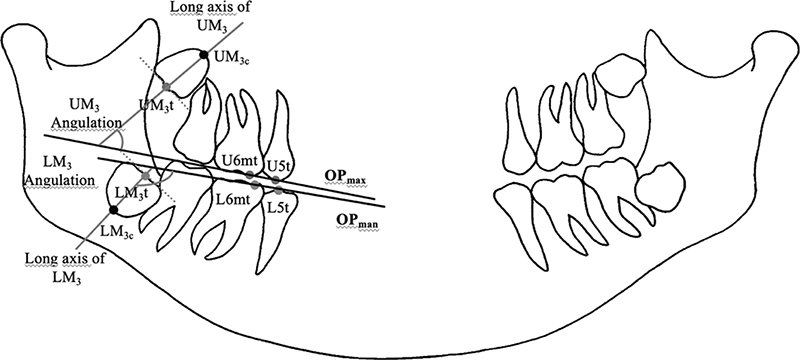
Figure 4. Points and reference lines for measurement of OPG. Points: 1. UM3t, Upper third molar occlusal surface midpoint 2. U6mt, Upper first molar mesial tubercule top 3. UM3c Upper first molar midpoint of apex. 4. U5t, upper second premolar tubercule top, 5. LM3t, lower third molar occlusal surface midpoint 6. L6mt, lower first molar mesial tubercule top 8. LM3c lower first molar midpoint of apex. 9. L5t, lower second premolar tubercule top. Reference Lines: 1. OPmax,maxillary occlusal plan which is constracted between U5t and U6mt. 2. OPman, mandibular occlusal plan which constracted between L5t and L6mt, 3. Long axis of UM3, line between UM3c and UM3t. 4. Long axis of LM3, line between LM3c and LM3t. Measurements: 1. UM3 angulation, angle between OP max and long axis of UM3. 2.LM3 angulation, angle between OP man and long axis of LM3.
Study design
To evaluate sagittal changes in the positions of incisors and molars and M3 spaces in both dental arches, a Cartesian coordinate system was used. The X-axis was created between the anterior nasal spine (ANS) and posterior nasal spine (PNS) points, and was used for both upper and lower dental arches. The Y-axis for the maxilla was constructed from the PNS point to the X-axis. Sagittal changes in the positions of the upper incisors, molars, and M3 spaces were measured relative to the Y-axis. The Y-axis for the mandible was constructed from the point where the anterior border of the ascending ramus joins the end of the corpus mandible to the X-axis. Sagittal changes in the positions of the lower incisors, molars, and M3 spaces were measured relative to the Y-axis. M3 spaces for upper and lower dental arches were measured as the distances between the U6d and Y-axis (maxilla), and L6d and Y-axis (mandible). Reference landmarks and lines for measurement on lateral cephalograms are presented in Figure 3. Positional changes of the M3s were evaluated on OPG. Points and reference lines used on OPG are shown in Figure 4.
Statistical method
SPSS (Statistical Package for the Social Sciences) 26 was used for data analysis. The intraclass correlation coefficient was used to measure reliability. Because of the limited data for comparing the beginning and end of the treatment using lateral cephalogram and OPG measurements (N = 30), the nonparametric Wilcoxon signed-rank test was used as an alternative to the dependent group t-test for comparing two different measurements within a single group [20]. The nonparametric Brown correlation method was used instead of the Pearson correlation method to assess measurement differences between the beginning (T0) and end (T1) of treatment. The correlation coefficient (r) was considered low if below 0.40, medium if between 0.40 and 0.70, and high if equal to or greater than 0.70 [21]. A significance level of p < 0.05 was used for statistical analyses.
Measurements were conducted twice with a 20-day interval to determine the repeatability of landmark identification and measurement techniques. All angular and linear variables exhibited a coefficient of intra-rater reliability between 0.82 and 1.00, indicating negligible variation.
Sagittal position changes of the incisors and molars, M3 spaces, and mesioangular M3s were analysed using a paired-t test at T1. The relationship between changes in the positions of incisors/first molars and changes in M3 positions and M3 spaces were tested using correlation analysis.
Results
At the end of orthodontic treatment, a statistically significant retraction of the upper incisors and mesialization of the molars was observed (p < 0.01), along with a significant increase in the upper M3 space (p < 0.01, Table 1). Although there was no change in the lower incisor position, a statistically significant mesialization of the lower molars and an increase in the lower M3 space were found (p < 0.01, Table 1).
| n = 30 | Before Treatment (T0) | After Treatment (T1) | p |
| X ± Sx | X ± Sx | ||
| U1 position (mm) | 51.52 ± 4.22 | 48.68 ± 4.17 | ** |
| U6 position (mm) | 22.42 ± 4.12 | 24.95 ± 3.42 | ** |
| M3 max space (mm) | 10.13 ± 3.18 | 12.65 ± 3. 55 | ** |
| L1 position (mm) | 50.68 ± 3.47 | 50.25 ± 4.61 | Ns |
| L6 position (mm) | 27.37 ± 3.15 | 30.23 ± 3.79 | ** |
| M3 man space (mm) | 13.20 ± 3.17 | 16.06 ± 6. 53 | ** |
| Significance level: Ns: Not significant; **p < 0.01. | |||
A statistically significant decrease in the right lower M3 angulation was detected at the end of the treatment (p < 0.05, Table 2), while no significant changes were observed in the positions of other M3s.
| Before Treatment (T0) | After treatment (T1) | p | |
| x ± S× | x ± S× | ||
| Long axis of URM3/OPmax | 63.04 ± 20.49 | 65.54 ± 11.42 | Ns |
| Long axis of LRM3/OPman | 146.72 ± 18.32 | 141.10 ± 21.14 | * |
| Long axis of ULM3/OPmax | 61.52 ± 18.31 | 64.48 ± 15.10 | Ns |
| Long axis of LLM3/OPman | 134.88 ± 19.05 | 131.52 ± 21.11 | Ns |
| Significance level: Ns: Not significant; *p < 0.05, **p < 0.01. | |||
A statistically significant positive correlation was found between treatment and changes in the positions of U6 and L6 (p < 0.05, Table 3). Additionally, a statistically significant positive correlation was found between the lower right M3 position and the positions of the lower incisors and lower first molars (p < 0.01, Table 3).
| U6 | L1 | L6 | |
| L6 | 0.417* | Ns | Ns |
| Long axis of LRM3/OPman | Ns | 0.480** | 0.484** |
| Significance level: *r0.05 = 0.374, **r0.01 = 0.479. | |||
Discussion
This study examined the changes in the required space for the eruption of mesioangular M3s and their angulation during fixed orthodontic treatment involving four premolar extractions and moderate anchorage requirements. While many studies have addressed this topic, controversies remain regarding craniofacial growth patterns, impaction detection methods, and orthodontic treatment planning. This study found that fixed orthodontic treatment with four premolar extractions, requiring moderate anchorage, led to upper incisor retraction, molar mesialization, an increase in the space necessary for M3 eruption in both the upper and lower dental arches, and an improvement in the mesioangular M3 position.
Anchorage requirements play a critical role in tooth movement during orthodontic treatments involving extractions. Different anchorage systems yield varying results in tooth positioning. In this study, patients were treated only with fixed orthodontic treatment, without the use of intraoral or extraoral anchorage systems. Increased anchorage requirements in the orthodontic treatment of Class II malocclusion, necessary for retracting the upper incisors and molars, may contribute to M3 impaction.
Orthopantomograms were utilised to assess M3 angulation in this study. Although cone-beam computed tomography (CBCT) has gained popularity, OPGs remain a standard practice because of their routine use and lower radiation doses in orthodontics [22, 23].
Various factors influence the space available for M3 eruption. Richardson noted that mesial molar movement can partially increase the space for M3 eruption [1]. Brash and Scott observed that anterior dentition movement contributes to creating space for M3 eruption [24, 25]. Premolar extraction has been found to increase the space required for M3 eruption [26]. Ricketts reported that in premolar extraction treatment, the space required for mandibular M3 eruption increases by 25%, necessitating early M3 prognosis evaluation [27]. In non-extraction orthodontic treatment, 45% of M3s must be extracted, compared to 15% – 20% in treatments involving first premolar extractions.
This study found that orthodontic treatment with premolar extractions positively influenced the mesioangular position of M3s. Moderate anchorage needs and skeletal growth were important factors. Conversely, some studies suggest that growth has little effect on changes in M3 angulation, and orthodontic treatment at the end of the growth period may not significantly affect M3 angulation [28, 29]. Although literature indicates that extraction-inclusive treatments positively impact the necessary space for M3 eruption, particularly in the lower jaw, factors such as growth model, treatment technique, and anchorage needs have not been fully considered [1, 9, 10, 30].
Orthodontic treatment with premolar extraction, requiring moderate anchorage, contributed positively to increasing the distance between the ascending ramus and the distal surface of the M2s, facilitating the eruption of M3s in a forward skeletal growth pattern. Based on these results, the decision for surgical extraction should be carefully evaluated, considering the potential risk of psychological trauma associated with the surgical extraction of third molars during adolescence, either before or after orthodontic treatment.
Declarations
Ethical Approval: Ethical approval for this study was obtained from the Ankara University Faculty of Dentistry Clinical Research Ethics Committee and in accordance with the 1964 Declaration of Helsinki and its subsequent changes or similar ethical standards.
Patient Consent: Written informed consent was obtained from all individual participants included in the study.
All authors have viewed and agreed to the submission.
Author Contributions
Conceptualisation: H.G. and M.K.E. Methodology: H.G. and M.K.E. Software: H.G. and M.K.E. Validation: H.G. and M.K.E. Formal analysis: H.G. and M.K.E. Investigation: H.G. and M.K.E. Resources: H.G. Data curation: H.G. and M.K.E. Writing – Original draft: H.G. and M.K.E. Writing – Review & Editing: H.G. and M.K.E. Visualisation: H.G. and M.K.E. Supervision: H.G. Project administration: H.G. and M.K.E. Funding acquisition: No funding
References
[1] Richardson ME. Lower third molar space. Angle Orthod. 1987;57(2):155–61.
[2] Becker A. Orthodontic treatment of impacted teeth. 3rd ed. Wiley-Blackwell; West Sussex, UK, 2012.
[3] Dachi SF, Howell FV. A survey of 3874 routine full-mouth radiographs. II. A study of impacted teeth. Oral Surg Oral Med Oral Pathol. 1961;14:1165–69.
[4] Grover PS, Lorton L. The incidence of unerupted permanent teeth and related clinical cases. Oral Surg Oral Med Oral Pathol. 1985;59:420–5.
[5] Henry CB, Morant GM. A preliminary study of the eruption of the mandibular third molar tooth in man based on measurements obtained from radiographs, with special reference to the problem of predicting cases of ultimate impaction of the tooth. Biometrika. 1936;28:378.
[6] Björk A, Jensen E, Palling M. Mandibular growth and third molar impaction. Acta Odontol Scand. 1956;14:231–72. http://dx.doi.org/10.3109/00016355609019762
[7] Reddy KVG. Distribution of third molar impactions among rural and urban dwellers in the age group of 22–30 years in South India: a comparative study. J Maxillofac Oral Surg. 2012;11(3):271–5.
[8] Proffit WR. Chapter 4, later stages of development. In: Contemporary orthodontics. St. Louis: Mosby-Year Book; 1992. p. 103.
[9] Elsey MJ, Rock WP. Influence of orthodontic treatment on development of third molars. Br J Oral Maxillofac Surg. 2000;38(4):350–3.
[10] Faubion B. The effect of extraction of premolars on the eruption of mandibular third molars. J Am Dent Assoc. 1968;76:316–20.
[11] Richardson ME. The relative effects of the extraction of various teeth on the development of mandibular third molars. Trans Europ Orthod Soc. 1976;6:79–85.
[12] Graber TM, Kaineg TF. The mandibular third molar-its predictive status and role in lower incisor crowding. Proc Finn Dent Soc. 1981;77:37–44.
[13] Bastos Ado C, de Oliveira JB, Mello KF, Le ã PB, Artese F, Normando D. The ability of orthodontists and oral/maxillofacial surgeons to predict eruption of lower third molar. Prog Orthod. 2016;17(1):21. https://doi.org/10.1186/s40510-016-0134-0
[14] Libdy MR, Rabello NM,Marques LS, Normando D. The ability of orthodontists and maxillofacial surgeons in predicting spontaneous eruption of mandibular third molar using panoramic serial radiographs. Dental Press J Orthod. 2020;25 (4):68–74. https://doi.org/10.1590/2177-6709.25.4.068-074.oar
[15] Anjrini AA, Kruger E, Tennant M. International bench marking of hospitalisations for impacted teeth: a 10-year retrospective study from the United Kingdom, France and Australia. Br Dent J. 2014;216(7):E16. https://doi.org/10.1038/sj.bdj.2014.251
[16] Qiao F, Huang X, Li B, Dong R, Huang X, Sun J. A validated model to predict postoperative symptom severity after mandibular third molar removal. J Oral Maxillofac Surg. 2020;78(6):893–901. https://doi.org/10.1016/j.joms.2020.02.007
[17] Archer WH. Oral and maxillofacial surgery. 5th ed. Phildelphia, London, Toronto: W.B. Saunders Company; 1975.
[18] Winter GB. Principles of exodontia as applied to the impacted third molar. St. Louis: American Medical Books; 1926.
[19] Andrews L.F. The six keys to normal occlusion. Am J Orthod. 1972;62:296–309.
[20] Tabachnick BG, Fidell LS. Using multivariate statistics (6th ed.). Boston: Allyn and Bacon; 2013.
[21] Pallant J. SPSS survival manual: a step by step guide to data analysis using SPSS for windows. 3rd ed. New York: McGraw Hill Open University Press; 2007.
[22] Shin HS, Nam KC, Park H, Choi HU, Kim HY, Park CS. Effective doses from panoramic radiography and CBCT (cone beam CT) using dose area product (DAP) in dentistry. Dentomaxillofac Radiol. 2014;43(5):20130439. https://doi.org/10.1259/dmfr.20130439
[23] Zuniga J. Quantification by quadrants of the distortion present in conventional panoramic radiograph. Int J Morphol. 2017;35(1):265–72. https://doi.org/10.4067/S0717-95022017000100043
[24] Brash JC. Comparative anatomy of tooth movement during growth of the jaws. Dent Rec. 1953;73:460–6.
[25] Scott JH. The alveolar bulb. Dent Rec. 1953;73:693–9.
[26] Turkoz C, Ulusoy C. Effect of premolar extraction on mandibular third molar impaction in young adults. Angle Orthod. 2013;83:572–7.
[27] Ricketts RM. A principle racial growth of the mandible. Angle Orthod. 1972;42:368–86.
[28] Ribeiro GL, Jacob HB. Understanding the basis of space closure in orthodontics for a more efficient orthodontic treatment. Dental Press J Orthod. 2016;21(2):115–25. https://doi.org/10.1590/2177-6709.21.2.115-125.sar
[29] Saysel MY, Meral GD, Kocadereli I, Tasar F. The effects of first premolar extractions on third molar angulations. Angle Orthod. 2005;75(5):719–22.
[30] Kim TW, Artun J, Behbehani FF. Artese. Prevalance of third molar impaction in orthodontic patients treated nonextraction and with extraction of 4 premolars. Am J Orthod Dentofacial Orthop. 2003;123:138–45.