RESEARCH ARTICLE
Clinical efficacy of diode laser for pulpotomy in primary teeth: a meta-analysis of randomised controlled trials
Yue Gaoa#, Mina Hub# and Jian Xub
aDepartment of Periodontology, Shaoxing Stomatological Hospital, Shaoxing City, China; bDepartment of Endodontics, Shaoxing Stomatological Hospital, Shaoxing City, China
ABSTRACT
Objective: To systematically evaluate the efficacy of diode laser for pulpotomy in primary teeth using meta-analysis.
Methods: Electronic databases (PubMed, Web of Science, Cochrane Library, and Embase) were systematically searched to include randomised controlled trials (RCTs) evaluating the efficacy of diode laser pulpotomy in primary teeth, with the control group receiving conventional treatment without diode laser. This meta-analysis was based on a systematic literature search. Meta-analyses were conducted based on different follow-up times and control groups to assess the clinical and radiographic success rates of diode laser pulpotomy.
Results: A total of 17 RCTs met the inclusion criteria, involving 1,018 primary teeth. Meta-analysis results based on different follow-up times (≤ 3 months, 6 months, 9 months, ≥12 months) showed similar clinical success rates (total: relative risk [RR], 1.01; 95% confidence interval [CI], 0.99–1.03; I2 = 0%) and radiographic success rates (total: RR, 0.99; 95% CI, 0.97–1.02; I2 = 0%) between diode laser pulpotomy and conventional treatment. Meta-analysis based on different control groups (formocresol-zinc oxide eugenol [FC-ZOE], ferric sulfate-zinc oxide eugenol [FS-ZOE], mineral trioxide aggregate-zinc oxide eugenol [MTA-ZOE], simvastatin gel-resin modified glass ionomer cement [SG-REGIC]) showed similar clinical success rates between diode laser pulpotomy and FC-ZOE (RR: 1.00; 95% CI: 0.98–1.02), MTA-ZOE (RR: 1.01; 95% CI: 0.94–1.09), and SG-REGIC (RR: 0.99; 95% CI: 0.83–1.17), but it was more likely to achieve clinical success compared to FS-ZOE (RR: 1.04; 95% CI: 1.01–1.07). In addition, diode laser pulpotomy showed similar radiographic success rates with different control groups.
Conclusion: Diode laser can be considered as an alternative treatment method to current conventional pulpotomy. However, further high-quality trials are needed to confirm the accuracy and reliability of these findings.
KEYWORDS: diode laser; formocresol; pulpotomy; meta-analysis; randomized controlled trials
Citation: ACTA ODONTOLOGICA SCANDINAVICA 2025; VOL. 84: 332–340. DOI: https://doi.org/10.2340/aos.v84.43804.
Copyright: © 2025 The Author(s). Published by MJS Publishing on behalf of Acta Odontologica Scandinavica Society. This is an Open Access article distributed under the terms of the Creative Commons Attribution 4.0 International License (http://creativecommons.org/licenses/by/4.0/), allowing third parties to copy and redistribute the material in any medium or format and to remix, transform, and build upon the material, with the condition of proper attribution to the original work.
Received: 16 January 2025; Accepted: 05 May 2025; Published: 10 June 2025.
CONTACT: Jian Xu xuj2025j@163.com Department of Endodontics, Shaoxing Stomatological Hospital, No. 399, Yan’an East Road, Yuecheng District, Shaoxing City, Zhejiang Province, 312500, China
#These authors made equal contributions.
Supplemental data for this article can be accessed online at https://doi.org/10.2340/aos.v84.43804
Competing interests and funding: The authors of this manuscript separately declared no personal, financial, commercial, or academic conflicts of interest.
This study did not receive any funding in any form.
Introduction
Pulpotomy is a surgical procedure used to treat deep caries exposure, traumatic exposure, or partial pulp infection in primary teeth. When direct pulp capping is not feasible, this surgery involves the removal of coronal pulp tissue under local anaesthesia and the application of medication to the pulp wound to preserve the healthy pulp tissue in the root [1, 2]. Commonly used medications include formocresol (FC), ferric sulfate (FS), and mineral trioxide aggregate (MTA). FC was once considered the ‘gold standard’ for pulpotomy in primary teeth [3], but its clinical application is controversial due to the cytotoxicity, teratogenicity, and carcinogenicity of formaldehyde and cresol in its composition [4]. FS solution forms an iron-protein complex upon contact with blood, which mechanically seals the cut vessels, achieving haemostasis and reducing pulp infection and root resorption [5]. However, it has high technical requirements and similar toxicity to FC, gradually being replaced by other drugs with better biocompatibility. MTA, because of its excellent marginal adaptation, ability to induce pulp cell proliferation, and formation of high-quality hard tissue barriers, has become the mainstream choice [6]. However, its high cost and strict storage requirements limit its widespread clinical application [7].
In recent years, one of the most innovative and popular advancements in the field of dentistry has been the application of laser technology, including CO2, diode, Nd: YAG, and Erbium lasers, which are commonly used for pulp ablation. Among them, diode laser has received increasing attention in pulpotomy because of its excellent haemostatic effect, strong antibacterial ability, and promotion of wound healing [8, 9]. Diode laser is mainly composed of aluminium, gallium, and arsenic, three excitation elements, and is an efficient laser device. Compared with traditional laser devices, diode laser instruments have the advantages of small size, high cost-performance ratio, and easy operation, and have been widely used in oral medicine. According to literature review, we have observed the efficacy of various techniques and drugs used in pulp ablation. Previous studies have shown that the success rate of diode laser pulpotomy is significantly higher than that of FC pulpotomy [10, 11], but some scholars have also observed contradictory results [12]. Due to inconsistent clinical, radiological, and histo-pathological results, the clinical efficacy of diode laser pulpotomy still needs further confirmation. Therefore, this study aims to comprehensively evaluate the therapeutic effect and safety of diode laser in pulpotomy of primary teeth through meta-analysis, providing evidence-based medical basis for clinical decision-making.
Materials and methods
Search strategy
This meta-analysis was based on a systematic literature search. Following the Preferred Reporting Items for Systematic Reviews and Meta-Analyses (PRISMA) 2020 statement [13], a systematic search without any filters and limits was conducted in four electronic databases: PubMed, Web of Science, Cochrane Library, and Embase. The search period was from the inception of the database to December 30, 2024. The main search strategy was as follows: (‘ablation, laser’ [MeSH Terms] OR ‘laser’ OR ‘diode laser’) AND (‘pulpotomy’ [MeSH Terms] OR ‘pulpotomy’) AND (‘primary teeth’ [MeSH Terms] OR ‘deciduous teeth’ [MeSH Terms] OR ‘primary molars’). In addition, target literature was obtained by reviewing the references of the included studies. This study has been registered in https://inplasy.com/ with the registration number INPLASY202540034.
Inclusion and exclusion criteria
Inclusion criteria: (1) Studies published in peer-reviewed journals in Chinese and English; (2) Pulpotomy in primary teeth using diode laser; (3) The control group receives conventional treatment without diode laser; (4) Reports on the number of effective and ineffective teeth evaluated by clinical or radiographic examination results; (5) The study design is a randomised controlled trial (RCT).
Exclusion criteria: (1) Non-human studies; (2) Observational studies, conference articles, case reports, systematic reviews, and other types of studies; (3) Insufficient outcome information and inability to perform data analysis; (4) Duplicate reporting of literature studies; (5) Studies for which the full text cannot be obtained.
Literature screening and data extraction
Two researchers independently screened the literature according to the inclusion and exclusion criteria. Initially, the literature was screened by reading the titles and abstracts, and then the full text of potentially eligible studies was read. When there were disagreements between the two researchers, a third researcher was consulted to reach a consensus. After the literature screening was completed, two researchers independently extracted data according to the standard data extraction form, extracting information including literature information, demographic characteristics of the study subjects, tooth characteristics, diode laser information, study time, and outcome events. In case the information was missing, the researchers were contacted for the missing information. If they provided the data, then the study would be included, or it would be excluded. If the data were ratio, we would calculate the event numbers according to ratio and sample size.
Quality evaluation
The Cochrane collaboration risk assessment tool (ROB1.0) [14] was used to evaluate the quality of the literature. This method evaluates aspects such as random allocation methods, allocation concealment, blinding, completeness of outcome data, selective reporting of study results, and other sources of bias.
Statistical analysis methods
Revman5.3 software was used for statistical analysis. The effect size of the count data was represented by relative risk (RR), and the 95% confidence interval (CI) was used to estimate the range of the effect size. Heterogeneity was judged by I^2 statistics and Q test, with I^2<50% or p > 0.1 considered to have homogeneity among the included studies, and a fixed-effect model was used for analysis; if I^2>50% or p ≤ 0.1, it was considered that the homogeneity among the included studies was poor, and a random-effect model was used for analysis. Subgroup analysis was conducted based on different follow-up times and different control groups. Funnel plots were used to determine whether there was publication bias among the included studies. If heterogeneity was large, sensitivity analysis was conducted to explore the source of heterogeneity. Unless otherwise specified, the test level was set to 0.05.
Results
Characteristics of included studies
After searching public electronic databases, a total of 1,392 studies were included in the literature review process, and the literature screening process is shown in Figure 1. After excluding 701 duplicate studies and 590 irrelevant studies, 101 studies were reviewed in full, and ultimately 17 qualified studies were included [10–12, 15–28].
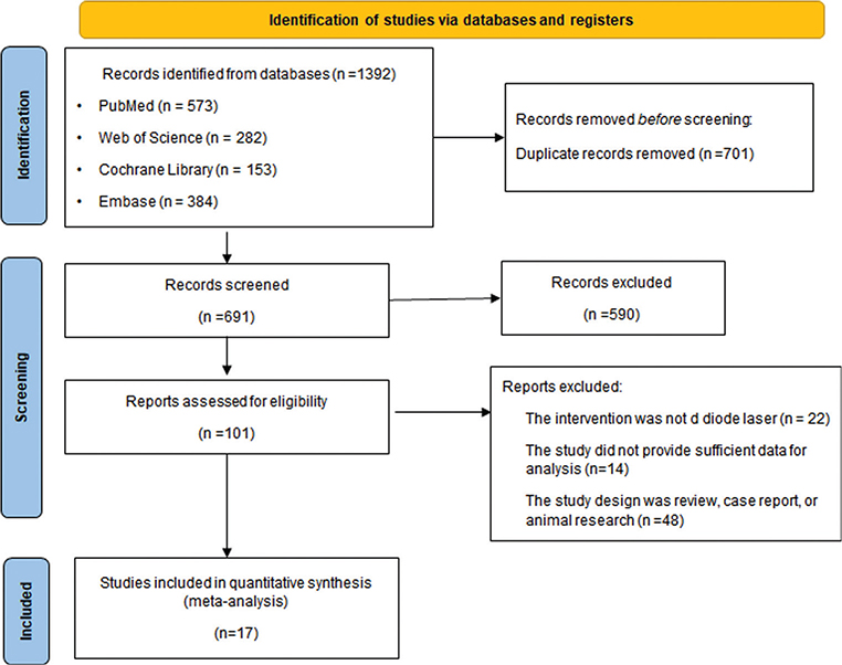
Figure 1. Flowchart of study screening.
Table 1 summarises the basic characteristics of the included studies. The publication time of the included studies ranged from 2005 to 2022, with studies from India (n = 8), Iran (n = 4), Brazil (n = 1), Canada (n = 1), Taiwan, China (n = 1), Egypt (n = 1), and Turkey (n = 1). All study subjects were children, aged between 2 and 10 years, involving 1,018 primary teeth. In terms of intervention, all studies used diode lasers, and the control groups included FC, FS, MTA, and simvastatin gel (SG). Zinc oxide eugenol (ZOE) was the commonly used pulp capping material. In different studies, the final restorative material for pulpotomy was mainly stainless steel crowns (SSC), with one study using resin-modified glass ionomer cement (RMGIC).
Quality assessment of literature
The quality of the RCT studies was assessed using the Cochrane collaboration risk of bias tool. The results showed that the included studies had certain biases in allocation concealment and blinding implementation, while they had lower biases in randomisation implementation, completeness of outcome data, selective reporting of study results, and other biases. Overall, the included studies had acceptable quality (Figures 2 and 3).
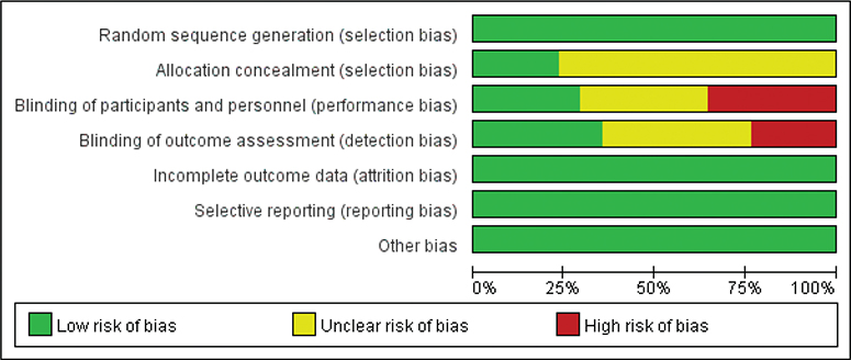
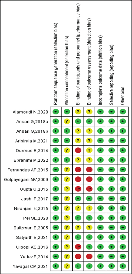
Figure 3. Risk of bias summary.
Clinical success rate by different follow-up duration
We evaluated the clinical success rate of diode laser pulpotomy based on different follow-up times. The heterogeneity assessment showed good homogeneity among the included studies (Total: I2 = 0%; ≤3 months: I2 = 0%; 6 months: I2 = 0%; 9 months: I2 = 0%; ≥12 months: I2 = 0%), and a fixed-effect model was used for meta-analysis. The overall analysis suggested that diode laser pulpotomy had a similar clinical success rate compared to the control group (RR: 1.01; 95% CI: 0.99–1.03). For different follow-up times, diode laser pulpotomy showed similar clinical success rates compared to the control group at ≤3 months (n = 672; RR: 1.01; 95% CI: 0.98–1.05), 6 months (n = 822; RR: 1.01; 95% CI: 0.98–1.04), 9 months (n = 449; RR: 1.02; 95% CI: 0.97–1.06), and ≥12 months (n = 847; RR: 1.01; 95% CI: 0.98–1.05), indicating similar short-term, medium-term, and long-term clinical success rates for diode laser pulpotomy (see Figure 4).
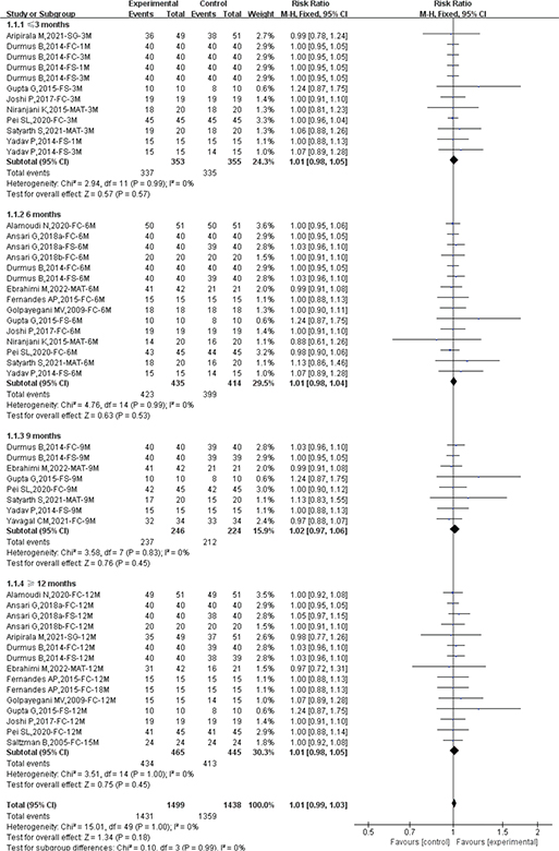
Figure 4. Forest plot comparing clinical success between diode laser and control by different follow-up duration.
Radiographic success rate by different follow-up duration
The heterogeneity assessment showed low heterogeneity among the included studies, suggesting the use of a fixed-effect model for efficacy evaluation (Total: I2 = 0%; ≤3 months: I2 = 0%; 6 months: I2 = 0%; 9 months: I2 = 48%; ≥12 months: I2 = 47%). Overall, there was no statistically significant difference in radiographic success rates between diode laser pulpotomy and the control group (RR: 0.99; 95% CI: 0.97–1.02). Subgroup analysis by different follow-up times found that compared to the control group, diode laser pulpotomy had similar radiographic success rates at ≤3 months (n = 714; RR: 1.01; 95% CI: 0.97–1.05), 6 months (n = 889; RR: 0.99; 95% CI: 0.95–1.03), 9 months (n = 470; RR: 1.06; 95% CI: 0.98–1.15), and ≥12 months (n = 950; RR: 0.95; 95% CI: 0.90–1.01) (see Figure 5).
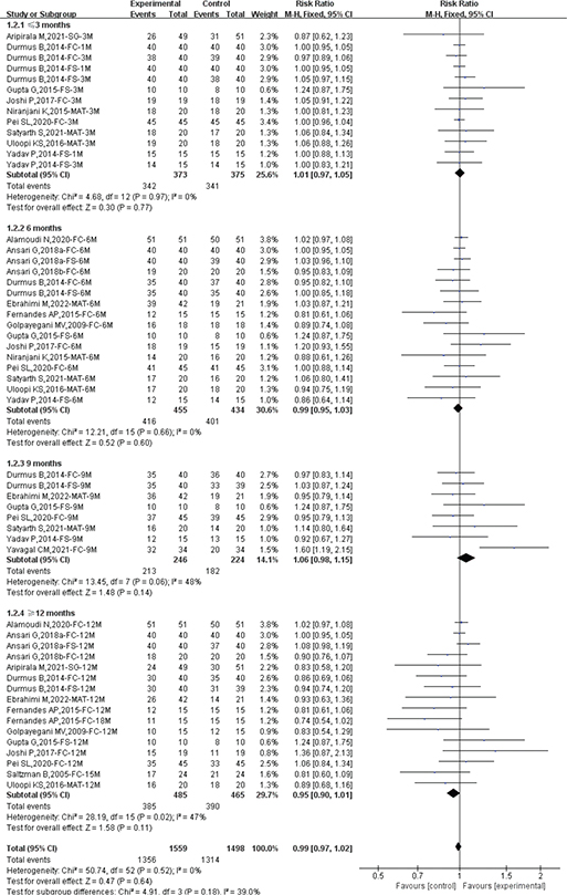
Figure 5. Forest plot comparing radiological success between diode laser and control by different follow-up duration.
Subgroup analysis by different control groups
Due to the differences in control groups used in the included studies, subgroup analyses were conducted based on different control groups to further determine whether there were significant differences in efficacy between diode laser pulpotomy and different controls.
Regarding clinical success rate, the heterogeneity assessment showed low heterogeneity among the included studies, and a fixed-effect model was used for efficacy evaluation (Total: I2 = 0%; FC-ZOE: I2 = 0%; FS-ZOE: I2 = 0%; MTA-ZOE: I2 = 0%; SG-REGIC: I2 = 0%). Compared to FC-ZOE (n = 1,590; RR: 1.00; 95% CI: 0.98–1.02), MTA-ZOE (n = 389; RR: 1.01; 95% CI: 0.94–1.09), and SG-REGIC (n = 200; RR: 0.99; 95% CI: 0.83–1.17), diode laser pulpotomy had similar clinical success rates. Compared to FS-ZOE, diode laser pulpotomy was more likely to achieve clinical success (n = 758; RR: 1.04; 95% CI: 1.01–1.07) (see Figure 6).
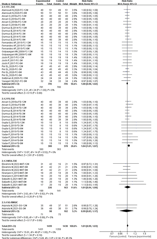
Figure 6. Forest plot of clinical success between diode laser and different control group.
Regarding radiographic success rate, the heterogeneity assessment showed low heterogeneity among the included studies, and a fixed-effect model was used for efficacy evaluation (Total: I2 = 0%; FC-ZOE: I2 = 29%; FS-ZOE: I2 = 0%; MTA-ZOE: I2 = 0%; SG-REGIC: I2 = 0%). Compared to FC-ZOE (n = 1,590; RR: 0.99; 95% CI: 0.96–1.02), FS-ZOE (n = 758; RR: 1.03; 95% CI: 0.99–1.08), MTA-ZOE (n = 509; RR: 0.99; 95% CI: 0.92–1.07), and SG-REGIC (n = 200; RR: 0.85; 95% CI: 0.66–1.10) (see Figure 7).
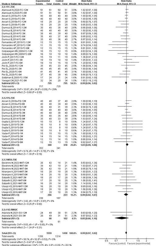
Figure 7. Forest plot of radiological success between diode laser and different control group.
Publication bias and certainty of evidence
We assessed the presence of significant publication bias among the included studies using funnel plots. The results showed that the included studies were evenly distributed in the upper part of the funnel, with no significant publication bias detected (Figure 8). In addition, the results of the certainty of evidence were summarised in Supplementary Table 1. In Section 2.4, we described the contents of the bias assessment. We found that only four studies had a high risk of blinding implementation. All included studies had a low risk of bias in random allocation, allocation concealment, completeness of outcome data, selective reporting of study results, and other sources of bias (Figures 2 and 3). In addition, the heterogeneity of included studies was evaluated using I2 statistics, and all results indicated the heterogeneity was modest, with I2 ranging from 0% to 48% (most results were 0%). Therefore, we considered the included studies to have an acceptable quality in risk of bias assessment.
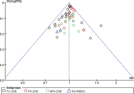
Figure 8. Funnel plot of included studies.
Discussion
This study included current research evaluating diode laser pulpotomy in primary teeth compared to conventional pulpotomy. We used meta-analysis to comprehensively assess the clinical and radiographic success rates of diode laser pulpotomy. A total of 17 RCTs met the inclusion criteria of this study, involving 1,018 primary teeth. Follow-up results in the short term (≤3 months), medium term (6–9 months), and long term (≥12 months) all showed similar clinical and radiographic success rates between diode laser pulpotomy and conventional pulpotomy. When comparing different control groups, diode laser pulpotomy showed similar efficacy with FC-ZOE, FS-ZOE, MTA-ZOE, and SG-REGIC. However, diode laser pulpotomy demonstrated a higher likelihood of clinical success compared to FS-ZOE, suggesting potential advantages in specific clinical scenarios.
Laser-assisted pulpotomy has gained widespread acceptance because of its notable advantages over traditional methods. Diode lasers, in particular, are favoured for their reliability and simplicity. However, concerns regarding peripheral thermal damage to pulp tissue have been raised. This issue can be mitigated by optimising laser parameters such as power, frequency, and application time, as well as employing water irrigation during the procedure [29]. Some researchers recommend an irradiation time of 1–3 seconds to minimise degenerative changes [30]. For instance, Pei et al. [24] utilised diode laser parameters set at 1.5w with a 2-second irradiation time, resulting in a thin laser-induced necrotic layer that prevented direct contact with the capping material, thereby reducing chemical or toxic effects. In this study, the included studies used diode laser with output wavelengths ranging from 632 nm to 980 nm, and power settings from 10 mW to 10W, using both contact and non-contact modes. Nevertheless, the impact of these parameter variations on treatment efficacy remains unclear and warrants further investigation. Notably, Saltzman et al. [15] reported lower radiographic success rates with a 980 nm wavelength and 3W power laser compared to the FC group, primarily because of periodontal lesions and pathological root resorption. While most included studies show comparable therapeutic outcomes between diode laser pulpotomy and FC, Gupta et al. [21] found that diode laser achieved a higher success rate than FS, aligning with our findings that diode laser exhibits similar clinical and radiographic success rates to FC and MTA but significantly outperforms FS in clinical success rates.
In vitro studies by Cannon et al. [31] demonstrated that diode laser treatment induced milder pulp inflammation compared to FC and FS after 28 days of observation in mechanically exposed pig teeth. Yamakawa et al. [32] further supported these findings by showing that diode laser enhances alkaline phosphatase activity, promoting the formation of dentin-like cells. In addition, Sivadas et al. [33] conducted an in vivo study on histological changes following pulpotomy in primary teeth and found that both diode laser and FS facilitated dentin bridge formation at the pulp interface, with the diode laser group having superior dentin bridge quality. These findings collectively suggest that diode laser pulpotomy not only results in less inflammation but also stimulates the mineralisation and repair capacity, making it a relatively safe and effective treatment modality.
Despite these promising results, the current evidence remains inconclusive, highlighting the need for further high-quality RCTs with standardised protocols to establish definitive conclusions regarding the efficacy and safety of diode laser pulpotomy. Future research should focus on optimising laser parameters, assessing long-term outcomes, and comparing diode laser pulpotomy with other emerging pulpotomy techniques to provide a more comprehensive understanding of its clinical applications.
Although this study comprehensively and scientifically explored the clinical and radiographic follow-up results of diode laser pulpotomy, several limitations should be acknowledged. Firstly, some of the included studies had relatively small sample sizes, which may limit the statistical power and generalisability of the findings. Furthermore, certain studies did not implement allocation concealment and blinding, potentially introducing bias risks that could affect the reliability of the results. Another significant limitation is the heterogeneity in diode laser parameter settings across the included studies. Variations in output power, energy density, wavelength, and the mode of application (contact vs. non-contact) were observed, making it challenging to draw definitive conclusions about the optimal parameters for diode laser pulpotomy. Due to insufficient data, it was not possible to perform a detailed subgroup analysis to further investigate the relationship between specific laser parameter settings and clinical or radiographic success rates. These limitations highlight the need for future research to standardise laser parameters, employ larger sample sizes, and adhere to rigorous methodological practices, such as allocation concealment and blinding, to minimise bias and enhance the validity of the findings. Addressing these issues in future studies will provide more robust evidence to guide clinical decision-making regarding diode laser pulpotomy in primary teeth.
Conclusion
In summary, diode laser pulpotomy in primary teeth is a viable alternative to conventional treatment methods. Compared to conventional treatment, diode laser treatment shows similar clinical and radiographic success rates in short-term (≤3 months), medium-term (6–9 months), and long-term (≥12 months) follow-ups. Therefore, diode laser pulpotomy can be considered a suitable alternative to conventional treatment. However, because of certain limitations in this study, future research should conduct more RCTs with larger sample sizes, consistent methodology, and standardised laser characteristics to further explore the clinical application efficacy of diode laser pulpotomy.
Declarations
Ethics approval and consent to participate
Not applicable.
Consent for publication
Not applicable.
Availability of data and materials
All data generated or analysed during this study are included in this article. Further enquiries can be directed to the corresponding author.
Authors’ contributions
Gao Y and Hu MN conceived and designed the study, and participated in data analysis and statistics. Gao Y, Hu MN, and Xu J helped in drafting the manuscript. All the authors read and approved the final manuscript.
Registering systematic review protocols
INPLASY202540034.
References
[1] Kazeminia M, Abdi A, Shohaimi S, Jalali R, Vaisi-Raygani A, Salari N, et al. Dental caries in primary and permanent teeth in children’s worldwide, 1995 to 2019: a systematic review and meta-analysis. Head Face Med. 2020;16(1):22. https://doi.org/10.1186/s13005-020-00237-z
[2] Nematollahi H, Sahebnasagh M, Parisay I. Comparison of electrosurgical pulpotomy with zinc oxide eugenol or zinc polycarboxylate cements sub-base. J Clin Pediatr Dent. 2011;36(2):133–7. https://doi.org/10.17796/jcpd.36.2.3527457504x6k880
[3] King SR, McWhorter AG, Seale NS. Concentration of formocresol used by pediatric dentists in primary tooth pulpotomy. Pediatr Dent. 2002;24(2):157–9.
[4] de Alencar Filho AV, Dos Santos Junior VE, da Silva Calixto M, Santos N, Heimer MV, Rosenblatt A. Evaluation of the genotoxic effects of formocresol application in vital pulp therapy of primary teeth: a clinical study and meta-analysis. Clin Oral Investig. 2018;22(7):2553–8. https://doi.org/10.1007/s00784-018-2352-y
[5] Yamasaki M, Nakamura H, Kameyama Y. Irritating effect of formocresol after pulpectomy in vivo. Int Endod J. 1994;27(5):245–51. https://doi.org/10.1111/j.1365-2591.1994.tb00263.x
[6] Aeinehchi M, Eslami B, Ghanbariha M, Saffar AS. Mineral trioxide aggregate (MTA) and calcium hydroxide as pulp-capping agents in human teeth: a preliminary report. Int Endod J. 2003;36(3):225–31. https://doi.org/10.1046/j.1365-2591.2003.00652.x
[7] Naved N, Umer F, Khowaja AR. Cost-effectiveness analysis of regenerative endodontics versus MTA apexification. JDR Clin Trans Res. 2024;9(3):231–8. https://doi.org/10.1177/23800844231191515
[8] Nematollahi H, Sarraf Shirazi A, Mehrabkhani M, Sabbagh S. Clinical and radiographic outcomes of laser pulpotomy in vital primary teeth: a systematic review and meta-analysis. Eur Arch Paediatr Dent. 2018;19(4):205–20. https://doi.org/10.1007/s40368-018-0358-4
[9] Ansari G, Safi Aghdam H, Taheri P, Ghazizadeh Ahsaie M. Laser pulpotomy-an effective alternative to conventional techniques – a systematic review of literature and meta-analysis. Lasers Med Sci. 2018;33(8):1621–9. https://doi.org/10.1007/s10103-018-2588-4
[10] Joshi P, Baliga S, Rathi N, Thosar N, Dharmadhikari P, Chandak P. A comparative evaluation between formocresol and diode laser assisted pulpotomy in primary molars – an in vivo study. Eur J Pharm Med Res. 2017;4:569–75.
[11] Yavagal CM, Lal A, Chavan Patil VV, Yavagal PC, Neelakantappa KK, Hariharan M. Efficacy of laser photobiomodulation pulpotomy in human primary teeth: a randomized controlled trial. J Indian Soc Pedod Prev Dent. 2021;39(4):436–41. https://doi.org/10.4103/jisppd.jisppd_150_21
[12] Ansari G, Chitsazan A, Fekrazad R, Javadi F. Clinical and radiographic evaluation of diode laser pulpotomy on human primary teeth. Laser Ther. 2018;27(3):187–92. https://doi.org/10.5978/islsm.27_18-OR-17
[13] Page MJ, McKenzie JE, Bossuyt PM, Boutron I, Hoffmann TC, Mulrow CD, et al. The PRISMA 2020 statement: an updated guideline for reporting systematic reviews. BMJ. 2021;372:n71. https://doi.org/10.1136/bmj.n71
[14] Higgins J, Altman DG. Chapter 8: assessing risk of biasin included studies. In: Higgins J, Green S, editors Cochrane handbook for systematic reviews of interventions version 5.0 [Internet]. [cited 2010 Jul 20]. Available from: http://www.cochrane-handbook.org
[15] Saltzman B, Sigal M, Clokie C, Rukavina J, Titley K, Kulkarni GV. Assessment of a novel alternative to conventional formocresol-zinc oxide eugenol pulpotomy for the treatment of pulpally involved human primary teeth: diode laser-mineral trioxide aggregate pulpotomy. Int J Paediatr Dent. 2005;15(6):437–47. https://doi.org/10.1111/j.1365-263X.2005.00670.x
[16] Golpayegani MV, Ansari G, Tadayon N, Shams S, Mir M. Low-level laser therapy for pulpotomy treatment of primary molars. Tehran Univ Med J. 2009;6:168–74.
[17] Durmus B, Tanboga I. In vivo evaluation of the treatment outcome of pulpotomy in primary molars using diode laser, formocresol, and ferric sulphate. Photomed Laser Surg. 2014;32(5):289–95. https://doi.org/10.1089/pho.2013.3628
[18] Yadav P, Indushekar K, Saraf B, Sheoran N, Sardana D. Comparative evaluation of Ferric Sulfate, Electrosurgical and Diode Laser on human primary molars pulpotomy: an ‘in-vivo’ study. Laser Ther. 2014;23(1):41–7. https://doi.org/10.5978/islsm.14-OR-05
[19] Fernandes AP, Lourenço Neto N, Teixeira Marques NC, Silveira Moretti AB, Sakai VT, Cruvinel Silva T, et al. Clinical and radiographic outcomes of the use of low-level laser therapy in vital pulp of primary teeth. Int J Paediatr Dent. 2015;25(2):144–50. https://doi.org/10.1111/ipd.12115
[20] Niranjani K, Prasad MG, Vasa AA, Divya G, Thakur MS, Saujanya K. Clinical evaluation of success of primary teeth pulpotomy using mineral trioxide aggregate(®), laser and biodentine(TM) – an in vivo study. J Clin Diagn Res. 2015;9(4):ZC35–7. https://doi.org/10.7860/JCDR/2015/13153.5823
[21] Gupta G, Rana V, Srivastava N, Chandna P. Laser pulpotomy – an effective alternative to conventional techniques: a 12 months clinicoradiographic study. Int J Clin Pediatr Dent. 2015;8(1):18–21. https://doi.org/10.5005/jp-journals-10005-1277
[22] Uloopi KS, Vinay C, Ratnaditya A, Gopal AS, Mrudula KJ, Rao RC. Clinical evaluation of low level diode laser application for primary teeth pulpotomy. J Clin Diagn Res. 2016;10(1):ZC67–70. https://doi.org/10.7860/JCDR/2016/13218.7140
[23] Ansari G, Morovati SP, Asgary S. Evaluation of four pulpotomy techniques in primary molars: a randomized controlled trial. Iran Endod J. 2018;13(1):7–12.
[24] Pei SL, Shih WY, Liu JF. Outcome comparison between diode laser pulpotomy and formocresol pulpotomy on human primary molars. J Dent Sci. 2020;15(2):163–7. https://doi.org/10.1016/j.jds.2020.03.005
[25] Alamoudi N, Nadhreen A, Sabbagh H, El Meligy O, Al Tuwirqi A, Elkhodary H. Clinical and radiographic success of low-level laser therapy compared with formocresol pulpotomy treatment in primary molars. Pediatr Dent. 2020;42(5):359–66.
[26] Satyarth S, Alkhamis AM, Almunahi HF, Abdulaziz Alsuhaymi MO, Vadde HB, Senapathi SN, et al. Comparative evaluation of mineral trioxide aggregate pulpotomy and laser-assisted mineral trioxide aggregate pulpotomy: an original research article. J Microsc Ultrastruct. 2021;9(1):7–11. https://doi.org/10.4103/JMAU.JMAU_54_19
[27] Aripirala M, Bansal K, Mathur VP, Tewari N, Gupta P, Logani A. Comparative evaluation of diode laser and simvastatin gel in pulpotomy of primary molars: a randomized clinical trial. J Indian Soc Pedod Prev Dent. 2021;39(3):303–9. https://doi.org/10.4103/jisppd.jisppd_60_21
[28] Ebrahimi M, Changiz S, Makarem A, Ahrari F. Clinical and radiographic effectiveness of mineral trioxide aggregate (MTA) partial pulpotomy with low power or high power diode laser irradiation in deciduous molars: a randomized clinical trial. Lasers Med Sci. 2022;37(4):2293–303. https://doi.org/10.1007/s10103-022-03503-5
[29] Movaniya PN, Desai NN, Makwana TR, Matariya RG, Makwana KG, Patel HB, et al. Effectiveness of diode laser in intraoral soft tissue surgeries – an evaluative study. Ann Maxillofac Surg. 2023;13(2):167–72. https://doi.org/10.4103/ams.ams_140_23
[30] Mareddy A, Mallikarjun SB, Shetty PV, Rao VVN, Chandru TP. Histological evaluation of diode laser pulpotomy in dogs. J Oral Laser Appl. 2010;10:7–16.
[31] Cannon M, Wagner C, Thobaben JZ, Jurado R, Solt D. Early response of mechanically exposed dental pulps of swine to antibacterial-hemostatic agents or diode laser irradiation. J Clin Pediatr Dent. 2011;35(3):271–6. https://doi.org/10.17796/jcpd.35.3.73621w8l447r3887
[32] Yamakawa S, Niwa T, Karakida T, Kobayashi K, Yamamoto R, Chiba R, et al. Effects of Er:YAG and diode laser irradiation on dental pulp cells and tissues. Int J Mol Sci. 2018;19(8):2429. https://doi.org/10.3390/ijms19082429
[33] Sivadas S, Rao A, Natarajan S, Shenoy R, Srikrishna SB. Pulpal response to ferric sulfate and diode laser when used as pulpotomy agent: an in vivo study. J Clin Diagn Res. 2017;11(6):ZC87–91. https://doi.org/10.7860/JCDR/2017/25180.10121