ORIGINAL ARTICLE
miR-1246 regulates odontogenic differentiation of human dental pulp stem cells in an inflammatory microenvironment by targeting AXIN2: an in vitro study
Shunying Wu, Yueyan Wang and Jiayi Yang
Department of Endodontics, Wuxi Stomatological Hospital, Wuxi City, Jiangsu Province, China
ABSTRACT
Objective: This study aimed to investigate the modulatory function of microRNA-1246 (miR-1246) in the odontogenic differentiation of human dental pulp stem cells (hDPSCs) under inflammatory conditions in vitro and to elucidate its underlying molecular mechanism.
Methods: An inflammatory state was replicated in vitro by treating hDPSCs with lipopolysaccharide (LPS). miR-1246 expression was modulated via transfection with its mimics or inhibitors. Cell proliferation, movement, and apoptosis were assessed. Odontogenic differentiation capacity was assessed via alkaline phosphatase (ALP) activity, Alizarin Red S (ARS) staining, and transcript levels by quantitative real-time quantitative real-time polymerase chain reaction (qRT-PCR), along with Western blotting. To validate the direct interaction between miR-1246 and AXIN2, a dual-luciferase reporter assay was employed. All experiments were performed in three independent biological replicates.
Results: miR-1246 overexpression significantly reduced inflammatory cytokine levels (IL-1β, P < 0.05; IL-6, P < 0.05; TNF-α, P < 0.001) and partially restored hDPSC proliferation (P < 0.01), migration (P < 0.001), and survival (reduced apoptosis rate, P < 0.001). It also significantly increased ALP activity (P < 0.001), mineralized nodule formation (P < 0.001), and the expression of odontogenic markers (NRP1, DSPP, DMP1). Mechanistically, miR-1246 directly targeted AXIN2, preventing AXIN2-mediated β-catenin degradation and promoting the Wnt/β-catenin pathway, elevating downstream factors such as Cyclin D1 and c-Myc. AXIN2 knockdown reproduced the differentiation-promoting effects of miR-1246, whereas AXIN2 overexpression diminished them.
Conclusion: miR-1246 promotes the odontogenic differentiation of hDPSCs and partially counteracts the detrimental effects of inflammation in vitro by directly targeting and inhibiting AXIN2 expression, thereby stimulating the Wnt/β-catenin signaling cascade.
KEYWORDS: Dental pulp stem cells; inflammatory microenvironment; miR-1246; odontogenic differentiation
Citation: ACTA ODONTOLOGICA SCANDINAVICA 2026; VOL. 85: 635–653. DOI: https://doi.org/10.2340/aos.v85.46788.
Copyright: © 2026 The Author(s). Published by MJS Publishing on behalf of Acta Odontologica Scandinavica Society. This is an Open Access article distributed under the terms of the Creative Commons Attribution 4.0 International License (http://creativecommons.org/licenses/by/4.0/), allowing third parties to copy and redistribute the material in any medium or format and to remix, transform, and build upon the material, with the condition of proper attribution to the original work.
Received: 22 January 2026; Accepted: 18 August 2026; Published: 17 September 2026.
CONTACT: Jiayi Yang jiayiyang00@163.com Wuxi Stomatological Hospital, No. 6 Jiankang Road, Liangxi District, Wuxi City, Jiangsu Province, 214000, China
Competing interests and funding: All authors declare no conflict of interest.
The authors received no financial support for the research, authorship, and/or publication of this article.
Introduction
The dental pulp, the sole soft tissue within the tooth, is housed in the pulp chamber and surrounded by dense dentin. It plays an irreplaceable role in maintaining tooth physiology, including nutrient supply, sensory perception, and immune defense [1, 2]. Following pulp tissue injury, resident odontoblasts synthesize and secrete a dentin-matrix to initiate repair. If these cells are lost, undifferentiated mesenchymal stem cells within the pulp can differentiate into new odontoblasts, forming an ‘odontoblast bridge’ to facilitate regeneration [3–5]. This endogenous repair mechanism is crucial for maintaining tooth integrity.
Clinically, deep carious lesions allow bacteria and their metabolic byproducts to invade the pulp via dentinal tubules, triggering inflammatory responses of varying degrees [5–7]. In the early stages, a moderate inflammatory response may promote repair by modulating the cytokine microenvironment. However, persistent or excessive inflammation leads to the destruction of pulp tissue architecture, impaired blood supply, and cell necrosis, culminating in irreversible pulpitis or pulp necrosis [5]. Current mainstream treatments for irreversible pulpitis, root canal therapy and tooth extraction, can eliminate infection and control symptoms but compromise the tooth’s structure and function, leading to adverse outcomes such as increased fragility, discoloration, and secondary infection [1]. Therefore, exploring strategies to inhibit detrimental reactions during early inflammation and promote pulp repair is vital for preserving pulp vitality and achieving functional regeneration.
Human dental pulp stem cells (hDPSCs), a neural crest-derived population of mesenchymal stem cells, possess high self-renewal capacity and multidirectional differentiation potential, including into odontoblasts, osteoblasts, neurons, and adipocytes [8, 9]. In pulp injury repair, hDPSCs are the core cell population for odontogenic regeneration. However, the abundance of pro-inflammatory factors (e.g. TNF-α, IL-1β) in the inflammatory microenvironment significantly impairs the biological characteristics of hDPSCs, leading to restricted proliferation, increased apoptosis, and diminished odontogenic differentiation capacity, thereby weakening their regenerative potential [10–14]. This presents a challenge for the clinical application of hDPSCs in pulp regeneration and underscores the need to identify molecular pathways that protect or enhance hDPSC function under inflammatory conditions.
In recent years, non-coding RNAs, particularly microRNAs (miRNAs), have drawn considerable interest due to their crucial functions in intercellular communication and cell fate regulation [15, 16]. Research has demonstrated that miR-1246 expression is markedly elevated within exosomes derived from hDPSCs under odontogenic induction compared to those from conventionally cultured cells [17]. miR-1246 is a highly conserved small RNA molecule that modulates the expression of target genes through interaction with the 3’-untranslated region (3’-UTR) of corresponding mRNAs. Furthermore, database predictions suggest that miR-1246 may induce odontogenic development of dental pulp stem cells through its interaction with AXIN2 [17]. As a crucial negative regulator within the Wnt/β-catenin signaling cascade, AXIN2 promotes β-catenin degradation and thereby suppresses the transcription of downstream target genes [18]. This signaling pathway is considered a critical signaling axis for promoting odontoblast differentiation and mineralization during pulp and the formation of dentin [19]. Therefore, it is plausible that miR-1246 relieves the inhibition of this signaling pathway by targeting and suppressing AXIN2, leading to the nuclear translocation of β-catenin and the subsequent activation of odontogenic differentiation-related genes.
Despite clues suggesting a potential link between miR-1246 and odontogenic differentiation, its specific effects on hDPSC function and the associated molecular regulatory mechanisms within the complex inflammatory microenvironment of pulpitis remain unclear. In the present study, we aimed to investigate the modulatory function of miR-1246 in the odontogenic differentiation of hDPSCs under inflammatory conditions in vitro and to elucidate its underlying molecular mechanism, specifically focusing on whether miR-1246 exerts its effects by targeting AXIN2 to activate the Wnt/β-catenin signaling cascade. These results should offer a solid theoretical foundation for developing novel pulp regeneration therapies targeting miR-1246.
Methods
Cell culture and inflammatory model establishment
HDPSCs were acquired from ScienCell Research Laboratories (Carlsbad, CA, USA). These cells were maintained in α-MEM medium (Gibco, USA) fortified with 10% fetal bovine serum (FBS; Gibco, USA), in addition to 100 U/mL penicillin and 100 μg/mL streptomycin (Beyotime, China). All cells were kept at 37°C within a humidified chamber containing 5% CO₂. To simulate an inflammatory microenvironment in vitro, hDPSCs were treated for 24 h with a culture medium including 1 μ g/mL LPS (Sigma-Aldrich, St. Louis, MO, USA) from Escherichia coli. This concentration was selected based on its established efficacy in inducing a robust and reproducible inflammatory response in hDPSCs without causing excessive cytotoxicity, consistent with previous in vitro pulpitis models [12, 20]. A set of non-treated cells was used for the control experiment.
Cell transfection and grouping
For functional studies of miR-1246, hDPSCs were assigned to the following groups: (1) Control groups: untreated cells; (2) Model groups: cells treated with 1 μg/mL LPS for 24 h; (3) miR-1246-OE groups: cells were subjected to LPS treatment followed by transfection with miR-1246 mimics; (4) miR-1246-OE-NC groups: cells were subjected to LPS treatment followed by transfection with negative control mimic; (5) miR-1246-KD groups: cells were subjected to LPS treatment followed by transfection with miR-1246 inhibitor; (6) miR-1246-KD-NC groups: cells were subjected to LPS treatment followed by transfection with negative control inhibitor. GenePharma (Shanghai, China) was the source for all mimics, inhibitors, and their corresponding negative controls.
For functional studies of AXIN2, hDPSCs were assigned to the following groups: (1) Control groups: untreated cells; (2) Model groups: cells treated with 1 μg/mL LPS for 24 h; (3) AXIN2-OE groups: cells were subjected to LPS treatment followed by transfection with the pcDNA3.1-AXIN2 plasmid; (4) AXIN2-OE-NC groups: cells were subjected to LPS treatment followed by transfection with the empty pcDNA3.1 vector; (5) AXIN2-KD groups: cells were subjected to LPS treatment followed by transfection with a small interfering RNA that targets AXIN2 (si-AXIN2); (6) AXIN2-KD-NC groups: cells were subjected to LPS treatment followed by transfection with a negative control siRNA (si-NC). The pcDNA3.1-AXIN2 overexpression plasmid and the corresponding empty vector (pcDNA3.1) were procured from YouBio (Changsha, China). The design and synthesis of si-AXIN2 and si-NC were carried out by GenePharma.
The transfection procedures were executed with Lipofectamine 3000 (Invitrogen, Carlsbad, CA, USA), adhering to the instructions provided by the manufacturer. Transfection efficiency was confirmed by quantitative real-time PCR (qRT-PCR) 48 h post-transfection.
Cell counting Kit-8 (CCK-8) assay
The evaluation of cell proliferation was conducted with the CCK-8 (Dojindo, Kumamoto, Japan). For this procedure, cells were first placed into 96-well plates at a concentration of 2 × 10³ cells for each well. After a 24 h, every well received an addition of 10 μL of the CCK-8 solution, which was succeeded by a 2 h incubation. The optical density was subsequently recorded at a wavelength of 450 nm utilizing a microplate reader (Bio-Rad, Hercules, CA, USA).
Cell migration assay
The migratory ability of cells was assessed with 24-well Transwell chambers (Corning, NY, USA). Specifically, a suspension of 5 × 10⁴ cells in serum-free medium was introduced into the top compartment. The bottom compartment contained medium supplemented with 20% FBS, acting as the chemotactic agent. After incubating for 24 h, non-migratory cells persisting on the upper surface of the membrane were meticulously removed with a cotton applicator. Cells that had traversed to the bottom surface were fixed with 4% paraformaldehyde (PA) for a duration of 30 min and subsequently stained using 0.1% crystal violet for another 20 min. For quantification, images from five randomly selected areas were captured and the cells were enumerated using a microscope.
Apoptosis assay
To evaluate apoptosis, the Annexin V-FITC/PI Apoptosis Detection Kit (Abcam, Cambridge, UK) was utilized. For this assay, cells were gathered, rinsed twice using cold PBS, and subsequently resuspended within 1× binding buffer. Subsequently, a staining solution composed of 5 μL of Annexin V-FITC and 5 μL of PI was added to the cell suspension. The mixture was then maintained for a 15-min incubation period at ambient temperature while protected from light. The analysis was conducted on a FACSCalibur flow cytometer (BD Biosciences, San Jose, CA, USA), and all acquired data were processed using FlowJo software.
Odontogenic differentiation
Following 48 h of culturing, hDPSCs were harvested, resuspended, and then seeded into 24-well plates (2.5 × 10⁵ cells/well). Once approximately 80% confluence was achieved, the existing culture medium was replaced with an odontogenic induction medium. This induction medium consisted of α-MEM fortified with 15% FBS, 50 μmol/L L-ascorbic acid, 10 mmol/L β-glycerophosphate, 10–8 mol/L dexamethasone, 100 U/mL penicillin, and 100 μg/mL streptomycin. The medium was refreshed every 48 h.
Alkaline phosphatase activity assay
After 7 days of induction, cells were lysed with an inhibitor-free cell lysis buffer. The collected lysate was assayed for alkaline phosphatase (ALP) activity using an ALP quantitative assay kit (Beyotime, China). The overall protein content, which was quantified via the BCA assay, served as the basis for normalization.
Alizarin Red S staining
Following a 15-day induction, the cells were subjected to fixation with 4% PA for a duration of 30 min, followed by staining with a 1% Alizarin Red S (ARS) solution (pH 4.2; Sigma-Aldrich, St. Louis, MO, USA) for 10 min. After removing the excess stain by washing with deionized water, the presence of mineralized deposits was visually inspected and documented through photography. For quantification, the stain was eluted by adding 10% cetylpyridinium chloride (Sigma-Aldrich, St. Louis, MO, USA) to each well, and the optical density of the resulting solution was determined at a wavelength of 562 nm.
qRT-PCR
The extraction of total cellular RNA was accomplished using the TRIzol reagent (Invitrogen, Thermo Fisher Scientific, Inc.). For miRNA analysis, reverse transcription was carried out with the Mir-X miRNA First-Strand Synthesis Kit (Takara, Dalian, China). Following this, qRT-PCR was executed on a CFX96 Real-Time PCR Detection System (Bio-Rad, Hercules, CA, USA), with U6 serving as the endogenous reference for miRNA quantification. The sequences of the specific primers employed in this research are detailed below: miR-1246 mimic, 5’-AAUGGAUUUUUGGAGCAGG-3’; mimic negative control, 5’-UUCUCCGAACGUGUCACGUTT-3’; miR-1246 inhibitor, 5’-CCUG CUCCAAAAAUCCAUU-3’; inhibitor negative control, 5’-CAGUACU UUUGUGUAGUACAA-3’ (GenePharma, Shanghai, China).
For the analysis of mRNA, the synthesis of cDNA from RNA was performed utilizing the PrimeScript RT Reagent Kit. Following this, qPCR was conducted with the SYBR Premix Ex Taq II (Takara, Dalian, China). The protocol for thermal cycling involved an initial step at 95°C for 10 s, which was succeeded by 35 cycles of 95°C for 5 s and 60°C for 30 s. For data normalization, GAPDH was employed as the endogenous control. Relative gene expression levels were calculated using the 2-ΔΔCt formula. All primer sequences can be found in Table 1.
Western Blot
Total proteins were extracted from cells using a RIPA lysis buffer (Beyotime, China), and its concentration determined with the BCA assay (Beyotime, China). Following this, the protein lysates were separated by SDS-PAGE and then transferred onto a PVDF membrane (Millipore, USA). To prevent non-specific binding, the membrane was treated with 5% non-fat milk for 1 h at ambient temperature, followed by an overnight incubation at 4°C with a panel of primary antibodies: NRP1 (1:1000, Abcam), DSPP (1:1000, Abcam), DMP1 (1:1000, Abcam), AXIN2 (1:1000, Cell Signaling Technology, USA), GSK-3β (1:1000, CST), p-GSK-3β (1:1000, CST), β-catenin (1:1000, CST), Cyclin D1 (1:1000, CST), c-Myc (1:1000, CST), and GAPDH (1:5000, CST). On the next day, the membrane underwent washing with TBST and was probed with an HRP-labeled secondary antibody (1:5000, CST) for 1 h at ambient temperature. The protein signals were visualized utilizing an ECL chemiluminescence detection kit (Millipore, USA), and the intensity of the bands was analyzed using ImageJ software.
Enzyme-linked immunosorbent assay
Following the collection of cell culture supernatants, the concentrations of TNF-α, IL-1β, and IL-6 were measured using enzyme-linked immunosorbent assay (ELISA) kits (R&D Systems, Minneapolis, MN, USA), in strict accordance with the manufacturer’s instructions. The absorbance values were subsequently determined at 450 nm; the final concentrations were calculated based on a standard curve.
Immunofluorescence staining
Cells were initially placed onto confocal dishes. Following treatment, they underwent fixation with 4% PA for a duration of 20 min. Subsequently, permeabilization was carried out using 0.5% Triton X-100 for a duration of 10 min. After a 1-h blocking step, the cells were blocked with 5% BSA and then kept overnight at 4°C with the designated primary antibodies (AXIN2, 1:200, CST; GSK-3β, 1:200, CST; p-GSK-3β,1:200, CST; β-catenin, 1:200, CST; Cyclin D1,1:200, CST, c-Myc,1:200, CST). On the following day, the cells were then incubated in the dark at room temperature for 1 h with fluorescent secondary antibodies (Alexa Fluor 488 or 594, 1:500, Invitrogen). DAPI (Beyotime, China) was employed for nuclear counterstaining. A laser confocal microscope (Leica, Wetzlar, Germany) was utilized to capture the images.
Dual-luciferase reporter assay
DNA fragments that encompassed either the wild-type (WT) or a mutant (MUT) version of the 3’-UTR of AXIN2 were inserted into the pmirGLO Dual-Luciferase Reporter Vector (Promega, Madison, WI, USA). The generated reporter vectors were then jointly introduced into HEK293T cells along with either the miR-1246 mimic or a mimic-NC. Forty-eight hours later, firefly and Renilla luciferase activities were measured by a dual-luciferase reporter detection system (Promega, Madison, WI, USA). For normalization purposes, Renilla luciferase activity functioned as the endogenous control.
Statistical analysis
All data processing was conducted with GraphPad Prism 8.0, and the results are displayed as the mean ± standard deviation (SD). All experiments were performed in three independent biological replicates. Student’s t-test was applied for comparisons between two groups. For evaluating differences among multiple groups, one-way analysis of variance (ANOVA) was utilized, followed by Tukey’s post hoc test for multiple comparisons. P-value < 0.05 was deemed to be statistically significant.
Results
miR-1246 attenuates inflammatory response and promotes proliferation in an LPS-induced hDPSCs model
To investigate the role of miR-1246 in pulpitis, an in vitro inflammatory model was established by treating hDPSCs with 1 µg/mL LPS. We then modulated miR-1246 levels via transfection. qRT-PCR confirmed that miR-1246 expression markedly elevated in the miR-1246-OE group relative to the miR-1246-OE-NC group (P < 0.001) and notably reduced in the miR-1246-KD group versus the miR-1246-KD-NC group (P < 0.05) (Figure 1A). Furthermore, the Model group (LPS-treated, without transfection) showed significantly decreased miR-1246 expression compared to the Control group (P < 0.05) (Figure 1B). A CCK-8 assay revealed that LPS treatment (Model group) resulted in a significant reduction in hDPSC viability when relative to the non-treated control (P < 0.001). In the inflammatory model, miR-1246 overexpression partially reversed this inhibitory effect, significantly restoring cell viability (P < 0.01), whereas miR-1246 knockdown further exacerbated the decline in viability (P < 0.01) (Figure 1C).
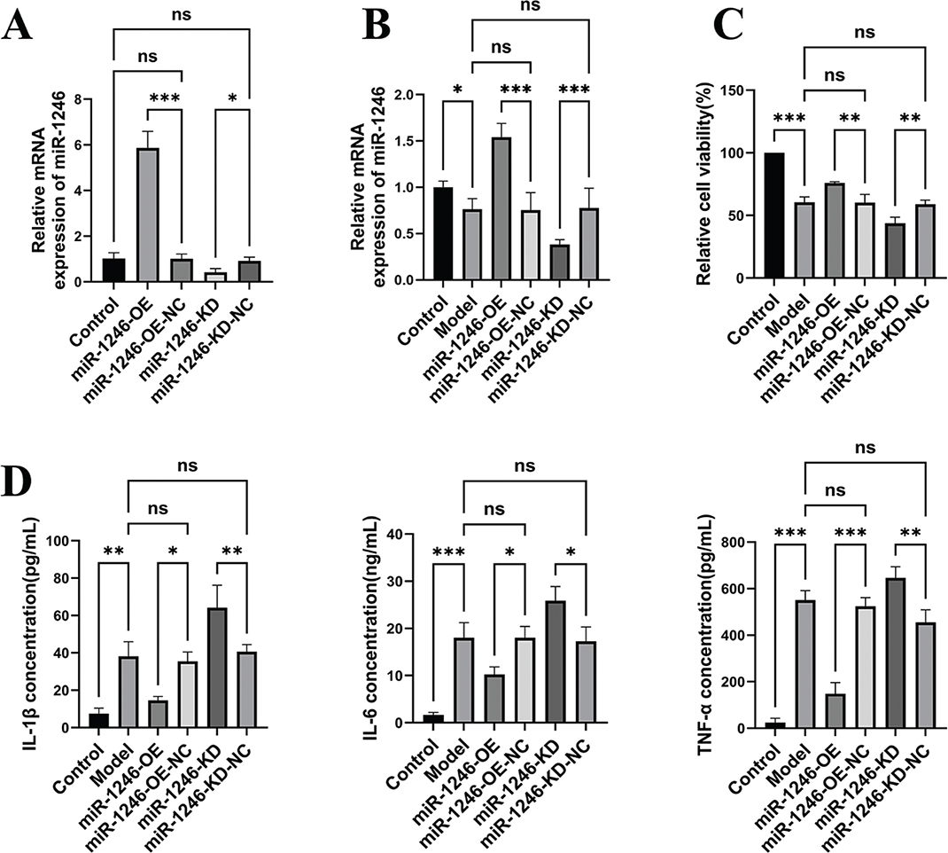
Figure 1. Effects of miR-1246 on LPS-induced proliferation and inflammatory response in hDPSCs. (A) The relative expression levels of miR-1246 in different groups of cells were detected by qRT-PCR. (C) The relative viability of cells in different groups was measured using the CCK-8 assay. (D) The concentrations of inflammatory cytokines IL-1β, IL-6, and TNF-α in the cell culture supernatants of different groups were determined by ELISA. Data are presented as the mean ± SD from three independent experiments. *P < 0.05, **P < 0.01, ***P < 0.001. ns indicates no significant difference.
The ELISA analysis revealed that LPS treatment markedly enhanced the secretion of pro-inflammatory mediators IL-1β, IL-6, and TNF-α, which validated the creation of the inflammatory model. Nevertheless, in cells exposed to LPS, an elevation in miR-1246 expression produced a notable reduction in the amounts of these mediators (P < 0.05, P < 0.05, P < 0.001, correspondingly). In contrast, reducing miR-1246 levels led to an additional rise in their release (P < 0.01, P < 0.05, P < 0.01, respectively) (Figure 1D). These data indicate that miR-1246 protects the proliferative capacity of hDPSCs and partially suppresses the inflammatory response in an LPS-induced inflammatory environment.
miR-1246 regulates migration and apoptosis of LPS-treated hDPSCs
To further investigate the biological functions of miR-1246, we conducted Transwell migration and flow cytometry apoptosis assays. The Transwell assay showed that LPS treatment (Model group) markedly suppressed the migratory capacity of hDPSCs (P < 0.001). Under LPS stimulation, miR-1246 overexpression significantly restored cell migration (P < 0.001), while its knockdown further impaired it (P < 0.01) (Figure 2A, B).
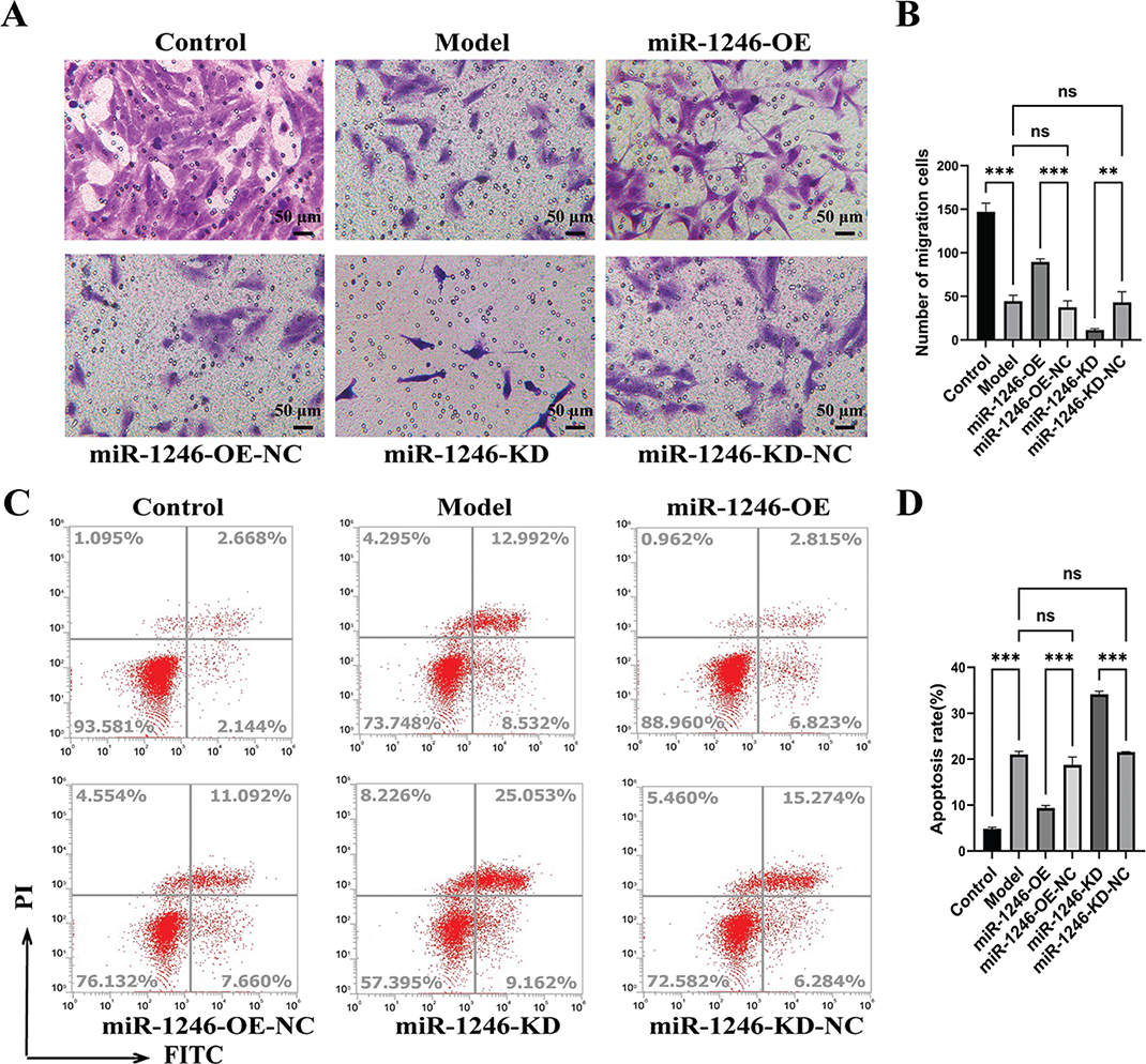
Figure 2. Effects of miR-1246 on LPS-induced migration and apoptosis in hDPSCs. (A) Representative images of the Transwell migration assay (crystal violet staining, scale bar = 50 μm). (B) Quantitative statistics of the number of migrated cells in the Transwell assay. (C) Representative flow cytometry scatter plots of cell apoptosis detected by Annexin V-FITC/PI double staining. (D) Quantitative analysis of the apoptosis rate in different groups of cells detected by flow cytometry. Data are presented as the mean ± SD from three independent experiments. **P < 0.01, ***P < 0.001. ns indicates no significant difference.
Flow cytometry analysis revealed that LPS treatment significantly induced hDPSC apoptosis (P < 0.001). In the inflammatory model, miR-1246 overexpression effectively reduced the apoptosis rate (P < 0.001). Conversely, miR-1246 knockdown resulted in a further significant increase in apoptosis (P < 0.001) relative to the respective negative control (Figure 2C, D). These findings indicate that miR-1246 enhances the migratory capacity and inhibits the apoptosis of hDPSCs in an inflammatory setting.
miR-1246 promotes odontogenic differentiation of hDPSCs in an inflammatory microenvironment
To investigate miR-1246’s function in modulating odontogenic differentiation of hDPSCs during inflammatory conditions, we evaluated their differentiation and mineralization capacity in our LPS-induced model. ARS staining and subsequent quantification revealed that LPS treatment significantly inhibited mineralized nodule formation relative to the control group (P < 0.001). Notably, miR-1246 overexpression (miR-1246-OE) effectively attenuated this inhibition, with a significant recovery in mineralization levels relative to the negative control group (miR-1246-OE-NC) (P < 0.001). Conversely, miR-1246 knockdown (miR-1246-KD) further exacerbated the decline in mineralization (P < 0.01) (Figure 3A, B).
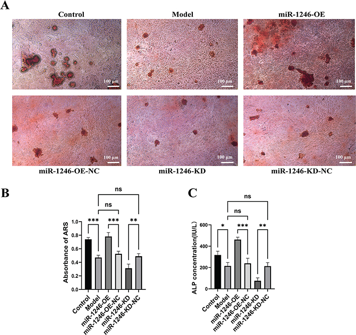
Figure 3. miR-1246 promotes the mineralization and odontogenic differentiation of hDPSCs in an in vitro inflammation model. (A) ARS staining shows the formation of mineralized nodules in different groups of hDPSCs after 15 days of odontogenic induction culture. Scale bar = 100 μm. (B) Quantitative analysis of ARS staining using 10% cetylpyridinium chloride. (C) Quantitative detection of ALP concentration in different groups of cells after 7 days of odontogenic induction culture using an ALP activity assay kit. Data are presented as the mean ± SD from three independent experiments. *P < 0.05, **P < 0.01, ***P < 0.001. ns indicates no significant difference.
Consistent with these findings, the activity of ALP, an early marker of odontogenic differentiation, was significantly reduced following LPS treatment (P < 0.05). Overexpression of miR-1246 substantially increased ALP activity (P < 0.001), whereas its knockdown led to further inhibition (P < 0.01) (Figure 3C). These data indicate that miR-1246 effectively promotes odontogenic differentiation and matrix mineralization in hDPSCs, thereby partially counteracting the inhibitory effects of the inflammatory microenvironment.
To further validate the pro-differentiative role of miR-1246, we assessed the expression of key odontogenic markers, including DSPP, DMP1, and NRP1. Both Western blot and qRT-PCR analyses demonstrated that the expression of these three proteins was markedly suppressed at both protein and mRNA levels in the LPS-induced inflammatory model. However, their expression was significantly upregulated following miR-1246 overexpression. Conversely, miR-1246 knockdown resulted in further downregulation of these markers (Figure 4A–C). Collectively, these results confirm the crucial role of miR-1246 for facilitating odontogenic differentiation of hDPSCs under inflammatory environment.
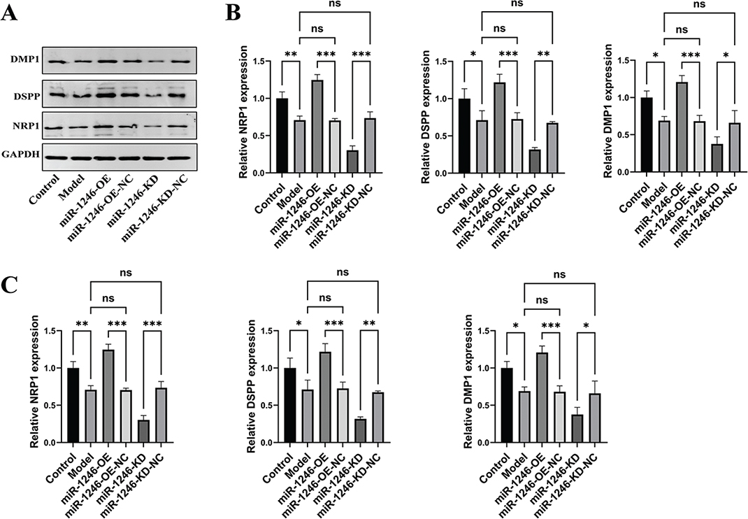
Figure 4. miR-1246 regulates the expression of odontogenic differentiation-related proteins in hDPSCs. (A) Western blot analysis of the expression levels of DMP1, DSPP, and NRP1 proteins in different groups of cells, with GAPDH used as the internal control. (B) Quantitative analysis of the Western blot bands by densitometry. (C) qRT-PCR analysis of the relative mRNA expression levels of DMP1, DSPP, and NRP1 in different groups of cells. Data are presented as the mean ± SD from three independent experiments. *P < 0.05, **P < 0.01, ***P < 0.001. ns indicates no significant difference.
AXIN2 is a direct target of miR-1246 in regulating odontogenic differentiation
Previous studies have indicated that AXIN2 is a target of miR-1246 and that this interaction may induce hDPSCs differentiation. To test this hypothesis, we first conducted a dual-luciferase reporter assay. Co-transfection of miR-1246 mimics with a reporter construct harboring the wild-type AXIN2 3’-UTR (AXIN2 WT) markedly suppressed luciferase activity relative to the control (P < 0.01). This inhibition was not observed with a reporter containing a mutated AXIN2 3’-UTR (AXIN2 mut) (Figure 5A), confirming that AXIN2 is a direct target of miR-1246.
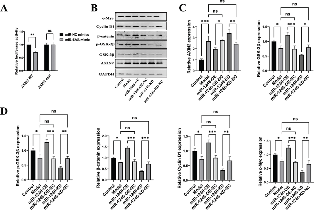
Figure 5. AXIN2 is a Direct Target of miR-1246 in Regulating Odontogenic Differentiation. (A) Dual-luciferase reporter assay confirming the targeting relationship between miR-1246 and the 3’-UTR of AXIN2. HEK293T cells were co-transfected with luciferase reporter plasmids containing either wild-type (WT) or mutated (mut) AXIN2 3’-UTR, along with miR-1246 mimics or miR-NC mimics. (B) Western blot analysis of the expression levels of Wnt/β-catenin pathway-related proteins (AXIN2, GSK-3β, p-GSK-3β, β-catenin, Cyclin D1, c-Myc) in different groups of cells, with GAPDH used as the internal control. (C-D) Quantitative analysis of the Western blot bands for AXIN2, GSK-3β, p-GSK-3β, β-catenin, Cyclin D1, and c-Myc proteins. Data are presented as the mean ± SD from three independent experiments. *P < 0.05, **P < 0.01, ***P < 0.001. ns indicates no significant difference.
Next, we investigated the levels of proteins involved in the Wnt/β-catenin pathway. Western blot and qRT-PCR analyses revealed that both protein and mRNA abundance of AXIN2 were markedly elevated in the LPS-induced inflammatory model. Overexpression of miR-1246 effectively suppressed AXIN2 expression, while its knockdown had the opposite effect (Figures 5B–D and 6A, B). As a negative regulator of the Wnt pathway, the downregulation of AXIN2 is expected to activate it. Indeed, in cells overexpressing miR-1246, we observed a significant increase in the p-GSK-3β (Ser9) and a corresponding accumulation of total β-catenin protein. Furthermore, the expression of downstream target genes Cyclin D1 and c-Myc was also significantly upregulated. Conversely, knockdown of miR-1246 resulted in decreased p-GSK-3β levels and suppressed abundance for β-catenin, Cyclin D1, and c-Myc (Figures 5B–D and 6A, B). These results strongly suggest that miR-1246 promotes the Wnt/β-catenin signaling cascade through direct binding and repression of AXIN2, thereby disrupting its role in the β-catenin degradation complex.
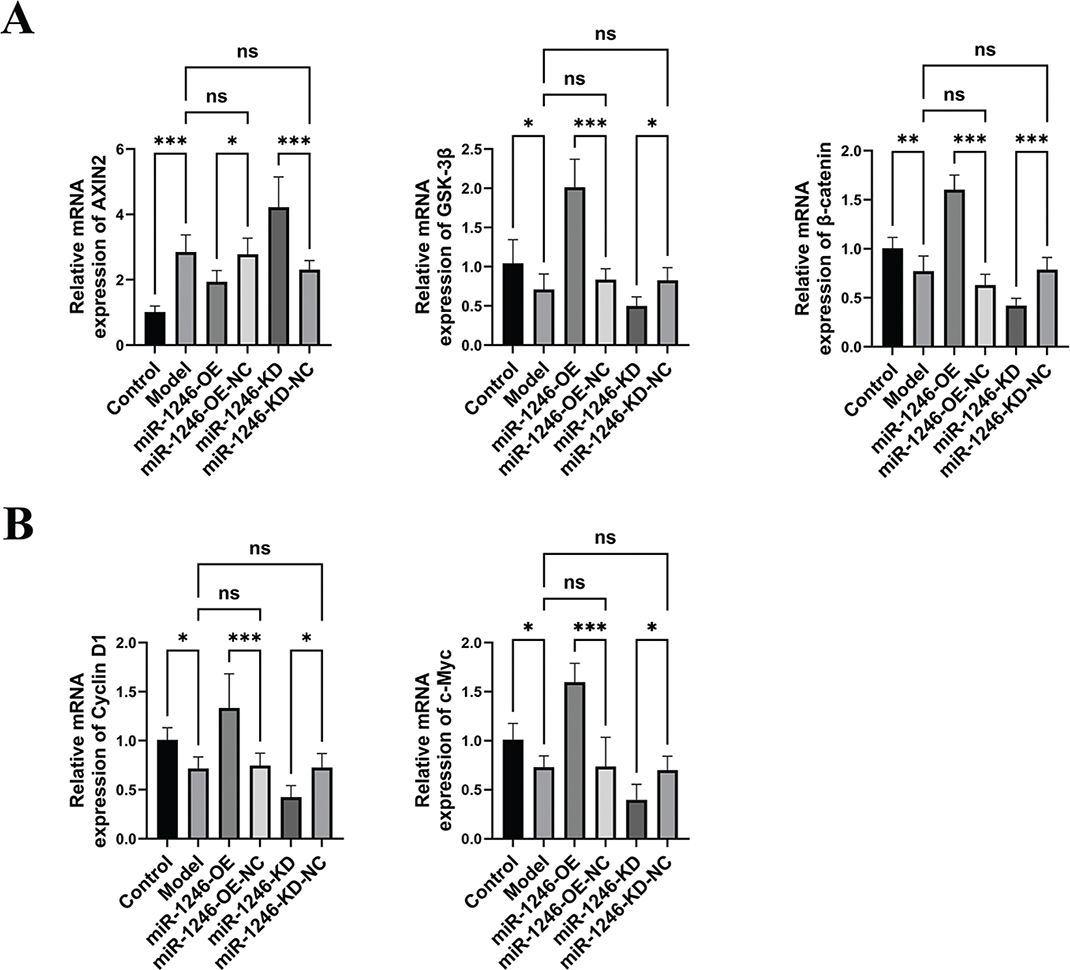
Figure 6. miR-1246 directly targets AXIN2 and regulates the transcription of Wnt pathway-related genes. (A) qRT-PCR analysis of the relative mRNA expression levels of AXIN2, GSK-3β, and β-catenin in different groups of cells. (B) qRT-PCR analysis of the relative mRNA expression levels of Wnt pathway downstream target genes Cyclin D1, and c-Myc. Data are presented as the mean ±SD from three independent experiments. *P < 0.05, ***P < 0.001. ns indicates no significant difference.
To further confirm miR-1246’s impact regarding expression and localization of key proteins within the AXIN2 and Wnt/β-catenin signaling pathway, we conducted immunofluorescence staining experiments. As demonstrated via Figure 7A–F, relative to the control group, the fluorescence intensity of AXIN2 was markedly increased in the LPS-treated model group, while the β-catenin fluorescence signal was mainly distributed in the cytoplasm with weak nuclear signals. In the LPS-induced inflammation model, overexpression of miR-1246 (miR-1246-OE group) significantly inhibited the expression of AXIN2 and resulted in a marked β-catenin nuclear enrichment as evidenced by the increased nuclear fluorescence intensity. Additionally, the fluorescence intensity of p-GSK-3β, Cyclin D1, and c-Myc was correspondingly upregulated. Conversely, knockdown of miR-1246 (miR-1246-KD group) led to further enhancement of AXIN2 expression and blocked nuclear translocation of β-catenin as well as abundance for p-GSK-3β, Cyclin D1, and c-Myc. The quantitative fluorescence analysis of protein expression in each group (Figure 8A, B) confirmed the trends seen in the Western Blot results (Figure 5B–D), further confirming that miR-1246 facilitates nuclear enrichment β-catenin and promotes the Wnt/β-catenin signaling pathway by targeting and inhibiting AXIN2.
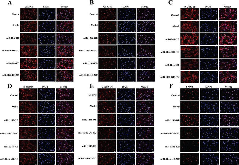
Figure 7. Immunofluorescence staining for AXIN2 and Wnt/β-catenin pathway-related proteins in hDPSCs with modulated miR-1246 expression under inflammatory conditions. Representative immunofluorescence images showing the expression and localization of AXIN2, GSK-3β, p-GSK-3β, β-catenin, Cyclin D1, and c-Myc in the Control, Model, miR-1246-OE, miR-1246-OE-NC, miR-1246-KD, and miR-1246-KD-NC groups. Nuclei were counterstained with DAPI (blue). Scale bar = 20 μm.
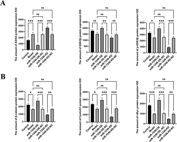
Figure 8. Quantitative analysis of immunofluorescence intensity for Wnt/β-catenin pathway-related proteins following miR-1246 modulation. (A-B) Quantification of the integrated optical density (IOD) of AXIN2, GSK3β, p-GSK3β, β-catenin, CyclinD1, and cMyc protein expression. Data are presented as mean ± SD (n = 3). *P < 0.05, *P < 0.05, **P < 0.01, ***P < 0.001. ns indicates no significant difference.
AXIN2 negatively regulates cell viability and inflammatory response in the hDPSCs inflammatory model
To elucidate the role of AXIN2 in the inflammatory microenvironment of hDPSCs, we modulated its expression using plasmid transfection and siRNA transfection in our LPS-induced model. qRT-PCR confirmed successful transfection, showing significantly increased AXIN2 mRNA levels in the AXIN2-OE group (P < 0.001) and significantly decreased levels in the AXIN2-KD group (P < 0.05) relative to their respective controls (Figure 9A). CCK-8 assays revealed that AXIN2 overexpression exacerbated the LPS-induced inhibition of cell viability, while AXIN2 knockdown partially restored it (Figure 9B). Furthermore, ELISA results showed that the LPS-induced increase in IL-1β, IL-6, and TNF-α secretion (P < 0.001) was enhanced by AXIN2 overexpression and significantly attenuated by its knockdown (Figure 9C). These findings indicate that AXIN2 negatively regulates cell viability and inflammatory responses in the LPS-induced inflammatory microenvironment of hDPSCs.
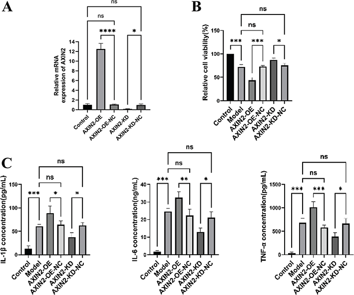
Figure 9. Effects of AXIN2 on cell viability and inflammatory response in LPS-induced hDPSCs. (A) The relative expression levels of AXIN2 in different groups of cells were detected by qRT-PCR. (B) Cell viability assessed by CCK-8 assay in different groups. (C) Concentrations of inflammatory cytokines IL-1β, IL-6, and TNF-α in the cell culture supernatant measured by ELISA. Data are presented as mean ± SD from three independent experiments. *P < 0.05, **P < 0.01, ***P < 0.001, ****P < 0.001. ns indicates no significant difference.
AXIN2 knockdown promotes migration and inhibits apoptosis of hDPSCs in an inflammatory microenvironment
We next assessed the impact of AXIN2 on hDPSC migration and apoptosis under LPS stimulation. Transwell assays confirmed that LPS treatment impaired hDPSC migration. Consistent with our previous observations, AXIN2 overexpression further inhibited cell migration, whereas its knockdown significantly restored migratory potential (Figure 10A, B).
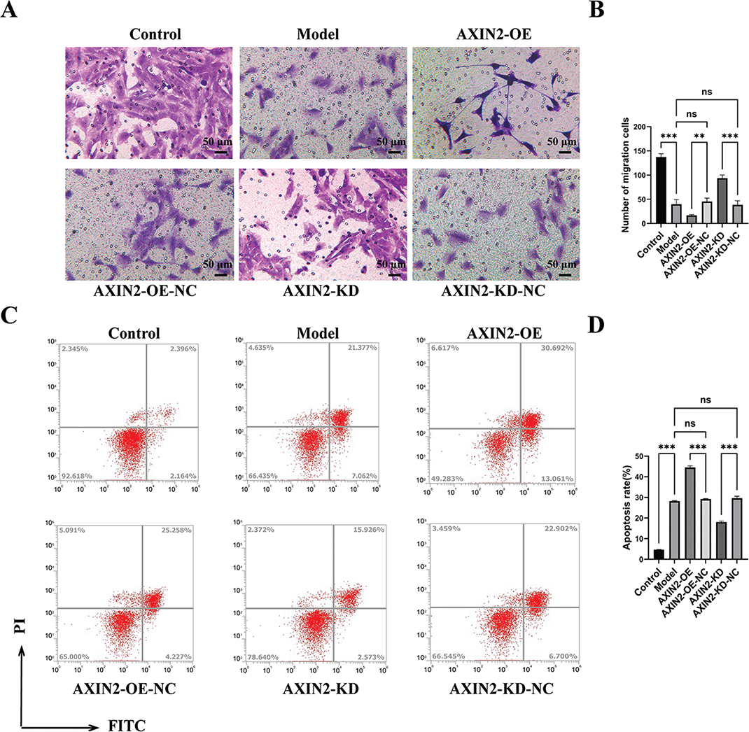
Figure 10. Effects of AXIN2 on cell migration and apoptosis in LPS-induced hDPSCs. (A-B) Representative images and quantitative analysis of hDPSC migration assessed by Transwell assay (crystal violet staining, scale bar = 50 μm). (C-D) Representative flow cytometry plots and quantitative analysis of apoptosis using Annexin V-FITC/PI staining. Data are presented as mean ± SD from three independent experiments. **P < 0.01, ***P < 0.001. ns indicates no significant difference.
An analysis via flow cytometry using Annexin V-FITC/PI staining revealed a notable rise in apoptosis within the model group exposed to LPS treatment relative to the control group (P < 0.001). This pro-apoptotic effect was amplified in the AXIN2-OE group. In contrast, silencing AXIN2 significantly reduced the apoptosis rate of LPS-treated hDPSCs (Figure 10C, D). These results indicate that, in an inflammatory context, AXIN2 negatively impacts the regenerative functions of hDPSCs by inhibiting migration and promoting apoptosis.
AXIN2 attenuates odontogenic differentiation of hDPSCs under inflammatory conditions
To determine AXIN2’s function in hDPSC odontogenic differentiation capacity, we induced differentiation in the presence of LPS. ARS staining after 15 days showed that LPS treatment significantly reduced mineralized nodule formation. AXIN2 knockdown markedly reversed this inhibition, leading to a significant increase in mineral deposition, whereas AXIN2 overexpression further suppressed mineralization (Figure 11A, B). Similarly, ALP activity, an early marker of differentiation, was reduced in the model group after 7 days of induction. AXIN2 knockdown effectively restored ALP activity, while its overexpression caused a further decline (Figure 11C). To further validate these results, we assessed the expression levels of crucial odontogenic markers. Analyses using both Western blot and qRT-PCR indicated that the expression of DMP1, DSPP, and NRP1 was diminished in the group treated with LPS. AXIN2 knockdown rescued the expression of these markers at both protein and mRNA levels, whereas AXIN2 overexpression had the opposite effect (Figure 12A–C). Taken together, this evidence suggests that AXIN2 functions as a negative modulator of hDPSC odontogenic differentiation within an inflammatory context.
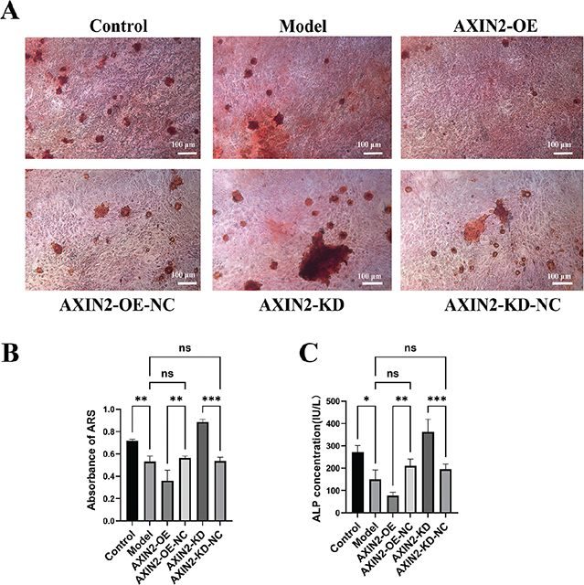
Figure 11. The effect of AXIN2 on the mineralization capacity of LPS-induced hDPSCs. (A-B) Representative images and corresponding quantitative analysis of ARS staining for mineralized nodule formation after 15 days of odontogenic induction. Scale bar = 100 μm. (C) Quantitative analysis of ALP activity after 7 days of induction. Data are presented as the mean ± standard deviation from three independent experiments. *P < 0.05, **P < 0.01, ***P < 0.001. ns indicates no significant difference.
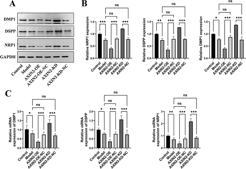
Figure 12. Effects of AXIN2 on the expression of odontoblastic differentiation markers in LPS-induced hDPSCs. (A) Western Blot analysis of DMP1, DSPP, and NRP1 protein expression. GAPDH was used as a loading control. (B) Densitometric quantification of the protein bands from (A). (C) qRT-PCR analysis of DMP1, DSPP, and NRP1 mRNA expression. Data are presented as mean ± SD from three independent experiments. *P < 0.05, **P < 0.01, ***P < 0.001. ns indicates no significant difference.
AXIN2 regulates odontogenic differentiation of hDPSCs via the Wnt/β-catenin signaling pathway
Given that AXIN2 is a key negative regulator of the Wnt/β-catenin pathway, we investigated its involvement in the observed effects. Western blot analysis confirmed that, consistent with mRNA levels, AXIN2 protein levels were elevated in the LPS-induced model group. AXIN2 silencing resulted in a marked elevation of p-GSK-3β (Ser9), indicating GSK-3β inactivation, which in turn reduced β-catenin phosphorylation and degradation, leading to β-catenin accumulation and upregulation of its downstream targets, including c-Myc as well as Cyclin D1 (Figure 13A–C). Correspondingly, qRT-PCR findings indicated that AXIN2 knockdown markedly elevated the mRNA levels of β-catenin, c-Myc, and Cyclin D1 in LPS-treated cells, whereas AXIN2 overexpression significantly reduced these levels (Figure 14A, B).
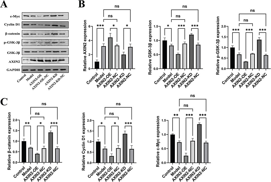
Figure 13. AXIN2 modulates the Wnt/β-catenin signaling pathway at the protein level. (A) Western Blot analysis for the expression of AXIN2, GSK-3β, p-GSK-3β, β-catenin, c-Myc, and Cyclin D1. GAPDH served as the loading control. (B, C) Densitometric quantification of the protein bands. Data are presented as mean ± SD from three independent experiments. *P < 0.05, **P < 0.01, ***P < 0.001. ns indicates no significant difference.
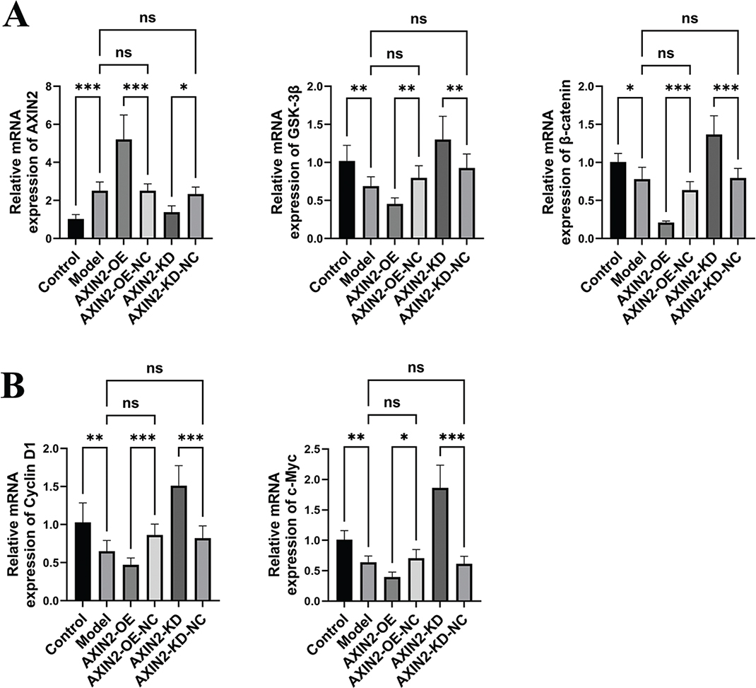
Figure 14. AXIN2 modulates the Wnt/β-catenin signaling pathway at the mRNA level. (A) qRT-PCR analysis of the mRNA expression of AXIN2, GSK-3β, and β-catenin. (B) qRT-PCR analysis of the mRNA expression of downstream targets Cyclin D1, and c-Myc. Data are presented as mean ± SD from three independent experiments. *P < 0.05, **P < 0.01, ***P < 0.001. ns indicates no significant difference.
Additionally, immunofluorescence was employed to observe alterations in the levels of pathway-related proteins following AXIN2 overexpression or knockdown. Figure 15A–F illustrates that within the LPS-stimulated model, elevated AXIN2 expression (AXIN2-OE group) resulted in significantly lower fluorescence intensities of p-GSK-3β, β-catenin, Cyclin D1, and c-Myc compared to the negative control group (AXIN2-OE-NC group), with β-catenin primarily localized in the cytoplasm. Conversely, knockdown of AXIN2 (AXIN2-KD group) significantly upregulated the expression of p-GSK-3β, β-catenin, Cyclin D1, and c-Myc, and clearly demonstrated the translocation of β-catenin from the cytoplasm to the nucleus. The quantitative fluorescence analysis results of protein expression in each group (Figure 16A, B) were highly consistent with the Western Blot results (Figure 13A–C). These findings visually demonstrate the role of AXIN2 as an inhibitory modulator within the Wnt/β-catenin signaling cascade under inflammatory conditions, with its downregulation being a critical step for promoting this pathway and facilitating β-catenin nuclear translocation. The data strongly indicate that AXIN2 exerts its suppressive influence on hDPSC odontogenic differentiation under inflammatory conditions through inhibition of the Wnt/β-catenin signaling cascade. Knockdown of AXIN2 alleviates this suppression, thereby promoting the pathway and promoting differentiation.
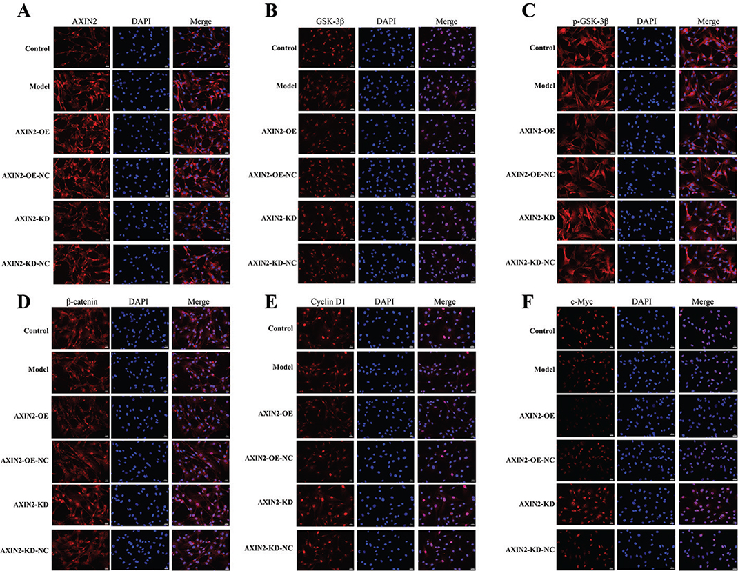
Figure 15. Immunofluorescence staining for Wnt/β-catenin pathway-related proteins in hDPSCs with modulated AXIN2 expression under inflammatory conditions. Representative immunofluorescence images showing the expression and localization of AXIN2, GSK-3β, p-GSK-3β, β-catenin, Cyclin D1, and c-Myc in the Control, Model, AXIN2-OE, AXIN2-OE-NC, AXIN2-KD, and AXIN2-KD-NC groups. Nuclei were counterstained with DAPI (blue). Scale bar = 20 μm.
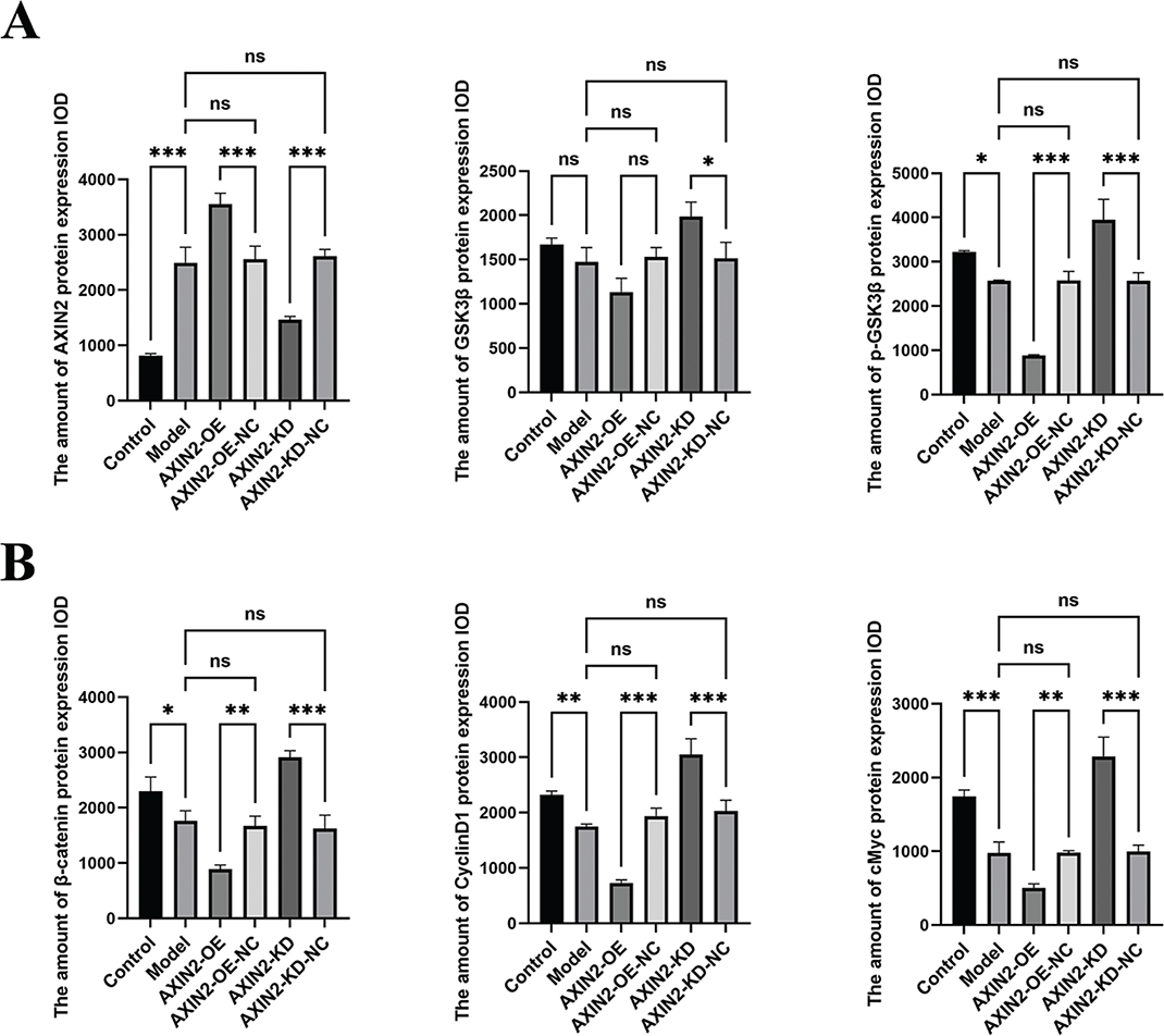
Figure 16. Quantitative analysis of immunofluorescence intensity for Wnt/β-catenin pathway-related proteins following AXIN2 modulation. (A, B) Quantification of the IOD of AXIN2, GSK3β, p-GSK3β, β-catenin, CyclinD1, and cMyc protein expression. Data are presented as mean ± SD (n = 3). *P < 0.05, **P < 0.01, ***P < 0.001. ns indicates no significant difference.
Discussion
Pulp regeneration is a current research hotspot in endodontics. The key to this process is the mobilization of endogenous or exogenous DPSCs to differentiate into odontoblasts within an inflammatory microenvironment, form reparative dentin, and reconstruct the pulp-dentin complex [21, 22]. However, the influence of the inflammatory microenvironment on DPSC proliferation, migration, and differentiation is complex, with the potential for both inhibition and promotion of their regenerative capacity, and the precise mechanisms are not fully understood [23, 24]. This research sought to examine AXIN2’s function regarding miR-1246 within an LPS-induced inflammatory model and its molecular mechanism, to provide new targets and a theoretical basis for pulp regeneration therapy.
This study first confirmed that in an LPS-induced hDPSC inflammatory model, upregulation of miR-1246 markedly enhances cellular proliferation and migratory capabilities, inhibits apoptosis, and reduces pro-inflammatory mediator levels (IL-1β, IL-6, TNF-α). This indicates that miR-1246 plays an important protective and anti-inflammatory role in the pulp’s inflammatory microenvironment. This is consistent with previous studies showing that miR-1246 has anti-inflammatory and tissue-protective functions in various disease models. For instance, one study showed that exosomes from mesenchymal stem cells can modulate their Th17/Treg cell balance by transferring miR-1246, thereby ameliorating periodontitis [25]. Our results further reveal a direct protective effect of miR-1246 in local pulp tissue inflammation. It improves hDPSC survival and regenerative ability by suppressing the inflammatory reaction. Consequently, this establishes a conducive setting for subsequent odontogenic differentiation.
More importantly, this study found that miR-1246 can effectively counteract the suppressive impact of LPS on the odontogenic development of hDPSCs. Under LPS-induced inflammatory conditions, miR-1246 overexpression markedly elevated the levels of crucial markers for odontogenic differentiation (DMP1, DSPP, NRP1) and promoted mineralized nodule formation. This suggests that miR-1246 is a key regulator for promoting odontogenic differentiation of hDPSCs. Inflammation’s impact on odontogenic differentiation is still debated. Some studies propose that severe inflammation inhibits differentiation; for example, Sonmez Kaplan et al. revealed that elevated of TNF-α, IL-1β, and IL-6 suppress the capacity for multidirectional differentiation in DPSCs [10]. Other studies propose that moderate inflammatory stimuli may actually promote reparative dentin formation. For example, Kim et al. found that LPS-induced inflammation can enhance DPSC odontogenic differentiation via the C5aR/p38 pathway [26], while Yu et al. also showed that the inflammatory milieu associated with moderate pulpitis boosts the differentiation potential of DPSCs [27]. Our findings support the latter view and suggest that miR-1246 may be a key molecule mediating this ‘pro-reparative’ inflammatory response. In early inflammation, the upregulation of miR-1246 may act as an endogenous protective mechanism, initiating cellular repair and differentiation programs while suppressing excessive inflammation. It should be noted, however, that the present study was conducted exclusively in an in vitro LPS-induced model, which cannot fully recapitulate the complex multicellular inflammatory microenvironment of pulpitis in vivo. Whether miR-1246 exerts similar pro-differentiative effects under the dynamic interactions of immune cells, the vascular system, and neural networks in vivo warrants further investigation.
To explore this molecular mechanism, our research verified through a dual-luciferase reporter analysis, which confirmed AXIN2 as a direct molecular target for miR-1246. AXIN2 functions as a negative regulator within the Wnt/β-catenin signaling cascade, inhibiting it actively through promoting the phosphorylation and degradation of β-catenin [28]. Our study found that in LPS-induced hDPSCs, miR-1246 overexpression significantly downregulates AXIN2 expression, which in turn leads to phosphorylation and activation for β-catenin and activation of its downstream target genes (Cyclin D1, c-Myc), ultimately activating the Wnt/β-catenin signaling pathway. Conversely, miR-1246 knockdown produced the opposite effect. This finding is similar to that of Xie et al., who found that within ischemia-reperfusion injury model, exosome-derived miR-1246 also activates Wnt/β-catenin signaling through targeting GSK3β (another key member of β-catenin’s degradation complex) [29]. This suggests that the miR-1246-Wnt/β-catenin axis may be a conserved regulatory pathway in the repair of various tissue injuries.
The Wnt/β-catenin signaling cascade plays a crucial role within tooth development and pulp regeneration [30, 31]. Activation of this pathway is widely confirmed for promoting odontogenic differentiation within DPSCs [32]. This study further confirmed the importance of the miR-1246/AXIN2/Wnt axis through functional rescue experiments. We found that in the LPS-induced inflammatory model, knocking down AXIN2 expression alone could mimic the effects of miR-1246 overexpression, namely, promoting hDPSC proliferation, migration, and odontogenic differentiation, while inhibiting the inflammatory response and apoptosis. This strongly demonstrates that AXIN2 is the primary downstream effector molecule through which miR-1246 exerts its biological functions. Notably, some studies have shown that during tooth injury repair, the AXIN2-expressing cell population itself serves as a progenitor for reparative odontoblasts, which differentiate via autocrine Wnt signaling [33]. Our study reveals an upstream regulator of this cell population’s activity – miR-1246 – providing a new avenue for more precise targeted regulation of pulp regeneration.
In summary, this study systematically elucidates the key role and molecular mechanism of miR-1246 in an LPS-induced hDPSC inflammatory model. We have demonstrated the important role of the ‘miR-1246-AXIN2-Wnt/β-catenin’ signaling axis in regulating the response of dental pulp stem cells to inflammatory stimuli and initiating the process of odontogenic differentiation in vitro. These results not only enhance our comprehension regarding mechanisms of pulpitis and repair but also provide a significant experimental basis for developing new pulp regeneration strategies based on miR-1246. From a translational perspective, local delivery of miR-1246 or its agonists could be incorporated into vital pulp therapy protocols or direct pulp capping materials to enhance the regenerative capacity of inflamed dental pulp, potentially improving clinical outcomes of pulp preservation strategies [34, 35].
However, our research has specific constraints. First, all experiments were conducted in vitro using an LPS-induced model. While LPS treatment is a well-established approach for simulating bacterial infection-induced inflammation, it cannot fully recapitulate the complex, multicellular microenvironment of pulpitis in vivo, which involves dynamic interactions among immune cells, the vascular system, and neural networks. Future in vivo studies using animal models of pulpitis are therefore warranted to validate the translational relevance of these findings. Second, while we have clearly elucidated the miR-1246/AXIN2/Wnt pathway, we have not excluded the possibility that miR-1246 may act synergistically by targeting other genes or pathways. Future research should focus on validating the therapeutic efficacy of miR-1246 in an in vivo animal model of pulpitis. Additionally, exploring the upstream mechanisms that regulate miR-1246 expression during inflammation is another valuable research direction.
Conclusion
Our research demonstrates the molecular mechanism by which miR-1246 promotes the odontogenic differentiation of hDPSCs and partially counteracts the detrimental effects of inflammation in an LPS-induced inflammatory model in vitro. It achieves this by targeting and inhibiting AXIN2, which in turn activates the Wnt/β-catenin signaling pathway. Our results suggest that miR-1246 is a key regulatory factor for initiating endogenous repair and regeneration programs in pulp tissue within an inflammatory microenvironment. While these in vitro findings provide a new perspective for understanding the regulatory network of pulp inflammation and repair, further in vivo studies are required to confirm the therapeutic potential of targeting the miR-1246/AXIN2/Wnt signaling axis for future pulp vitality preservation and regeneration therapies.
Authors’ contributions
S.Y.W.: Conceptualization, Data curation, Formal analysis, Investigation, Methodology, Writing – original draft.
Y.Y.W.: Data curation, Formal analysis, Investigation.
J.Y.Y.: Supervision, Validation, Writing – review & editing.
Data availability
The data that support the findings of this study are not publicly available due to privacy reasons, but are available from the corresponding author upon request.
Ethics declarations
Not applicable.
Patient consent statement
Not applicable.
Clinical trial number
Not applicable.
References
[1] Bar JK, Lis-Nawara A, Grelewski PG. Dental pulp stem cell-derived secretome and its regenerative potential. Int J Mol Sci. 2021;22:12018. https://doi.org/10.3390/ijms222112018
[2] Sloan AJ, Smith AJ. Stem cells and the dental pulp: potential roles in dentine regeneration and repair. Oral Dis. 2007;13:151–7. https://doi.org/10.1111/j.1601-0825.2006.01346.x
[3] Bugueno IM, Alastra G, Balic A, Stadlinger B, Mitsiadis TA. Limited adipogenic differentiation potential of human dental pulp stem cells compared to human bone marrow stem cells. Int J Mol Sci. 2024;25:11105. https://doi.org/10.3390/ijms252011105
[4] Chmilewsky F, Liang R, Kanazawa M, About I, Cooper LF, George A. C5L2 regulates DMP1 expression during odontoblastic differentiation. J Dent Res. 2019;98:597–604. https://doi.org/10.1177/0022034518820461
[5] Galler KM, Weber M, Korkmaz Y, Widbiller M, Feuerer M. Inflammatory response mechanisms of the dentine-pulp complex and the periapical tissues. Int J Mol Sci. 2021;22:1480. https://doi.org/10.3390/ijms22031480
[6] Colombo JS, Moore AN, Hartgerink JD, D’Souza RN. Scaffolds to control inflammation and facilitate dental pulp regeneration. J Endod. 2014;40(4 Suppl):S6–12. https://doi.org/10.1016/j.joen.2014.01.019
[7] Arora S, Cooper PR, Friedlander LT, Rizwan S, Seo B, Rich AM, et al. Potential application of immunotherapy for modulation of pulp inflammation: opportunities for vital pulp treatment. Int Endod J. 2021;54:1263–74. https://doi.org/10.1111/iej.13524
[8] Gronthos S, Mankani M, Brahim J, Robey PG, Shi S. Postnatal human dental pulp stem cells (DPSCs) in vitro and in vivo. Proc Natl Acad Sci U S A. 2000;97:13625–30. https://doi.org/10.1073/pnas.240309797
[9] Yang X, Li L, Xiao L, Zhang D. Recycle the dental fairy’s package: overview of dental pulp stem cells. Stem Cell Res Ther. 2018;9:347. https://doi.org/10.1186/s13287-018-1094-8
[10] Sonmez Kaplan S, Sazak Ovecoglu H, Genc D, Akkoc T. TNF-α, IL-1B and IL-6 affect the differentiation ability of dental pulp stem cells. BMC Oral Health. 2023;23:555. https://doi.org/10.1186/s12903-023-03288-1
[11] Pezelj-Ribaric S, Anic I, Brekalo I, Miletic I, Hasan M, Simunovic-Soskic M. Detection of tumor necrosis factor alpha in normal and inflamed human dental pulps. Arch Med Res. 2002;33:482–4. https://doi.org/10.1016/s0188-4409(02)00396-x
[12] Bucchi C, Bucchi A, Martínez-Rodríguez P. Biological properties of dental pulp stem cells isolated from inflamed and healthy pulp and cultured in an inflammatory microenvironment. J Endod. 2023;49:395–401.e6. https://doi.org/10.1016/j.joen.2023.02.002
[13] Inostroza C, Vega-Letter AM, Brizuela C, Castrillón L, Saint Jean N, Duran CM, et al. Mesenchymal stem cells derived from human inflamed dental pulp exhibit impaired immunomodulatory capacity in vitro. J Endod. 2020;46:1091–8.e2. https://doi.org/10.1016/j.joen.2020.05.003
[14] Morsczeck C. Cellular senescence in dental pulp stem cells. Arch Oral Biol. 2019;99:150–5. https://doi.org/10.1016/j.archoralbio.2019.01.012
[15] Chang C, Weiping L, Jibing C. Exosomal MiRNA therapy for central nervous system injury diseases. Cell Mol Neurobiol. 2024;45:3. https://doi.org/10.1007/s10571-024-01522-0
[16] Ramprosand S, Govinden-Soulange J, Ranghoo-Sanmukhiya VM, Sanan-Mishra N. miRNA, phytometabolites and disease: connecting the dots. Phytother Res. 2024;38:4570–91. https://doi.org/10.1002/ptr.8287
[17] Ye YY, Yue L, Zou XY, Wang XY. [Characteristics and microRNA expression profile of exosomes derived from odontogenic dental pulp stem cells]. Beijing Da Xue Xue Bao Yi Xue Ban. 2023;55:689–96. https://doi.org/10.19723/j.issn.1671-167X.2023.04.020
[18] Liu L, Silke J. AXIN2 is a non-redundant regulator of AXIN1 stability and β-catenin in colorectal cancer cells. FEBS J. 2025;292:990–4. https://doi.org/10.1111/febs.17336
[19] Choi H, Jeong J-K, Adasooriya D, Cho S-W, Cho E-S. Notum as a crucial regulator of matrix integrity in dentinogenesis. J Cell Physiol. 2025;240:e70070. https://doi.org/10.1002/jcp.70070
[20] Lan C, Chen S, Jiang S, Lei H, Cai Z, Huang X. Different expression patterns of inflammatory cytokines induced by lipopolysaccharides from Escherichia coli or Porphyromonas gingivalis in human dental pulp stem cells. BMC Oral Health. 2022;22:121. https://doi.org/10.1186/s12903-022-02161-x
[21] Liang C, Liao L, Tian W. Stem cell-based dental pulp regeneration: insights from signaling pathways. Stem Cell Rev Rep. 2021;17(4):1251–1263. https://doi.org/10.1007/s12015-020-10117-3
[22] Amir M, Jeevithan L, Barkat M, Fatima SH, Khan M, Israr S, et al. Advances in regenerative dentistry: a systematic review of harnessing Wnt/β-Catenin in dentin-pulp regeneration. Cells. 2024;13:1153. https://doi.org/10.3390/cells13131153
[23] Ma Y, Liu X, Dai R, Li Q, Cao CY. LL-37 regulates odontogenic differentiation of dental pulp stem cells in an inflammatory microenvironment. Stem Cell Res Ther. 2024;15:469. https://doi.org/10.1186/s13287-024-04075-7
[24] He W, Wang Z, Luo Z, Yu Q, Jiang Y, Zhang Y, et al. LPS promote the odontoblastic differentiation of human dental pulp stem cells via MAPK signaling pathway. J Cell Physiol. 2015;230:554–61. https://doi.org/10.1002/jcp.24732
[25] Xia Y, Cheng T, Zhang C, Zhou M, Hu Z, Kang F, et al. Human bone marrow mesenchymal stem cell-derived extracellular vesicles restore Th17/Treg homeostasis in periodontitis via miR-1246. FASEB J. 2023;37:e23226. https://doi.org/10.1096/fj.202300674RR
[26] Kim J-H, Irfan M, Hossain MA, Shin S, George A, Chung S. LPS-induced inflammation potentiates dental pulp stem cell odontogenic differentiation through C5aR and p38. Connect Tissue Res. 2023;64:505–15. https://doi.org/10.1080/03008207.2023.2218944
[27] Yu S, Liu X-M, Liu Y, Tang L, Lei S, Geng C, et al. Inflammatory microenvironment of moderate pulpitis enhances the osteo-/odontogenic potential of dental pulp stem cells by autophagy. Int Endod J. 2024;57:1465–77. https://doi.org/10.1111/iej.14108
[28] Kornsuthisopon C, Photichailert S, Nowwarote N, Tompkins KA, Osathanon T. Wnt signaling in dental pulp homeostasis and dentin regeneration. Arch Oral Biol. 2022;134:105322. https://doi.org/10.1016/j.archoralbio.2021.105322
[29] Xie K, Liu L, Chen J, Liu F. Exosomes derived from human umbilical cord blood mesenchymal stem cells improve hepatic ischemia reperfusion injury via delivering miR-1246. Cell Cycle. 2019;18:3491–501. https://doi.org/10.1080/15384101.2019.1689480
[30] Wu S, Xu X, Gao S, Huo S, Wan M, Zhou X, et al. MicroRNA-93-5p regulates odontogenic differentiation and dentin formation via KDM6B. J Transl Med. 2024;22:54. https://doi.org/10.1186/s12967-024-04862-z
[31] Liu P, Zhang Q, Mi J, Wang S, Xu Q, Zhuang D, et al. Exosomes derived from stem cells of human deciduous exfoliated teeth inhibit angiogenesis in vivo and in vitro via the transfer of miR-100-5p and miR-1246. Stem Cell Res Ther. 2022;13:89. https://doi.org/10.1186/s13287-022-02764-9
[32] Liu S, Sun J, Yuan S, Yang Y, Gong Y, Wang Y, et al. Treated dentin matrix induces odontogenic differentiation of dental pulp stem cells via regulation of Wnt/β-catenin signaling. Bioact Mater. 2022;7:85–97. https://doi.org/10.1016/j.bioactmat.2021.05.026
[33] Babb R, Chandrasekaran D, Vitor CMN, Sharpe PT. Axin2-expressing cells differentiate into reparative odontoblasts via autocrine Wnt/β-catenin signaling in response to tooth damage. Sci Rep. 2017;7:3102. https://doi.org/10.1038/s41598-017-03145-6
[34] Godoi-Lopes J, Ribeiro-Silva VHA, Candido Do Prado LG, Petean IBF, Magri LV, Lopes-Olhê FC, et al. Efficacy of dental stem cell–derived exosomes for pulp regeneration: a systematic review of clinical, animal, and in vitro studies. Mol Biol Rep. 2026;53:426. https://doi.org/10.1007/s11033-026-11547-x
[35] Nasiri K, Jahri M, Kolahdouz S, Soleimani M, Makiya A, Saini RS, et al. MicroRNAs function in dental stem cells as a promising biomarker and therapeutic target for dental diseases. Mol Diagn Ther. 2023;27:703–22. https://doi.org/10.1007/s40291-023-00675-w