ORIGINAL ARTICLE
Medication-related osteonecrosis of the jaw in cancer and osteoporosis patients: a retrospective seven-year single-center Norwegian cohort study of clinical profiles and documented soft tissue healing
Safiyye Süslüa, Håvard J. Haugena, Tormod B. Krügerb, Janne Elin Reselanda and Bente Brokstad Herlofsonb
aDepartment of Biomaterials, Institute of Clinical Dentistry, University of Oslo, Oslo, Norway; bDepartment of Oral Medicine and Oral Surgery, Institute of Clinical Dentistry, University of Oslo, Oslo, Norway
ABSTRACT
Objective: To compare systemic profiles, oral findings, and documented soft tissue healing between cancer and osteoporosis patients with medication-related osteonecrosis of the jaw (MRONJ).
Material and methods: This retrospective single-center cohort included 105 cases treated in 2013–2019 (56 cancer; 49 osteoporosis). Healing at last follow-up was analyzed using adjusted logistic regression and Firth sensitivity analysis.
Results: Of 102 cases with known final status, 68 had documented healing. Osteoporosis cases were older, predominantly female, and had longer antiresorptive exposure than cancer cases (76.7 ± 11.1 vs. 70.0 ± 8.5 years; 102.5 ± 77.8 vs. 34.6 ± 28.5 months; both p < 0.001). Stage II predominated and Stage 0 was uncommon (3/105, 2.9%). Previous tooth extraction at the MRONJ lesion site was more frequent in osteoporosis (95.9% vs. 71.4%; p < 0.001). Surgical treatment was associated with higher healing odds (adjusted odds ratio [OR]: 11.12, 95% confidence interval [CI]: 4.12 to 33.35; p < 0.001), whereas cancer indication was associated with lower odds (adjusted OR: 0.23, 95% CI: 0.07 to 0.68; p = 0.011).
Conclusions: Systemic profiles differed; oral patterns were similar. Non-random treatment and missing healing dates limit interpretation. Previous tooth extraction was the most frequent antecedent event; its indication and timing were unverified.
KEYWORDS: Osteonecrosis of the jaw; antiresorptive agents; neoplasms; osteoporosis; tooth extraction; retrospective studies
Citation: ACTA ODONTOLOGICA SCANDINAVICA 2026; VOL. 85: 713–724. DOI: https://doi.org/10.2340/aos.v85.46983.
Copyright: © 2026 The Author(s). Published by MJS Publishing on behalf of Acta Odontologica Scandinavica Society. This is an Open Access article distributed under the terms of the Creative Commons Attribution 4.0 International License (http://creativecommons.org/licenses/by/4.0/), allowing third parties to copy and redistribute the material in any medium or format and to remix, transform, and build upon the material, with the condition of proper attribution to the original work.
Received: 23 February 2026; Accepted: 26 September 2026; Published: 09 October 2026.
CONTACT: Håvard J. Haugen haavarjh@uio.no Department of Biomaterials, Institute of Clinical Dentistry, University of Oslo, Geitmyrsveien 71, 0455 Oslo, Norway.
Supplemental data for this article can be accessed online at https://doi.org/10.2340/aos.v85.46983
Competing interests and funding: The authors report there are no competing interests to declare.
This research received no specific grant from any funding agency in the public, commercial, or not-for-profit sectors.
Introduction
Medication-related osteonecrosis of the jaw (MRONJ) is the current umbrella term for jaw osteonecrosis associated with antiresorptive and other relevant bone-modifying or antiangiogenic therapies. Historically, the entity was first described as bisphosphonate-related osteonecrosis of the jaw (BRONJ) in early American Association of Oral and Maxillofacial Surgeons (AAOMS) position papers, before the terminology broadened to MRONJ as denosumab and other non-bisphosphonate agents entered routine use [1–4]. Because the present cohort spans that terminology transition, we use MRONJ consistently in this paper and refer to BRONJ only when discussing historical definitions.
Antiresorptive therapy is used across a wider clinical spectrum than osteoporosis alone. Lower-dose regimens are used for osteoporosis and other metabolic bone disorders, and bone-targeted treatment is also used for reducing the risk of cancer recurrence and for cancer treatment-induced bone loss in selected patients receiving endocrine or other anticancer therapy [2, 5–7]. Higher-intensity regimens are most commonly used in malignant diseases with skeletal involvement, but the distinction between “low-dose” and “high-dose” therapy is better understood as a continuum of treatment intensity rather than an absolute disease-based separation [8, 9].
Current pathobiology models emphasize interactions between suppressed bone remodeling, altered oral barrier repair, local infection/inflammation, and treatment-related changes in host response, rather than a single isolated mechanism [10–13]. Consistent with that framework, periodontal or periapical infection and dental extractions are repeatedly reported in MRONJ case series, but extraction often occurs in already diseased teeth, making causal interpretation difficult [14–16].
MRONJ management remains controversial. CContemporary position papers and consensus documents recognize both non-surgical and surgical approaches to MRONJ management, but comparisons of their outcomes remain vulnerable to confounding by indication [6, 7, 17, 18]. Cohort studies and systematic reviews report favorable outcomes with appropriately planned surgery [19–22]. Structured non-surgical wound care may also resolve MRONJ and remains an option for patients unable to undergo surgery [23], although other cohorts have reported limited healing and progression during non-surgical management [24]. Seluki et al.’s systematic review supports consideration of disease stage when selecting treatment [19]; contemporary position papers also emphasize the patient’s systemic condition and local disease extent [6, 7]. Prospective studies have additionally assessed quality of life following MRONJ treatment [25, 26].
Published studies address different aspects of MRONJ, including pharmacological management followed by removal of bone sequestra [27], MRONJ after tooth extraction in patients treated for osteoporosis [28], surgical outcomes [20], and survival among patients with cancer [29]. These studies differ in populations, treatment protocols, and outcome definitions, limiting direct comparison across indications. Consequently, well-characterized MRONJ cohorts are needed with comprehensive systemic and local data, clearly defined clinical outcomes, transparent follow-up reporting, and appropriately adjusted analyses.
This retrospective single-center Norwegian cohort study aimed to compare systemic profiles, oral findings, AAOMS stage distribution, recorded antecedent dental events, treatment patterns, follow-up characteristics, and documented clinical soft tissue healing status at the last recorded follow-up between cancer-related and osteoporosis-related MRONJ cases.
Materials and methods
Study design and patient selection
This retrospective single-center case-only cohort study was conducted at the Department of Oral Surgery and Oral Medicine, University of Oslo, Norway. All patients referred between July 2013 and December 2019 with a verified MRONJ diagnosis according to the contemporaneous AAOMS criteria were included in the analytical cohort; no patients with diagnosed MRONJ were excluded. All included patients consented to their inclusion in the study and the registration of their clinical data for research. The cohort therefore comprised 105 consecutive MRONJ cases, including 56 cancer-related and 49 osteoporosis-related cases. The study was approved by the Regional Committee for Medical and Health Research Ethics South-East B, Norway (REK 2013/1053).
Case definition and terminology
Cases were diagnosed during the study period according to contemporaneous AAOMS criteria. For this manuscript, all cases are described as MRONJ, with historical BRONJ terminology retained only when discussing earlier AAOMS position papers [1, 2]. The 2022 AAOMS position paper [7] informed the terminology and conceptual framing of this revision, but cases were not retrospectively re-adjudicated beyond harmonized staging. Staging was harmonized as Stage 0, I, II, or III; non-exposed disease in the dataset was treated as Stage 0. Because Stage 0 is diagnostically non-specific and no structured radiographic dataset was available for systematic retrospective re-evaluation, Stage 0 analyses were interpreted descriptively and examined in sensitivity analyses.
Exposure classification
The primary clinical grouping was cancer-related versus osteoporosis-related MRONJ, based on the recorded indication for bone-modifying agent (BMA) treatment. A proxy high-dose versus low-dose BMA classification could be derived by combining indication, agent, route, and dosing frequency. Exact administered dose, cumulative exposure, adherence, and detailed switching chronology were not available in a sufficiently complete form. In this dataset, the proxy was concordant with the indication grouping and therefore provided no independent information; it was retained only as a transparency check and was not included in the same adjusted model as the indication (Supplementary Table S11).
Variables and data collection
The dataset included age, sex, indication for antiresorptive therapy, cancer subtype where applicable, antiresorptive agent, route, dosing frequency, and antiresorptive therapy (ART) duration. Oral variables included MRONJ stage, jaw location, lesion-site dental findings, tooth attrition/bruxism, a recorded history of previous tooth extraction at the MRONJ lesion site, denture trauma, and prior endodontic treatment. Pain (visual analogue scale [VAS]) was recorded but was not retained in the final comparative table because the coding range was inconsistent. Among the 87 patients with an extraction recorded at the MRONJ lesion site, tooth mobility, local endodontic treatment, tooth or crown fracture, previous deep caries, apical periodontitis, and generalized marginal bone loss were recorded as possible clinical reasons or dental conditions associated with extraction. However, these non-mutually-exclusive variables were not labelled in the dataset as verified extraction indications, and their timing relative to extraction and MRONJ recognition was unavailable. Implant involvement was recorded separately as local implant-related context (Figure 1; Supplementary Table S8).
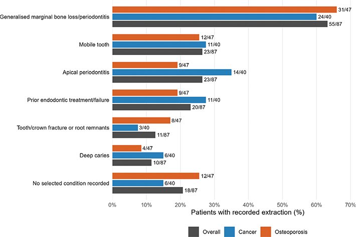
Figure 1. Recorded possible reasons or local dental conditions associated with extraction among patients with an extraction recorded at the MRONJ lesion site. Bars show the proportions of multiple responses overall (n = 87), in cancer-related cases (n = 40), and in osteoporosis-related cases (n = 47). Individual patients could have more than one recorded condition, so percentages do not sum to 100%.
Systemic variables included predefined comorbidities and comedications recorded at the time of MRONJ diagnosis. However, a valid full Charlson derivation was not possible because several required diagnostic categories were absent, aggregated, or insufficiently specific in the dataset.
Outcomes, treatment definitions, and follow-up
Treatment was classified into two groups: non-surgical management and surgical treatment. Surgical treatment included sequestrectomy, debridement, or marginal bone resection followed by primary soft tissue closure using local flaps. This patient cohort underwent no segmental resections. According to the clinic’s general treatment protocol, antibiotics and antimicrobial mouth rinses formed part of surgical care and were used when clinically indicated during non-surgical management. Patient-level regimen, duration, and local wound-care data were not available in a sufficiently structured form for analysis. All included patients discontinued ART after their MRONJ diagnosis, at the discretion of the treating oncologist or osteologist. This represented post-diagnosis ART discontinuation. Bedogni et al. distinguish this therapeutic context from a prophylactic drug holiday before dentoalveolar surgery in patients without established or suspected MRONJ [6]. Exact timing and duration were unavailable, and the independent effect of post-diagnosis discontinuation could not be evaluated.
Clinical soft tissue healing was defined as documented mucosal coverage in the area of previously exposed bone or a final recorded status of soft tissue healed at the affected site. The structured dataset captured the follow-up duration, in months, from the start of MRONJ treatment to the last recorded follow-up visit. In contrast, the exact date of the first documented healing was unavailable. Accordingly, the primary analysis evaluated documented clinical soft tissue healing status at the last recorded follow-up rather than a conventional time-to-first-healing endpoint. No predefined minimum follow-up threshold was applied; the primary final-status analysis was restricted to cases with a known final clinical outcome. Mortality was summarized descriptively because the timing of death was recorded relative to MRONJ diagnosis rather than treatment start.
Panoramic radiography was routinely used, and cone-beam computed tomography (CBCT) was obtained for MRONJ diagnosis and thereafter when clinically indicated. Serial radiographic findings were not available in a standardized analytical dataset. Consequently, all reported healing outcomes refer to documented clinical soft tissue status and do not establish radiographic healing or complete disease resolution.
Statistical methods
Group comparisons used Student’s t-test or Wilcoxon rank-sum test for continuous variables. Categorical variables were compared using Pearson’s chi-square test or Fisher’s exact test, as appropriate. P-values are reported to three decimal places, with p < 0.001 used for values below 0.001. To characterize differences between cancer-related and osteoporosis-related MRONJ within this case-only cohort, we first used descriptive comparisons. Then we fitted a multivariable logistic regression model with indication group (cancer vs. osteoporosis) as the dependent variable. Covariates were selected a priori from clinically relevant demographic, treatment, systemic, and oral variables. Model discrimination was assessed by the area under the receiver operating characteristic curve (AUC), and calibration was assessed by comparing observed and predicted probabilities across deciles of fitted probability. Because some variables showed quasi-complete separation, a Firth-penalized logistic regression was fitted as a sensitivity analysis. A reduced-sensitivity model, excluding chemotherapy and antiangiogenic therapy, assessed whether the contrast between indication groups persisted without explicitly including diagnosis-driven oncological treatment variables. Multiple correspondence analysis followed by k-means clustering was used as an exploratory descriptive visualization; because indication was included among the input variables, it was not interpreted as independent confirmation of indication-specific profiles.
Documented clinical soft tissue healing at the last recorded follow-up was analyzed as a binary endpoint. The primary multivariable model included treatment group, indication group, and baseline stage group (Stage 0/I, Stage II, Stage III). Standard logistic regression was fitted first, and Firth-penalized logistic regression was treated as a small-sample sensitivity analysis. Additional sensitivity analyses added sex, exploratory comedication burden, or both to the primary model; classified unknown final status as no documented healing; excluded Stage 0 cases; or added age or ART duration. A secondary model also accounted for the duration of documented clinical observation. The high-dose versus low-dose BMA proxy was concordant with indication and was therefore not treated as an independent sensitivity analysis. Because the number of non-healed outcomes was limited, no single model was interpreted causally or as definitive for all measured covariates; residual confounding by performance status and other unmeasured factors remains possible. Model diagnostics and analytic sample flow are provided in Supplementary Tables S9 and S10.
Exploratory comorbidity burden was examined using Poisson regression adjusted for age, with a second model additionally adjusted for sex. Follow-up duration was reported descriptively as months from treatment start to the last recorded follow-up visit. It was included only in secondary ascertainment analyses because it may be outcome-dependent. Exact binomial confidence intervals were used for key healed proportions. Supplementary Table S6 summarizes variable-level missingness.
Results
Study population and antiresorptive therapy characteristics
The cohort comprised 105 MRONJ cases: 56 cancer-related and 49 osteoporosis-related. Osteoporosis patients were older than cancer patients (76.7 ± 11.1 vs. 70.0 ± 8.5 years) and were more often female (43/49, 87.8% vs. 25/56, 44.6%; both p < 0.001). Antiresorptive exposure was markedly longer in osteoporosis than in cancer (102.5 ± 77.8 vs. 34.6 ± 28.5 months, p < 0.001), whereas cancer-related MRONJ clustered around monthly subcutaneous or intravenous regimens and osteoporosis-related MRONJ around oral weekly or other lower-frequency regimens (Table 1; Supplementary Figure S2).
The exploratory comorbidity score was higher in osteoporosis than in cancer (2.0 ± 1.6 vs. 1.1 ± 1.1, p = 0.002), whereas the exploratory comedication score was higher in cancer than in osteoporosis (2.5 ± 1.1 vs. 1.9 ± 1.4, p = 0.012) (Table 1). Supplementary Tables S2 and S3 provide detailed distributions of variable-level systemic comorbidity and concomitant medications.
In supplementary indication models, shorter antiresorptive treatment duration remained the clearest marker of cancer indication after exclusion of chemotherapy and antiangiogenic therapy, and the Firth-penalized sensitivity model yielded directionally concordant estimates (Supplementary Tables S4 and S5). Model discrimination and calibration were assessed in-sample and were good (Supplementary Figures S3 and S4). Multiple correspondence analysis provided a descriptive visualization of partially overlapping profiles; because indication was included in the analysis, it was not interpreted as independent confirmation of the two groups (Supplementary Figure S1).
Oral findings, antecedent dental events, lesion distribution, and stage
A recorded history of previous tooth extraction at the MRONJ lesion site was present in 87/105 cases (82.9%) overall, including 47/49 osteoporosis cases (95.9%) and 40/56 cancer cases (71.4%; p < 0.001) (Table 2). Among these 87 extraction cases, the most frequently recorded possible reason or associated local dental condition was generalized marginal bone loss/periodontitis (55/87, 63.2%), followed by mobile teeth and apical periodontitis (both 23/87, 26.4%) and prior endodontic treatment/failure (20/87, 23.0%); individual patients could have more than one recorded condition. No selected condition was recorded in 18/87 cases (20.7%) (Figure 1; Supplementary Table S8; Supplementary Figure S7). Tooth attrition/bruxism was more frequent in cancer than in osteoporosis patients (19/56, 33.9% vs. 8/49, 16.3%; p = 0.040) (Table 2).
| Characteristic | Osteoporosis (n = 49) | Cancer (n = 56) | P | ||
| MRONJ stage: Stage 0 | 2 (4.1%) | 1 (1.8%) | 0.380 | ||
| MRONJ stage: Stage I | 13 (26.5%) | 12 (21.4%) | |||
| MRONJ stage: Stage II | 26 (53.1%) | 26 (46.4%) | |||
| MRONJ stage: Stage III | 8 (16.3%) | 17 (30.4%) | |||
| Lesion site: Maxilla | 14 (28.6%) | 23 (41.1%) | 0.208 | ||
| Lesion site: Mandible | 27 (55.1%) | 29 (51.8%) | |||
| Lesion site: Both jaws | 8 (16.3%) | 4 (7.1%) | |||
| Previous tooth extraction at MRONJ lesion site | 47 (95.9%) | 40 (71.4%) | < 0.001 | ||
| Mobile tooth at lesion site | 13 (26.5%) | 19 (33.9%) | 0.411 | ||
| Endodontic treatment/failure at lesion site | 9 (18.8%) | 16 (28.6%) | 0.243 | ||
| Tooth fracture at lesion site | 9 (18.4%) | 5 (8.9%) | 0.156 | ||
| Deep caries at lesion site | 4 (8.5%) | 6 (10.9%) | 0.749 | ||
| Apical periodontitis at lesion site | 10 (20.8%) | 19 (33.9%) | 0.138 | ||
| General marginal bone loss/periodontitis | 32 (65.3%) | 33 (58.9%) | 0.502 | ||
| Tooth attrition/bruxism | 8 (16.3%) | 19 (33.9%) | 0.040 | ||
| Overall p-values are shown once for the stage and lesion-site blocks. Categorical variables were compared using Pearson’s chi-square test or Fisher’s exact test, as appropriate. Percentages are based on available observations; variable-level missingness is summarized in Supplementary Table S6. | |||||
Lesion-site distribution was recorded as mandible-only in 56/105 cases (53.3%), maxilla-only in 37/105 cases (35.2%), and both jaws in 12/105 cases (11.4%). Posterior premolar/molar involvement was recorded in 93/105 cases (88.6%; 95% CI: 80.9–94.0%) (Figure 2). Individual patients could contribute more than one affected tooth site to the tooth-position plot.
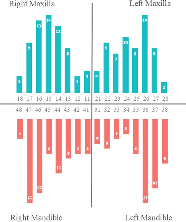
Figure 2. Dental sites of MRONJ lesions. Bars show affected tooth sites by region in the total study population (n = 105), separately for the mandible and maxilla; individual patients could contribute more than one affected tooth site. Patient-level exclusive jaw categories showed mandible-only involvement in 56/105 cases (53.3%), maxilla-only involvement in 37/105 cases (35.2%), and both-jaw involvement in 12/105 cases (11.4%).
Overall, Stage 0 occurred in 3/105 cases (2.9%), Stage I in 25/105 (23.8%), Stage II in 52/105 (49.5%), and Stage III in 25/105 cases (23.8%). Stage II predominated in both indication groups, and stage distributions did not materially differ between osteoporosis-related and cancer-related MRONJ (Figure 3; Table 2; Supplementary Table S7). Stage 0 or non-exposed disease was uncommon in both groups, accounting for 2/49 cases (4.1%) in osteoporosis and 1/56 cases (1.8%) in cancer. Supplementary Table S1 provides an expanded descriptive summary of stage, jaw localization, treatment, follow-up, and clinical outcome.
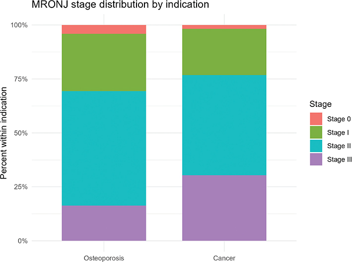
Figure 3. MRONJ stage distribution by indication group. Stacked bars show the percentage distribution of MRONJ stages within the osteoporosis and cancer groups. Stage II was the most frequent presentation in both groups, while Stage 0/non-exposed disease was uncommon. Cancer-related MRONJ showed a somewhat higher proportion of Stage III cases, whereas osteoporosis-related MRONJ showed a higher proportion of Stage I cases.
Treatment, follow-up, and healing
Treatment distribution did not differ materially between indication groups (Table 3; Supplementary Table S1). Overall, 70/105 patients (66.7%) received surgical treatment and 35/105 (33.3%) received non-surgical management. Follow-up from treatment start was available for 103/105 cases, with a median of 10.0 months (interquartile range [IQR]: 5.5–17.0). No predefined minimum follow-up threshold was applied. Final clinical status was known for 102/105 cases; among these, documented clinical soft tissue healing was observed in 68/102 cases (66.7%), including 37/46 osteoporosis-related cases (80.4%) and 31/56 cancer-related cases (55.4%; p = 0.008) (Table 3). Among patients with a known final clinical outcome, documented soft tissue healing was recorded in 11/34 non-surgically managed patients (32.4%) and 57/68 surgically treated patients (83.8%; p < 0.001) (Supplementary Table S1B; Supplementary Figure S6). ART was discontinued in all patients after MRONJ diagnosis; exact timing and duration were not available. Death during follow-up was recorded more often in cancer than in osteoporosis (40/55, 72.7% vs. 15/46, 32.6%; p < 0.001) (Table 3).
When treatment groups were compared directly, patients receiving surgical treatment were more often female and had a lower exploratory comedication score than those receiving non-surgical management. Age, indication group, stage group, antiresorptive duration, exploratory comorbidity score, chemotherapy, and antiangiogenic therapy were similar on the measured baseline variables (Table 4). The exploratory treatment-selection model did not identify a dominant measured predictor, but residual confounding by clinical judgement, performance status, prognosis, and surgical suitability remains possible (Supplementary Table S12). Observed healing proportions by treatment group are shown in Supplementary Figure S6.
In the primary multivariable binary final-status model (n = 102; 68 healed; 34 not healed), surgical treatment was associated with higher odds of documented clinical soft tissue healing at the last recorded follow-up than non-surgical management (adjusted odds ratio [OR]: 11.12, 95% confidence interval [CI]: 4.12 to 33.35; p < 0.001), while cancer indication was associated with lower odds of documented healing than osteoporosis (adjusted OR: 0.23, 95% CI: 0.07 to 0.68; p = 0.011) (Table 5; Figure 4). The stage group was not independently associated with documented healing in this parsimonious model. Supplementary Figure S5 shows model-based predicted probabilities.
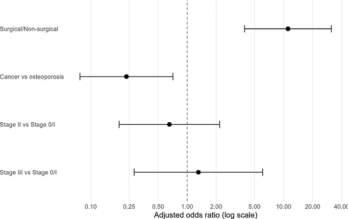
Figure 4. Adjusted associations with documented clinical soft tissue healing at the last recorded follow-up. Odds ratios and 95% confidence intervals are from the primary multivariable logistic regression model. Estimates describe associations with final documented clinical soft tissue status and not time to first healing.
Sensitivity analyses were directionally similar (Supplementary Tables S11 and S11B). After additional adjustment for sex, the treatment association remained positive (adjusted OR: 9.91, 95% CI: 3.56 to 30.57; p < 0.001). After adjustment for exploratory comedication burden, the corresponding estimate was 11.63 (95% CI: 4.23 to 35.81; p < 0.001), and after adjustment for both sex and exploratory comedication burden, it was 10.56 (95% CI: 3.73 to 33.28; p < 0.001). Excluding Stage 0 cases and Firth penalization also yielded directionally consistent estimates. When unknown outcomes were classified as no documented healing, the treatment estimate remained positive, whereas the cancer estimate attenuated and crossed the null. A secondary model accounting for duration of documented clinical observation yielded similar estimates for treatment and indication, while follow-up duration itself was not independently associated with documented healing.
In an additional exploratory analysis, the difference in comorbidity burden persisted after age adjustment, with cancer cases showing a lower count than osteoporosis cases (incidence rate ratio [IRR]: 0.59, 95% CI: 0.40–0.85; p = 0.005) (Table 6).
Discussion
This retrospective single-center study describes systemic profiles, oral findings, treatment patterns, and documented clinical soft tissue outcomes in a Norwegian cohort of 105 patients with MRONJ managed in a specialized university setting. Cancer- and osteoporosis-related MRONJ occurred in distinct systemic and pharmacological contexts, while showing broadly similar local oral manifestations, consistent with contemporary MRONJ position papers and the oncology bone-modifying-agent literature [1, 7, 14]. This study contributes a structured characterization of indication-specific case profiles and multivariable analysis, not an estimate of disease incidence or causal treatment effects.
Within this case-only cohort, shorter recorded antiresorptive treatment duration and diagnosis-driven oncological therapies distinguished cancer-related from osteoporosis-related MRONJ. At the same time, final documented clinical status and mortality differed by indication. These findings cannot be interpreted as incidence or relative risk estimates for treated populations because the cohort included only patients who had already developed MRONJ. Rather, they describe differences between two clinical profiles of MRONJ. Local dental disease and previous tooth extraction at the MRONJ lesion site were frequently recorded in both groups and should be interpreted as antecedent dental context rather than proof of a single causal trigger [30–32].
Tooth extraction and other dentoalveolar procedures were among the most frequently recorded antecedent dental events in this cohort, consistent with numerous retrospective and prospective reports [30–35]. Contemporary evidence also indicates that extraction undertaken within a structured preventive protocol, including control of local infection, atraumatic technique, alveoplasty where indicated, antimicrobial measures, and tension-free primary closure, may be associated with a low risk of subsequent MRONJ [36–39]. Many cases recorded possible clinical reasons or local dental conditions associated with extraction, although several conditions could coexist and completeness varied. Experimental studies also report that pre-existing dental infections lead to impaired post-extraction healing during antiresorptive exposure [40]. Reverse causality cannot be excluded; extraction may therefore indicate treatment of diseased teeth or early clinically unrecognized MRONJ rather than an independent primary cause [2, 16, 30–35]. Non-extraction presentations, including denture trauma and minor mucosal injury, are also compatible with a multifactorial local disease context [41, 42]. Studies of infection and bacterial diversity further support the relevance of the local disease environment [43, 44].
Systemically, the two indication groups differed in clinically predictable ways. Previous studies have examined systemic and treatment-related factors in oncology and osteoporosis populations, although their designs and populations differ [45–52]. Among cases with known final status, 68/102 (66.7%) had documented clinical soft tissue healing at the last recorded follow-up, within the range reported in heterogeneous clinical series [21, 53–62]. Patient-reported quality of life has also been evaluated separately from clinical healing [26, 63]. Documented healing was more frequent among patients receiving surgical treatment than among those managed non-surgically, and the adjusted association was directionally consistent across sensitivity analyses. However, these final-status analyses do not establish treatment efficacy. Treatment was not randomly allocated and likely reflected disease extent, systemic health, prognosis, clinician judgement, and suitability for surgery; therefore, it is important to interpret the findings as observational and hypothesis-generating [19, 22, 64–66]. At the same time, clinical worsening at the last recorded follow-up was uncommon among non-surgically managed patients, occurring in only 3 of 34 (8.8%) with known final outcome, and no worsening was recorded among 68 surgically treated patients with known final outcome. All patients in the present cohort had discontinued ART after their MRONJ diagnosis. The independent effect of this post-diagnosis discontinuation cannot be assessed because all patients followed this management, and exact timing and duration were unavailable. Ristow et al. also reported pausing antiresorptive therapy after MRONJ diagnosis [24]; differences in clinical course therefore cannot be attributed to withdrawal alone. The low proportion of documented worsening contrasts with reports of progression during conservative MRONJ management, including the study by Ristow et al. [24], and may reflect differences in case selection, local treatment protocols, follow-up duration, and outcome definitions (Supplementary Table S1B).
The exposure-duration patterns also require cautious interpretation. Cancer-related cases clustered around shorter but more intensive regimens, whereas osteoporosis-related cases had longer recorded treatment durations. These patterns are biologically plausible given the different schedules used in oncology and osteoporosis [48, 67, 68]. Nevertheless, this case-only cohort cannot estimate MRONJ risk in the underlying treated populations or separate the effects of indication, dose intensity, cumulative dose, adherence, and switching history [49–52]. The absence of validated performance-status measures further limits the interpretation of group differences in documented healing, as systemic fitness, survival, and opportunities for definitive treatment and follow-up may be interrelated [29, 62].
Stage 0 or non-exposed disease was uncommon (3/105, 2.9%). This likely reflects the referral of an already established MRONJ case to the Oral Surgery and Oral Medicine Department, as well as the diagnostic difficulty of non-specific Stage 0 presentations. Without exposed bone, early symptoms may be attributed to odontogenic or neuropathic conditions, and subtle radiographic changes may be overlooked or identified only after progression [69, 70]. Therefore, it is important to interpret stage-dependent conclusions cautiously. Comorbidity and comedication scores should likewise be understood as exploratory descriptive summaries rather than validated severity indices.
Mortality remains clinically relevant when interpreting final documented healing status. The lower proportion of documented healing in cancer-related cases may partly reflect patient death before mucosal closure or fewer opportunities for prolonged follow-up, rather than intrinsically poorer local repair. This may explain the pattern in our cohort; other studies have examined the adverse prognostic context of cancer-associated MRONJ and variation in its clinical course [29, 53, 71]. Treatment goals may therefore differ across indications. In oncology, early coordination between oncology and oral health services remains important for patients receiving high-intensity antiresorptive therapy with chemotherapy or antiangiogenic agents [14, 18, 72, 73]. In osteoporosis, systematic dental and periodontal assessment, patient education, and careful planning of invasive dental treatment remain important during long-term therapy [28].
This study has several limitations. Its retrospective, single-center, case-only design precludes causal inference and does not permit estimation of MRONJ incidence or risk among the treated populations. Possible clinical reasons or local dental conditions associated with extraction were recorded, but exact extraction dates, dentist-level decision documentation, and biopsy confirmation at extraction were unavailable; clinically unrecognized MRONJ at extraction cannot therefore be excluded. Treatment comparisons are vulnerable to confounding by indication. The dataset lacked validated performance-status measures and sufficiently structured patient-level details on antibiotics, antimicrobial mouth rinses, and the timing and duration of ART discontinuation after MRONJ diagnosis. All patients discontinued ART after diagnosis, so its independent effect could not be assessed. Serial radiographic findings were not available in a standardized analytical dataset; clinical mucosal coverage may therefore coexist with persistent radiographic abnormalities or residual non-exposed disease [6, 42, 70]. Stage 0 was uncommon and may have been under-ascertained.” Three Stage 0 cases were recorded; the limitation is possible under-ascertainment, not complete absence of captured cases. Finally, the exact date of the first healing was unavailable, so the study evaluated the final documented clinical soft tissue status rather than a conventional time-to-first-healing endpoint. Local findings and invasive procedures should therefore be interpreted as recorded clinical context, not as independently established causal factors [2, 16, 30–35].
The high proportion of cases with a recorded history of previous tooth extraction at the MRONJ lesion site, particularly in the osteoporosis group, indicates an opportunity to strengthen preventive oral-care pathways through coordinated dental assessment, management of local infections and diseases, and structured protocols for necessary invasive procedures before and during antiresorptive therapy [31, 36–39, 74].
Conclusions
Among patients with MRONJ, cancer-related and osteoporosis-related cases showed broadly similar local oral disease patterns but distinct systemic and treatment-exposure profiles. A recorded history of previous tooth extraction at the MRONJ lesion site was common, particularly in osteoporosis-related MRONJ, but the retrospective data did not permit verification of the extraction indication or precise temporal attribution. Documented clinical soft-tissue healing at the last recorded follow-up was less frequent in cancer-related cases, while surgical treatment was associated with a higher likelihood of documented healing. These findings should be interpreted cautiously because treatment allocation was non-random, exact first-healing dates were unavailable, and radiographic resolution was not assessed. Clinical worsening was uncommon among non-surgically treated patients and was not observed after surgical treatment; however, this observation remains descriptive and should not be interpreted as evidence of treatment superiority.
Acknowledgments
The authors wish to thank Katrine Oswald Bjørneset and Øystein Langeland for their assistance in compiling the dataset.
Author contributions
Conceptualization: BBH and JER. Data acquisition: SS, BBH, and TBK. Data analysis and interpretation: SS and HJH. Formal analysis: SS and HJH. Methodology: BBH and SS. Project administration: BBH. Supervision: BBH, JER, and HJH. Visualization: SS and HJH. Roles/Writing – original draft: SS. Writing – review & editing: SS, BBH, JER, HJH, and TBK. Approval of the revised version to be submitted: All authors.
Data availability statement
The data may be made available by the corresponding author upon reasonable request, subject to applicable Norwegian privacy legislation, institutional approval, and approval by the Regional Committee for Medical and Health Research Ethics.
Supplementary information
Supplementary Methods, Supplementary Results, Supplementary Tables S1-S12 (including S1B and S11B), and Supplementary Figures S1–S7 accompany this paper.
AI statement
Parts of the manuscript were edited for clarity and grammar using a language-model–assisted tool (MS Copilot and Google Gemini); the authors checked and approved all content.
References
[1] Ruggiero SL, Dodson TB, Fantasia J, Goodday R, Aghaloo T, Mehrotra B, et al. American Association of Oral and Maxillofacial Surgeons position paper on medication-related osteonecrosis of the jaw-2014 update. J Oral Maxillofac Surg. 2014;72(10):1938–56. https://doi.org/10.1016/j.joms.2014.04.031
[2] Nicolatou-Galitis O, Schiødt M, Mendes RA, Ripamonti C, Hope S, Drudge-Coates L, et al. Medication-related osteonecrosis of the jaw: definition and best practice for prevention, diagnosis, and treatment. Oral Surg Oral Med Oral Pathol Oral Radiol. 2019;127(2):117–35. https://doi.org/10.1016/j.oooo.2018.09.008
[3] Advisory Task Force on Bisphosphonate-Related Osteonecrosis of the Jaws, American Association of Oral and Maxillofacial Surgeons. American Association of Oral and Maxillofacial Surgeons position paper on bisphosphonate-related osteonecrosis of the jaws. J Oral Maxillofac Surg. 2007;65(3):369–76. https://doi.org/10.1016/j.joms.2006.11.003
[4] Ruggiero SL, Dodson TB, Assael LA, Landesberg R, Marx RE, Mehrotra B, et al. American Association of Oral and Maxillofacial Surgeons position paper on bisphosphonate-related osteonecrosis of the jaw – 2009 update. Aust Endod J. 2009;35(3):119–30. https://doi.org/10.1111/j.1747-4477.2009.00213.x
[5] Coleman R. The use of bisphosphonates in cancer treatment. Ann N Y Acad Sci. 2011;1218:3–14. https://doi.org/10.1111/j.1749-6632.2010.05766.x
[6] Bedogni A, Mauceri R, Fusco V, Bertoldo F, Bettini G, Di Fede O, et al. Italian position paper (SIPMO-SICMF) on medication-related osteonecrosis of the jaw (MRONJ). Oral Dis. 2024;30(6):3679–709. https://doi.org/10.1111/odi.14887
[7] Ruggiero SL, Dodson TB, Aghaloo T, Carlson ER, Ward BB, Kademani D. American Association of Oral and Maxillofacial Surgeons’ position paper on medication-related osteonecrosis of the jaws-2022 update. J Oral Maxillofac Surg. 2022;80(5):920–43. https://doi.org/10.1016/j.joms.2022.02.008
[8] Kajizono M, Sada H, Sugiura Y, Soga Y, Kitamura Y, Matsuoka J, et al. Incidence and risk factors of osteonecrosis of the jaw in advanced cancer patients after treatment with zoledronic acid or denosumab: a retrospective cohort study. Biol Pharm Bull. 2015;38(12):1850–5. https://doi.org/10.1248/bpb.b15-00385
[9] Terpos E, Confavreux CB, Clézardin P. Bone antiresorptive agents in the treatment of bone metastases associated with solid tumours or multiple myeloma. Bonekey Rep. 2015;4:744. https://doi.org/10.1038/bonekey.2015.113
[10] Lee K, Kim K, Kim JY, Kim JW, Kang YH, Kim YH, et al. Mechanisms underlying medication-related osteonecrosis of the jaw. Oral Dis. 2025;31(4):1073–83. https://doi.org/10.1111/odi.15198
[11] Kuroshima S, Al-Omari FA, Sasaki M, Sawase T. Medication-related osteonecrosis of the jaw: a literature review and update. Genesis. 2022;60(8–9):e23500. https://doi.org/10.1002/dvg.23500
[12] Jiang A, Zhang Z, Qiu X, Guo Q. Medication-related osteonecrosis of the jaw (MRONJ): a review of pathogenesis hypothesis and therapy strategies. Arch Toxicol. 2024;98(3):689–708. https://doi.org/10.1007/s00204-023-03653-7
[13] Zhao N, Li QX, Wang YF, Qiao Q, Huang HY, Guo CB, et al. Anti-angiogenic drug aggravates the degree of anti-resorptive drug-based medication-related osteonecrosis of the jaw by impairing the proliferation and migration function of gingival fibroblasts. BMC Oral Health. 2023;23(1):330. https://doi.org/10.1186/s12903-023-03034-7
[14] Otto S, Pautke C, Van den Wyngaert T, Niepel D, Schiødt M. Medication-related osteonecrosis of the jaw: prevention, diagnosis and management in patients with cancer and bone metastases. Cancer Treat Rev. 2018;69:177–87. https://doi.org/10.1016/j.ctrv.2018.06.007
[15] Saad F, Brown JE, Van Poznak C, Ibrahim T, Stemmer SM, Stopeck AT, et al. Incidence, risk factors, and outcomes of osteonecrosis of the jaw: integrated analysis from three blinded active-controlled phase III trials in cancer patients with bone metastases. Ann Oncol. 2012;23(5):1341–7. https://doi.org/10.1093/annonc/mdr435
[16] McGowan K, McGowan T, Ivanovski S. Risk factors for medication-related osteonecrosis of the jaws: a systematic review. Oral Dis. 2018;24(4):527–36. https://doi.org/10.1111/odi.12708
[17] Khan AA, Morrison A, Hanley DA, Felsenberg D, McCauley LK, O’Ryan F, et al. Diagnosis and management of osteonecrosis of the jaw: a systematic review and international consensus. J Bone Miner Res. 2015;30(1):3–23. https://doi.org/10.1002/jbmr.2405
[18] Yarom N, Shapiro CL, Peterson DE, Van Poznak CH, Bohlke K, Ruggiero SL, et al. Medication-related osteonecrosis of the jaw: MASCC/ISOO/ASCO clinical practice guideline. J Clin Oncol. 2019;37(25):2270–90. https://doi.org/10.1200/JCO.19.01186
[19] Seluki R, Seluki M, Vaitkeviciene I, Jagelaviciene E. Comparison of the effectiveness of conservative and surgical treatment of medication-related osteonecrosis of the jaw: a systematic review. J Oral Maxillofac Res. 2023;14(4):e1. https://doi.org/10.5037/jomr.2023.14401
[20] Choi NR, Lee JH, Park JY, Hwang DS. Surgical treatment of medication-related osteonecrosis of the jaw: a retrospective study. Int J Environ Res Public Health. 2020;17(23):8801. https://doi.org/10.3390/ijerph17238801
[21] El-Rabbany M, Lam DK, Shah PS, Azarpazhooh A. Surgical management of medication-related osteonecrosis of the jaw is associated with improved disease resolution: a retrospective cohort study. J Oral Maxillofac Surg. 2019;77(9):1816–22. https://doi.org/10.1016/j.joms.2019.03.040
[22] Fujimori M, Toriyabe Y, Sakakibara N, Nojima M, Makino S; Hokkaido Association of Hospital Dentistry Medication-Related Osteonecrosis of the Jaw Research Group. What Affects Healing rates in patients treated for medication-related osteonecrosis of the jaw? The role of operative therapy and other clinical factors. J Oral Maxillofac Surg. 2024;82(11):1441–55. https://doi.org/10.1016/j.joms.2024.06.176
[23] Hadaya D, Soundia A, Freymiller E, Grogan T, Elashoff D, Tetradis S, et al. Nonsurgical management of medication-related osteonecrosis of the jaws using local wound care. J Oral Maxillofac Surg. 2018;76(11):2332–9. https://doi.org/10.1016/j.joms.2018.05.025
[24] Ristow O, Rückschloß T, Müller M, Berger M, Kargus S, Pautke C, et al. Is the conservative non-surgical management of medication-related osteonecrosis of the jaw an appropriate treatment option for early stages? A long-term single-center cohort study. J Craniomaxillofac Surg. 2019;47(3):491–9. https://doi.org/10.1016/j.jcms.2018.12.014
[25] Rückschloß T, Smielowski M, Moratin J, Schnug G, Appel M, Muench P, et al. Comparing the influence of surgical and conservative therapy on quality of life in patients with early-stage medication-related osteonecrosis of the jaw – a prospective longitudinal study. Medicina (Kaunas). 2023;59(2):277. https://doi.org/10.3390/medicina59020277
[26] Winter A, Schulz SM, Schmitter M, Brands RC, Straub A, Kübler A, et al. Oral-health-related quality of life in patients with medication-related osteonecrosis of the jaw: a prospective clinical study. Int J Environ Res Public Health. 2022;19(18):11709. https://doi.org/10.3390/ijerph191811709
[27] Varoni EM, Lombardi N, Villa G, Pispero A, Sardella A, Lodi G. Conservative management of Medication-Related Osteonecrosis of the Jaws (MRONJ): a retrospective cohort study. Antibiotics (Basel). 2021;10(2):195. https://doi.org/10.3390/antibiotics10020195
[28] Seki K, Kaneko T, Kamimoto A, Wada M, Takeuchi Y, Furuchi M, et al. Medication-related osteonecrosis of the jaw after tooth extraction in patients receiving pharmaceutical treatment for osteoporosis: a retrospective cohort study. J Dent Sci. 2022;17(4):1619–25. https://doi.org/10.1016/j.jds.2022.03.014
[29] Corraini P, Heide-Jørgensen U, Schiødt M, Nørholt SE, Acquavella J, Sørensen HT, et al. Osteonecrosis of the jaw and survival of patients with cancer: a nationwide cohort study in Denmark. Cancer Med. 2017;6(10):2271–7. https://doi.org/10.1002/cam4.1173
[30] Hasegawa T, Hayashida S, Kondo E, Takeda Y, Miyamoto H, Kawaoka Y, et al. Medication-related osteonecrosis of the jaw after tooth extraction in cancer patients: a multicenter retrospective study. Osteoporos Int. 2019;30(1):231–9. https://doi.org/10.1007/s00198-018-4746-8
[31] Soutome S, Hayashida S, Funahara M, Sakamoto Y, Kojima Y, Yanamoto S, et al. Factors affecting development of medication-related osteonecrosis of the jaw in cancer patients receiving high-dose bisphosphonate or denosumab therapy: is tooth extraction a risk factor?. PLoS One. 2018;13(7):e0201343. https://doi.org/10.1371/journal.pone.0201343
[32] Otto S, Tröltzsch M, Jambrovic V, Panya S, Probst F, Ristow O, et al. Tooth extraction in patients receiving oral or intravenous bisphosphonate administration: a trigger for BRONJ development? J Craniomaxillofac Surg. 2015;43(6):847–54. https://doi.org/10.1016/j.jcms.2015.03.039
[33] Bodem JP, Kargus S, Eckstein S, Saure D, Engel M, Hoffmann J, et al. Incidence of bisphosphonate-related osteonecrosis of the jaw in high-risk patients undergoing surgical tooth extraction. J Craniomaxillofac Surg. 2015;43(4):510–4. https://doi.org/10.1016/j.jcms.2015.02.018
[34] Saia G, Blandamura S, Bettini G, Tronchet A, Totola A, Bedogni G, et al. Occurrence of bisphosphonate-related osteonecrosis of the jaw after surgical tooth extraction. J Oral Maxillofac Surg. 2010;68(4):797–804. https://doi.org/10.1016/j.joms.2009.10.026
[35] Soutome S, Otsuru M, Hayashida S, Murata M, Yanamoto S, Sawada S, et al. Relationship between tooth extraction and development of medication-related osteonecrosis of the jaw in cancer patients. Sci Rep. 2021;11(1):17226. https://doi.org/10.1038/s41598-021-96480-8
[36] Wang Y, Zhang Y, Li D, Zhang W, Wang S, Zheng X, et al. Potential role of the comprehensive tooth extraction procedure in preventing medication related osteonecrosis of the jaw (MRONJ): a prospective cohort study. Orphanet J Rare Dis. 2025;20(1):619. https://doi.org/10.1186/s13023-025-04138-9
[37] Chang YS, Nanayakkara S, Yaacoub A, Cox SC. Prevention of medication-related osteonecrosis of the jaw: institutional insights from a retrospective study. Aust Dent J. 2025;70(1):70–7. https://doi.org/10.1111/adj.13050
[38] Catania G, Monaco F, Limberti G, Alessio M, De Martino I, Barile C, et al. Osteonecrosis of the jaws (ONJ) after bisphosphonate treatment in patients with multiple myeloma: decreasing ONJ incidence after adoption of preventive measures. Dent J (Basel). 2016;4(4):45. https://doi.org/10.3390/dj4040045
[39] Di Fede O, Panzarella V, Mauceri R, Fusco V, Bedogni A, Lo Muzio L, et al. The Dental management of patients at risk of medication-related osteonecrosis of the jaw: new paradigm of primary prevention. Biomed Res Int. 2018;2018:2684924. https://doi.org/10.1155/2018/2684924
[40] Hadaya D, Soundia A, Gkouveris I, Dry SM, Aghaloo TL, Tetradis S. Development of medication-related osteonecrosis of the jaw after extraction of teeth with experimental periapical disease. J Oral Maxillofac Surg. 2019;77(1):71–86. https://doi.org/10.1016/j.joms.2018.08.010
[41] Yazdi PM, Schiodt M. Dentoalveolar trauma and minor trauma as precipitating factors for medication-related osteonecrosis of the jaw (ONJ): a retrospective study of 149 consecutive patients from the Copenhagen ONJ Cohort. Oral Surg Oral Med Oral Pathol Oral Radiol. 2015;119(4):416–22. https://doi.org/10.1016/j.oooo.2014.12.024
[42] Schiodt M, Reibel J, Oturai P, Kofod T. Comparison of nonexposed and exposed bisphosphonate-induced osteonecrosis of the jaws: a retrospective analysis from the Copenhagen cohort and a proposal for an updated classification system. Oral Surg Oral Med Oral Pathol Oral Radiol. 2014;117(2):204–13. https://doi.org/10.1016/j.oooo.2013.10.010
[43] Hallmer F, Bjørnland T, Andersson G, Becktor JP, Kristoffersen AK, Enersen M. Bacterial diversity in medication-related osteonecrosis of the jaw. Oral Surg Oral Med Oral Pathol Oral Radiol. 2017;123(4):436–44. https://doi.org/10.1016/j.oooo.2016.11.011
[44] Katsarelis H, Shah NP, Dhariwal DK, Pazianas M. Infection and medication-related osteonecrosis of the jaw. J Dent Res. 2015;94(4):534–9. https://doi.org/10.1177/0022034515572021
[45] Hallmer F, Andersson G, Götrick B, Warfvinge G, Anderud J, Bjørnland T. Prevalence, initiating factor, and treatment outcome of medication-related osteonecrosis of the jaw-a 4-year prospective study. Oral Surg Oral Med Oral Pathol Oral Radiol. 2018;126(6):477–85. https://doi.org/10.1016/j.oooo.2018.08.015
[46] Hallmer F, Bjarnadottir O, Götrick B, Malmström P, Andersson G. Incidence of and risk factors for medication-related osteonecrosis of the jaw in women with breast cancer with bone metastasis: a population-based study. Oral Surg Oral Med Oral Pathol Oral Radiol. 2020;130(3):252–7. https://doi.org/10.1016/j.oooo.2020.04.808
[47] Guarneri V, Miles D, Robert N, Diéras V, Glaspy J, Smith I, et al. Bevacizumab and osteonecrosis of the jaw: incidence and association with bisphosphonate therapy in three large prospective trials in advanced breast cancer. Breast Cancer Res Treat. 2010;122(1):181–8. https://doi.org/10.1007/s10549-010-0866-3
[48] Yang M, Yu X. Management of bone metastasis with intravenous bisphosphonates in breast cancer: a systematic review and meta-analysis of dosing frequency. Support Care Cancer. 2020;28(6):2533–40. https://doi.org/10.1007/s00520-020-05355-7
[49] Grbic JT, Black DM, Lyles KW, Reid DM, Orwoll E, McClung M, et al. The incidence of osteonecrosis of the jaw in patients receiving 5 milligrams of zoledronic acid: data from the health outcomes and reduced incidence with zoledronic acid once yearly clinical trials program. J Am Dent Assoc. 2010;141(11):1365–70. https://doi.org/10.14219/jada.archive.2010.0082
[50] Lo JC, O’Ryan FS, Gordon NP, Yang J, Hui RL, Martin D, et al. Prevalence of osteonecrosis of the jaw in patients with oral bisphosphonate exposure. J Oral Maxillofac Surg. 2010;68(2):243–53. https://doi.org/10.1016/j.joms.2009.03.050
[51] Chiu WY, Chien JY, Yang WS, Juang JM, Lee JJ, Tsai KS. The risk of osteonecrosis of the jaws in Taiwanese osteoporotic patients treated with oral alendronate or raloxifene. J Clin Endocrinol Metab. 2014;99(8):2729–35. https://doi.org/10.1210/jc.2013-4119
[52] Jung S, Kim J, Park JH, Kim KY, Kim HJ, Park W. A 5-year retrospective cohort study of denosumab induced medication related osteonecrosis of the jaw in osteoporosis patients. Sci Rep. 2022;12(1):8641. https://doi.org/10.1038/s41598-022-11615-9
[53] Martins AS, Correia JA, Salvado F, Caldas C, Santos N, Capelo A, et al. Relevant factors for treatment outcome and time to healing in medication-related osteonecrosis of the jaws – a retrospective cohort study. J Craniomaxillofac Surg. 2017;45(10):1736–42. https://doi.org/10.1016/j.jcms.2017.07.014
[54] Ruggiero SL, Kohn N. Disease stage and mode of therapy are important determinants of treatment outcomes for medication-related osteonecrosis of the jaw. J Oral Maxillofac Surg. 2015;73(12 Suppl):S94–100. https://doi.org/10.1016/j.joms.2015.09.024
[55] Lopes RN, Rabelo GD, Rocha AC, Carvalho PA, Alves FA. Surgical therapy for bisphosphonate-related osteonecrosis of the jaw: six-year experience of a single institution. J Oral Maxillofac Surg. 2015;73(7):1288–95. https://doi.org/10.1016/j.joms.2015.01.008
[56] Mücke T, Koschinski J, Deppe H, Wagenpfeil S, Pautke C, Mitchell DA, et al. Outcome of treatment and parameters influencing recurrence in patients with bisphosphonate-related osteonecrosis of the jaws. J Cancer Res Clin Oncol. 2011;137(5):907–13. https://doi.org/10.1007/s00432-010-0953-1
[57] Voss PJ, Joshi Oshero J, Kovalova-Müller A, Veigel Merino EA, Sauerbier S, Al-Jamali J, et al. Surgical treatment of bisphosphonate-associated osteonecrosis of the jaw: technical report and follow up of 21 patients. J Craniomaxillofac Surg. 2012;40(8):719–25. https://doi.org/10.1016/j.jcms.2012.01.005
[58] Choi WS, Lee JI, Yoon HJ, Min CK, Lee SH. Medication-related osteonecrosis of the jaw: a preliminary retrospective study of 130 patients with multiple myeloma. Maxillofac Plast Reconstr Surg. 2017;39(1):1. https://doi.org/10.1186/s40902-016-0099-4
[59] Wilde F, Heufelder M, Winter K, Hendricks J, Frerich B, Schramm A, et al. The role of surgical therapy in the management of intravenous bisphosphonates-related osteonecrosis of the jaw. Oral Surg Oral Med Oral Pathol Oral Radiol Endod. 2011;111(2):153–63. https://doi.org/10.1016/j.tripleo.2010.04.015
[60] Stockmann P, Vairaktaris E, Wehrhan F, Seiss M, Schwarz S, Spriewald B, et al. Osteotomy and primary wound closure in bisphosphonate-associated osteonecrosis of the jaw: a prospective clinical study with 12 months follow-up. Support Care Cancer. 2010;18(4):449–60. https://doi.org/10.1007/s00520-009-0688-1
[61] Ferlito S, Puzzo S, Palermo F, Verzì P. Treatment of bisphosphonate-related osteonecrosis of the jaws: presentation of a protocol and an observational longitudinal study of an Italian series of cases. Br J Oral Maxillofac Surg. 2012;50(5):425–9. https://doi.org/10.1016/j.bjoms.2011.08.004
[62] Favia G, Tempesta A, Limongelli L, Crincoli V, Maiorano E. Medication-related osteonecrosis of the jaw: surgical or non-surgical treatment? Oral Dis. 2018;24(1–2):238–42. https://doi.org/10.1111/odi.12764
[63] Oteri G, Trifirò G, Peditto M, Lo Presti L, Marcianò I, Giorgianni F, et al. Treatment of medication-related osteonecrosis of the jaw and its impact on a patient’s quality of life: a single-center, 10-year experience from Southern Italy. Drug Saf. 2018;41(1):111–23. https://doi.org/10.1007/s40264-017-0582-6
[64] Ramaglia L, Guida A, Iorio-Siciliano V, Cuozzo A, Blasi A, Sculean A. Stage-specific therapeutic strategies of medication-related osteonecrosis of the jaws: a systematic review and meta-analysis of the drug suspension protocol. Clin Oral Investig. 2018;22(2):597–615. https://doi.org/10.1007/s00784-017-2325-6
[65] Rollason V, Laverrière A, MacDonald LC, Walsh T, Tramèr MR, Vogt-Ferrier NB. Interventions for treating bisphosphonate-related osteonecrosis of the jaw (BRONJ). Cochrane Database Syst Rev. 2016;(2):CD008455. https://doi.org/10.1002/14651858.CD008455.pub2
[66] Goker F, Grecchi E, Grecchi F, Francetti L, Del Fabbro M. Treatment of medication-related osteonecrosis of the jaw (MRONJ). A systematic review. Eur Rev Med Pharmacol Sci. 2021;25(6):2662–73. https://doi.org/10.26355/eurrev_202103_25430
[67] Stopeck AT, Fizazi K, Body JJ, Brown JE, Carducci M, Diel I, et al. Safety of long-term denosumab therapy: results from the open label extension phase of two phase 3 studies in patients with metastatic breast and prostate cancer. Support Care Cancer. 2016;24(1):447–55. https://doi.org/10.1007/s00520-015-2904-5
[68] Raje N, Terpos E, Willenbacher W, Shimizu K, García-Sanz R, Durie B, et al. Denosumab versus zoledronic acid in bone disease treatment of newly diagnosed multiple myeloma: an international, double-blind, double-dummy, randomised, controlled, phase 3 study. Lancet Oncol. 2018;19(3):370–81. https://doi.org/10.1016/S1470-2045(18)30072-X
[69] Schiodt M, Otto S, Fedele S, Bedogni A, Nicolatou-Galitis O, Guggenberger R, et al. Workshop of European task force on medication-related osteonecrosis of the jaw-Current challenges. Oral Dis. 2019;25(7):1815–21. https://doi.org/10.1111/odi.13160
[70] Fedele S, Bedogni G, Scoletta M, Favia G, Colella G, Agrillo A, et al. Up to a quarter of patients with osteonecrosis of the jaw associated with antiresorptive agents remain undiagnosed. Br J Oral Maxillofac Surg. 2015;53(1):13–17. https://doi.org/10.1016/j.bjoms.2014.09.001
[71] Watters AL, Hansen HJ, Williams T, Chou JF, Riedel E, Halpern J, et al. Intravenous bisphosphonate-related osteonecrosis of the jaw: long-term follow-up of 109 patients. Oral Surg Oral Med Oral Pathol Oral Radiol. 2013;115(2):192–200. https://doi.org/10.1016/j.oooo.2012.05.017
[72] Srivastava A, Nogueras Gonzalez GM, Geng Y, Won AM, Myers J, Li Y, et al. Medication-related osteonecrosis of the jaw in patients treated concurrently with antiresorptive and antiangiogenic agents: systematic review and meta-analysis. J Immunother Precis Oncol. 2021;4(4):196–207. https://doi.org/10.36401/JIPO-21-14
[73] Sacco R, Woolley J, Patel G, Calasans-Maia MD, Yates J. Systematic review of medication related osteonecrosis of the jaw (MRONJ) in patients undergoing only antiangiogenic drug therapy: surgery or conservative therapy? Br J Oral Maxillofac Surg. 2022;60(2):e216–30. https://doi.org/10.1016/j.bjoms.2021.03.006
[74] Kawahara M, Kuroshima S, Sawase T. Clinical considerations for medication-related osteonecrosis of the jaw: a comprehensive literature review. Int J Implant Dent. 2021;7(1):47. https://doi.org/10.1186/s40729-021-00323-0