REVIEW ARTICLE
The clinical utility of circulating human papillomavirus across squamous cell carcinomas
Karen-Lise G. Spindlera,b, Anne V. Jakobsena,b, Jesper G. Eriksena,b, Lars Fokdala,b,c, Marianne Nordsmarka,b, Lise B.J. Thorsena,b, Karen L. Winda,b, Anna C. Lefevrea and Jens Overgaarda,b
aDepartment of Experimental Clinical Oncology, Aarhus University Hospital, Denmark; bDepartment of Clinical Medicine, Aarhus University, Denmark; cDepartment of Oncology, Vejle Hospital, Denmark
ABSTRACT
Background and purpose: The similarities in biology, treatment regimens and outcome between the different human papillomavirus (HPV) associated squamous cell carcinomas (SCCs) allow for extrapolation of results generated from one SC tumor type to another.
In HPV associated cancers, HPV is integrated into the tumor genome and can consequently be detected in the circulating fragments of the tumor DNA. Thus, measurement of HPV in the plasma is a surrogate for circulating tumor DNA (ctDNA) and holds promise as a clinically relevant biomarker in HPV associated cancers.
With the present overview we aim to present the status of circulating HPV studies in SCCs, the clinical potential and the gaps of knowledge, with the overall aim to facilitate the next steps into clinically relevant prospective trials.
Material and methods: We reviewed the literature and presented the data for each tumor type as well as analyses of the clinical utility across the SCC.
Results and interpretation: A total of 41 studies were identified in cervical, head and neck and anal SCC and we discuss the common signals from the results across the different tumor sites. Our results not only confirm the strong clinical potential but also emphasize an urgent need to coordinate studies to allow for relevant sample sizes and statistical validations.
KEYWORDS: Squamous cell carcinoma; circulating tumor DNA (ctDNA); Human papillomavirus
Citation: ACTA ONCOLOGICA 2025, VOL. 64, 1–12. https://doi.org/10.2340/1651-226X.2025.41288.
Copyright: © 2025 The Author(s). Published by MJS Publishing on behalf of Acta Oncologica. This is an Open Access article distributed under the terms of the Creative Commons Attribution 4.0 International License (http://creativecommons.org/licenses/by/4.0/).
Received: 14 August 2024; Accepted: 8 December 2024; Published: 2 January 2025
CONTACT: Karen-Lise Garm Spindler k.g.spindler@rm.dk Department of Experimental Clinical Oncology, Aarhus University Hospital, Denmark, Department of Clinical Medicine, Aarhus University, Denmark, Palle Juul-Jensens Boulevard 99, 8200 Aarhus N, Denmark
Supplemental data for this article can be accessed online at https://doi.org/10.2340/1651-226X.2025.41288
Competing interests and funding: Funding Health Research Foundation of Central Denmark Region, Danish Comprehensive Cancer Center Radiotherapy, The Danish Cancer Society.
Introduction to the biology of Squamous Cell Carcinomas
Squamous cell carcinoma is a frequent cancer type that develops from the squamous epithelium, which typically regenerates within 2–3 weeks and withstands external trauma and abrasions and has an ability to accelerate its repopulation to replace traumatized areas. Human papilloma virus (HPV) is a dominating and increasing etiological factor [1]. HPV-positive squamous cell carcinomas (SCCs) are frequently found in the uterine cervix (> 95%), head and neck region, especially the oropharynx (40–80%), upper part of the esophagus (20%), vulva (50%) and anal canal (> 80%).
All SCCs exhibit a characteristic pattern of behavior and response to treatment. The diseases are primarily loco-regional and the primary treatment is surgery and/or (chemo)radiotherapy (CRT) [2–4]. SCCs are characterized by showing a steep dose-response relationship and accelerated radiotherapy regimens can be relevant [5, 6]. Hypoxia, the EGFR biology and the immune environment are important common biological features [2, 7–14].
Human papillomavirus in squamous cell carcinomas
Human papillomavirus status plays a role in sensitivity to both radiotherapy and systemic treatment, but results are contradictory. The HPV relation seems to influence radiosensitivity in some (i.e. the oropharynx) tumors more than others [9, 15, 16]. The mechanism and the biological differences between HPV-positive and HPV-negative disease is unknown and they are currently treated with the same regimens and radiation doses.
There are several different subtypes of HPV, which can be classified into high-risk (oncogenic) and low-risk subtypes, but we have little knowledge of the role of the different HPV subtypes in relation to treatment sensitivity and outcome [16–19].
Pan SCC features
Most SCCs are sensitive to radiotherapy, platinum-based chemotherapy and, as recently reported, checkpoint inhibition, but biological features and predictive markers for response to these different treatment modalities are inadequately characterized. When treating primary, localized SCC with CRT, identification of biomarkers to guide personalized treatment is crucial to improve selection of ‘poor responders’ who may benefit from treatment intensification for a curative approach despite the risk of increasing toxicity, and to identify ‘good responders’, where decreasing radiation doses and potential side effects may be possible.
The optimal time-point for final assessment of complete response (CR) after CRT is not identified and with current practice, premature decisions may lead to unnecessary salvage surgery. In case of contradicting imaging and biopsy results, predictive markers could aid in the decision for salvage surgery. Finally, there is an urgent need for tools for early detection of recurrences with the possibility of cure through localized treatment.
Palliative chemotherapy for SSCs implies a high risk of side effects and limited benefit in some patients. In the palliative setting predictive markers will enable more rapid change of ineffective systemic treatment strategies. Pre-clinical data support that HPV-positive tumors are more susceptible to immunotherapy with checkpoint inhibitors, but optimal selection for therapy is warranted [20].
Circulating HPV as circulating tumor DNA measurement
Circulating tumor DNA (ctDNA) represents small DNA fragments with tumor specific characteristics and can be detected in a simple blood sample (Figure 1). This has gained considerable interest as a prognostic and predictive marker in both localized and metastatic cancer disease. The elimination half-life of ctDNA is only a few hours and biological clearance from the bloodstream is consequently expected immediately following curative removal of tumor tissue [21]. It is now established that the presence of ctDNA in plasma post-surgery indicates microscopic residual disease (MRD) and a subsequent very poor outcome [22, 23]. Fundamental aspects of detection and measurement of ctDNA comprises (1) biological knowledge of the individual tumor type (is the tumor cells likely to shed ctDNA or not? are there easily measurable and frequent well known tumor specific mutations or epigenetic alterations to measure?), (2) the laboratory methods (is a broad method that targets many different alterations necessary or can testing be covered by simple methods on few targets?) and (3) clinical need (is an ultra-high sensitivity or specificity needed? is it necessary to use a tumor informed approach or can a pragmatic tumor agnostic strategy be used?).
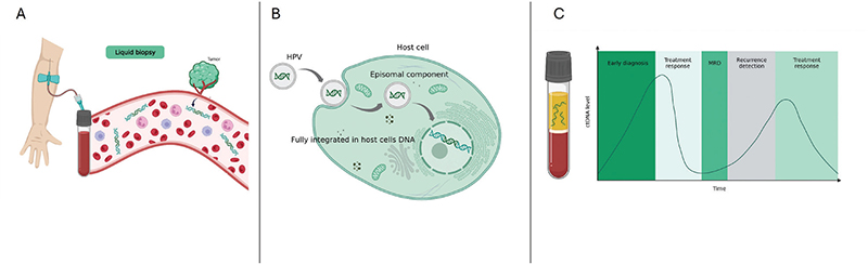
Figure 1. (A) Liquid biopsy. Circulating tumor DNA (ctDNA) represents small fragments of DNA from tumor cells that are released into the bloodstream and can be detected in a simple blood sample. In HPV associated cancers, HPV DNA can be detected in the circulating tumor DNA fragments. (B) HPV integration. The integration of HPV into the host cell can be full integration with the virus DNA being spliced into the host cell’s DNA or by partial or full episomal integration. Notably, recent data have suggested that measures of HPV integration in itself have a biological and prognostic impact [72]. (C) ctDNA levels during the disease and treatment course. The elimination half-life of ctDNA is less than a few hours, leading to rapid biological clearance from the bloodstream following the curative removal of tumor tissue. The presence of ctDNA in plasma following treatment indicates microscopic residual disease (MRD) and is generally associated with a poor outcome. Created with BioRender.com (Jakobsen, A. (2025) https://BioRender.com/w47u880)
In HPV associated cancers, HPV is integrated into the tumor genome (Figure 1) and can be detected in the circulating fragments of the tumor DNA [24]. Thus, measurement of HPV in the plasma is a surrogate for ctDNA and holds promise as a clinically relevant biomarker in HPV associated cancers.
The shared characteristics in biology, treatment approaches, and outcomes among HPV-associated SCCs allow for extrapolation of results from one squamous cell tumor type to another. This overview aims to present the current status of HPVctDNA research in SCCs, highlighting clinical potential and the gaps of knowledge.
Results
HPVctDNA in squamous cell carcinoma of the Anus
Nine studies were identified (2 case reports), comprising a total of less than 300 patients, most in the primary setting, and 3 in metastatic disease (Table 1). A study of HPV associated cancer included 15 patients with SCCA. HPVctDNA was present in 87–93% of HPV-positive tumors, in microinvasive carcinomas, but not in blood samples from patients with HPV associated high grade neoplasia [24]. Another study in SCCA showed that HPVctDNA could be detected before CRT in 29 of 33 patients with stage II-III disease, that levels dropped markedly during CRT, and further, that residual detectable HPVctDNA after CRT (3/18 patients) was strongly associated with shorter disease-free survival [25]. Bernard-Tessier et al. demonstrated that HPVctDNA was associated with prognosis during first line chemotherapy in advanced SCCA [26]. Recently, analysis in 88 SCCA patients revealed that pre-treatment level of HPVctDNA was associated with clinical stage and prognosis [19]. Furthermore, three distinct patterns of HPVctDNA elimination during CRT were observed with significantly different risks of local or distant failures. HPVctDNA measurements during FU indicated a strong potential for the prediction of recurrences with a clinically relevant lead time as marker of MRD (Table 2) [19, 27].
| Reference | Tumor site | Sample size | Stages included | Method | Material | Detected HPV subtypes | Concordance to tissue HPV status/p16 | Correlation to stage | Prognostic value |
| Cervical cancer | |||||||||
| Kedzia et al. 1992 [39] | CSCC CS |
5 4 |
Stage I-III | Southern blot hybridization | Whole blood | 16 | Sensitivity 80% | ND | ND |
| Pornthanakasem et al. 2001 [41] | CSCC Healthy |
63 20 |
Stages I-IV | PCR | Plasma | 16, 18 | Sensitivity 12% | + | Statistical tendency |
| Liu et al. 2001 [47] | CSCC | 60 | Stages I-IV | PCR | Serum | 16, 18 | Sensitivity 20% | ND | ND |
| Dong et al. 2002 [48] | CSCC CS Healthy |
175 57 60 |
Stages I-III(IV) | qPCR | Plasma | 16, 18 | Sensitivity 18% 66% samples collected post-treatment |
(+) | ND |
| Sathish et al. 2004 [49] | CSCC CS Healthy |
58 10 30 |
Stages I-IV | PCR | Plasma | 16, 58 | Sensitivity 15% | (+) | ND |
| Kay et al. 2005 [50] | CSCC CS Healthy |
45 32 77 |
Stage I-IV | PCR | Whole blood | 16, 18 | Sensitivity 24% | (+) | ND |
| Shimada et al. 2010 [51] | CSCC CS Healthy |
20 22+3 20 |
Stage I-IV | qPCR | Plasma | 16 | Sensitivity 30% | ND | ND |
| Campitelli et al. 2012 [44] | CSCC | 16 | Stage I-IV | qPCR | Serum | 16, 18 | Sensitivity 81% | (+) | ND |
| Kang et al. 2017 [52] | CSCC Healthy |
21 45 |
M+ | ddPCR | Serum | 16, 18 | Sensitivity 100% | ND | ND |
| Cheung et al. 2019 [42] | CSCC | 138 | Stage I-IV | ddPCR | Plasma | 16, 18 | Sensitivity 62% | (+) | Statistical tendency |
| Cabel et al. 2021 [40] | CC | 55 | Stage I-IV | ddPCR | Serum/plasma | 16, 18, 31, 33, 35, 45, 52, 58, 73 | Sensitivity 69% | + | No prognostic value |
| Jeannot et al. 2021 [43] | CC | 94 | Stage I-IV | ddPCR | Serum | 16, 18 | Sensitivity 63 % | + | No prognostic value |
| Bønløkke et al. 2022 [53] | CC CS Healthy |
60 8 15 |
Stage I-IV | ddPCR | Plasma | 16, 18 | Sensitivity 37% | + | ND |
| Head and neck cancer | |||||||||
| Cao et al. 2012 [37] | OPSCC Healthy |
64 10 |
Stage I-IV | qPCR | Plasma | 16, 18 | Sensitivity 65% | + | ND |
| Ahn et al. 2014 [54] | HNSCC | 93 | Stage I-IV | qPCR | Plasma Saliva |
16 | Sensitivity 67% | ND | ND |
| Wang et al. 2015 [55] | HNSCC | 47 | Stage I-IV | ddPCR | Plasma Saliva |
16 | Sensitivity 86% | (+) | ND |
| Dahlstrom et al. 2015 [30] | OPSCC | 262 | Stage I-IV | qPCR | Serum | 16 | Sensitivity 60% | + | No prognostic value |
| Mazurek et al. 2016 [56] | OPSCC Healthy |
200 15 |
Stage I-IV | PCR | Plasma | 16, 18 | Sensitivity 14% | ND | ND |
| Lee et al. 2017 [29] | HNSCC | 88 (test validation cohorts) | Stage III-IV | NGS | Plasma | 16 | Sensitivity 90–100% | ÷ | ND |
| Hanna et al. 2018 [31] | OPSCC | 22 | Stage IV or M+ | ddPCR | Plasma | 16, 18, 31, 33, 45 (investigated) | Sensitivity 71% | + | Prognostic value |
| Hanna et al. 2019 [32] | OPSCC | 21 | Stage IV or M+ | ddPCR | Plasma | 16, 18, 31, 33, 45 (investigated) | Sensitivity 76 % | + | Plasma ctHPV correlated with prognostic score. Salivary did not. |
| Chera et al. 2019 [33] | HNSCC Controls |
103 115 |
Stage I-IV | ddPCR | Plasma | 16, 31, 33, 35 | Sensitivity 89% | + | ND |
| Chera et al. 2020 [38] | HNSCC | 115* | Stage I-III | ddPCR | Plasma | 16, 18, 31, 33, 35 (investigated) | Sensitivity 99% | ND | ND |
| Rutkowski et al. 2020 [57] | OPSCC | 216 | Stage I-IV | PCR | Plasma | 16 | ND | + | ND |
| Reder et al. 2020 [58] | OPSCC | 50 | Stage I-IV or M+ | qPCR | Plasma | 16 | Sensitivity 87 % | + | ND |
| Tanaka et al. 2021 [28] | HNSCC | 35 | Stage II-IV | ddPCR | Plasma | 16 | Sensitivity 100% | ND | ND |
| Haring et al 2021 [59] | OPSCC | 16 | R/M+ | ddPCR | Plasma | 16, 18 | Sensitivity 75% | ND | ND |
| Siravegna et al. 2022 [60] | HNSCC Healthy |
70 70 |
Stage I-IV | ddPCR | Plasma | 16, 35, 45 | Sensitivity 98% | + | ND |
| Tanaka et al. 2022 [61] | HNSCC | 35 | Stage II-IV | ddPCR | Plasma | 16 | Sensitivity 100% | + | No correlation to failure. |
| Routman et al. 2022 [62] | OPSCC | 45 | Stage I-IV(III?) | ddPCR | Serum | 16, 18, 33, 35 | Sensitivity 89% (76–96%) |
ND | ND |
| Cao et al. 2022 [34] | OPSCC | 34 | Stage III | ddPCR | Plasma | 16, 18 | Sensitivity 82% | + | Higher levels were associated with progres-sion within 12 months |
| Adrian et al. 2023 [35] | OPSCC | 136 | Stage I-IV | qPCR Luminex multiplex |
Plasma | 16 +9 subtypes | Sensitivity 79% | + | Prognostic value |
| Califano et al. 2023 [36] | OPSCC | 233 | Stage III-IV | qPCR | Plasma Saliva | 16 | Sensitivity 75–87% | ND | No prognostic value |
| Anal cancer | |||||||||
| Cabel et al. 2017 [63] | SCCA | 1 | M+ | ddPCR | Plasma | 16 | ND | ND | ND |
| Cabel et al. 2018 [25] | SCCA | 33 | Stage II-III | ddPCR | Serum/plasma | 16, 18 | Sensitivity 88 % | + | No prognostic value |
| Bernard-Tessier et al. 2019 [26] | SCCA | 59 | Stage IV or M+ | ddPCR | Serum | 16 | Sensitivity 91 % | ÷ | Prognostic value |
| Veyer et al. 2019 [64] | SCCA | 1 | M+ | ddPCR | Plasma | 16 | ND | ND | ND |
| Lee et al. 2020 [65] | SCCA | 21 | Stage I-III | NGS | Plasma | 16, 18, 31, 33, 35, 45, 52, 58 (investigated) | Sensitivity 100 % | ÷ | ND |
| Lefevre et al. 2021 [19] | SCCA | 88 | Stage I-IV | ddPCR | Plasma | 18, 31, 33, 51, 58 | Sensitivity 82 % | + | Statistical tendency |
| Mazurek et al. 2023 [27] | SCCA | 62 | Stage I-IV | qPCR | Plasma | 16 | Sensitivity 87% | + | Prognostic value |
| Mixed tumor sites | |||||||||
| Jeannot et al. 2016 [24] | OPSCC CSCC SCCA |
8 47 15 |
Stage I-IV | qPCR ddPCR |
Serum | 16, 18 | Sensitivity 87% | + | ND |
| Damerla et al. 2019 [66] | HNSCC SCCA Healthy |
97 8 27 |
Stage I-IV | PCR | Plasma | 16, 33 | Sensitivity 96% | + | ND |
| HPV: human papilloma virus. HPVctDNA: circulating human papilloma virus DNA. CSCC: cervical squamous cell carcinoma. CS: cervical carcinoma in situ. HNSCC: head and neck squamous cell carcinoma. OPSCC: oropharyngeal squamous cell carcinomas. SCCA: squamous cell carcinoma of the anus. M+: metastatic disease. R: recurrent disease. PCR: polymerase chain reaction. qPCR: quantitative polymerase chain reaction. ddPCR: droplet digital polymerase chain reaction. NGS: next generation sequencing. ND: Not done. *115 included with post-treatment samples, 86 had pre-treatment samples available. | |||||||||
| Reference | Tumor site | Sample size total | Sample size Pre-treatment | Sample size Mid-therapy | Sample size EOT | Sample size FU | Conclusion |
| Cervical cancer | |||||||
| Dong et al. 2002 [48] | CSCC | 292 (175 invasive cancers) | 73 | 193 | No repeated measures. Either collected prior to treatment or after treatment. |
||
| Campitelli et al. 2012 [44] | CSCC | 16 | 16 | 2 | 2 | 2 | Two cases with recurrence had repeated measurements during palliative treatment. Dynamics in HPVctDNA correlated to outcome. |
| Cabel et al. 2021 [40] | CC | 55 | 14 (41) |
14 | 14 (25) |
Detectable HPVctDNA after treatment was associated with lower DFS: HR = 5.1, p = 0.05 Shorter OS: HR = 25.4, p < 0.01 |
|
| Jeannot et al. 2021 [43] | CC | 94 | 94 | 40 | 44 | Detectable HPVctDNA at end of treatment is associated with recurrence. | |
| Head and neck cancer | |||||||
| Cao et al. 2012 [37] | OPSCC | 64 | 64 | 14 | x | 3 | For 14 patients HPVctDNA was measured until elimination. Elimination pattern was rapid. HPVctDNA was detectable at the time of relapse in the three investigated patients. |
| Ahn et al. 2014 [54] | OPSCC | 93 | 35 | 35 | Shorter RFS if EOT pHPV was positive compared to pHPV negative, HR = 12.7 | ||
| Dahlstrom et al. 2015 [30] | OPSCC | 262 | 262 | x | x | ND | |
| Mazurek et al. 2016 [56] | OPSCC | 200 | 28 | 15 | 10 | 5–12 | HPVctDNA decreases during therapy. |
| Lee et al. 2017 [29] | HNSCC | 88 | 47 | 10 | 37 | 10 | HPVctDNA after treatment correlates to outcome in a case-based report. |
| Hanna et al. 2018 [31] | OPSCC (advanced) |
22 | 22 | x | x | x | HPVctDNA can capture dynamics in tumor burden and detect early treatment response. |
| Hanna et al. 2019 [32] | OPSCC (advanced) |
21 | 21 | x | x | x | Salivary HPVctDNA mirrors treatment response. Plasma HPVctDNA holds prognostic value. |
| Chera et al. 2019 [33] | OPSCC | 103 | 103 | 67 | 67 | 67 | Rapid HPVctDNA clearance during CRT (week 4) implied a low risk of treatment failure (p < 0.01) |
| Damerla et al. 2019 [66] | OPSCC | 97 | 97 | 28–(68*) | 28–(68)* | 28–(68)* | With few exceptions, the HPVctDNA declined rapidly during treatment with complete elimination after 7 weeks. |
| Chera et al. 2020 [38] | OPSCC | 115 | 86 | 115 | 11 | Two consecutive post-therapy tests: PPV of HPVctDNA for recurrence = 94% NPV of HPVctDNA for recurrence = 100% Mean lead time to biopsy proven recurrence 3.9 months |
|
| Rutkowski et al. 2020 [57] | OPSCC | 66 | 66 | 66 | Detectable HPVctDNA 12 weeks after treatment is strongly associated with later detected recurrence. PPV of HPVctDNA for recurrence = 83 % NPV of HPVctDNA for recurrence = 100% |
||
| Reder et al. 2020 [58] | OPSCC | 30 | 28 | 30 | Descriptive Four cases with recurrence all showed increasing HPVctDNA levels prior to clinical diagnosis of recurrence. |
||
| Tanaka et al. 2021 [28] | HNSCC | 35 | 30 | 30 | PPV of HPVctDNA at end of treatment for treatment failure = 100% NPV of HPVctDNA at end of treatment for treatment failure = 89.7% |
||
| Harring et al 2021 [59] | OPSCC (R/M+) |
12 | 12 | x | x | x | Samples were drawn at various time points. 60% increase and an early HPVctDNA progression after first cycle was prognostic for progression by RECIST. |
| Routman et al. 2022 [62] | OPSCC | 45 | 45 (32) | (32) 159* | Post-op HPVctDNA was associated with recurrence and survival. | ||
| Cao et al. 2022 [34] | OPSCC | 34 | 28 | x | 21 | 22 | Early changes in HPVctDNA were associated with freedom from progression. |
| Adrian et al. 2023 [35] | OPSCC | 136 | 136 | x | Prognostic value of AUC-ctHPV16DNA changes. | ||
| Califano et al. 2023 [36] | OPSCC | 233 | 207 | 204 | Post treatment HPVctDNA was associated with recurrence with a median lead time of 19 days, mean 122 days to failure. | ||
| Anal cancer | |||||||
| Cabel et al. 2017 [63] | SCAA | 1 | 1 | Consecutive samples during immunotherapy mirror response. | |||
| Cabel et al. 2018 [25] | SCCA | 33 | 33 | 18 | Most patients eliminate HPVctDNA after CRT. Residual HPVctDNA after CRT is strongly associated with shorter DFS. |
||
| Bernard Tessier et al. 2019 [26] | SCCA (metastatic) |
59 | 57 | 44 | Baseline levels were correlated to outcome. Responding patients had lower levels after chemotherapy and the median change correlated to radiologic response. Residual HPVctDNA after chemotherapy was correlated to outcome. |
||
| Lee et al. 2020 [65] | SCCA | 21 | 21 | 18 | Case-based description. Potential to predict disease response and recurrence. |
||
| Lefevre et al. 2021 [19] | SCCA | 88 | 73 | 72 | 64 | 41 | Elimination patterns significantly correlate to outcome, p < 0.01 |
| Mazurek et al. 2023 [27] | SCCA | 62 | 35 | X | x | x | Molecular detection of HPVctDNA correlated to recurrence. |
| HPV: human papilloma virus. HPVctDNA: circulating human papilloma virus DNA. FU: follow-up. EOT: end of treatment. CSCC: cervical squamous cell carcinoma. HNSCC: head and neck squamous cell carcinoma. OPSCC: oropharyngeal squamous cell carcinomas. SCCA, squamous cell carcinoma of the anus. x: no information on sample size. ND: Not done. DFS: disease free survival. HR: hazard ratio. RFS: recurrence free survival. CRT: chemoradiotherapy. PPV: positive prognostic value. NPV: negative prognostic value. AUC: area under the curve. *Metastatic/advanced setting. **Case-based description ***Retrospective cohort. ****Number unknown, 68 patients with multiple samples, 28 with complete weekly sample sets. *****45 pre-operative, 159 post-operative of which 32 had both samples available. | |||||||
HPVctDNA in Head and Neck Squamous Cell Carcinoma
A total of 22 studies comprising more than 1,000 patients were included (mostly OPSCC) and HNSCC is the HPV related SCC with the most data on HPVctDNA.
The sensitivity and specificity in the studies have increased over the years. Recently, Tanaka and colleagues reported sensitivity and specificity > 90% for the measurement of HPV16 [28]. A direct comparison of sensitivity and specificity between studies is difficult due to different cohorts and different methods used. The highest sensitivity was achieved for cases with matching tissue HPV classification compared to cases with matched p16 staining [29]. The number of investigated/detected HPV subtypes varies between studies. Nine studies reported only on HPV16 whereas six studies reported on two HPV subtypes (16 and 18, or 16 and 33). Others investigated a broad panel of subtypes, but the subtype distribution is still not established. A correlation between HPVctDNA and tumor burden was observed (Table 1).
The prognostic value of HPVctDNA prior to treatment was investigated in seven studies. Dahlstrom et al. found no correlation between the pre-treatment level of HPVctDNA and outcome after primary treatment [30]. Studies in metastatic OPSCC found a correlation between the level of HPVctDNA and the site of metastasis [31, 32], and Chera et al. developed a prognostic profile with a combination of the pre-treatment HPVctDNA level and the HPVctDNA elimination pattern during treatment [33]. There was a favorable outcome for patients with a high pre-treatment HPVctDNA level and a fast elimination. Of the three most recent publications, Cao et al. and Adrian et al. suggest a prognostic value of pre-treatment levels, in contrast to data presented by Califano et al. [34–36].
Eighteen studies reported serial HPVctDNA measurements, 15 studies during primary treatment and 3 studies during chemotherapy in metastatic HNSCC (Table 2). Across studies HPVctDNA dynamics correlated to treatment outcome. Clearance of HPVctDNA after primary treatment holds positive prognostic value for tumor control, whereas the presence of, or increase in HPVctDNA after treatment was related to poor outcome and a high risk of failure. However, data were often mentioned in descriptive terms only, or case-based reports and the statistical significance vary [29, 30, 33, 37]. Two studies mention a potential positive lead time from HPVctDNA detection to clinical diagnosis of recurrence [36, 38]. Overall, studies indicate a potential for HPVctDNA measurements, but none compared HPVctDNA monitoring to standard evaluation.
HPVctDNA in cervical squamous cell carcinoma
In the 1990s pioneering work of HPVctDNA measurement in CCSCs was described [39]. Today, the total number of studies is still limited (14 publications). Five studies include pre-cancer stages, and with few exceptions (2/125 cases) HPVctDNA was solely detectable in patients with invasive carcinomas. Mainly HPV16 and 18 were investigated, but recently Cabel et al. investigated and detected 8 different HPV subtypes. The rarer subtypes comprised around 10% in total [40].
The correlation between HPVctDNA level and tumor stage was described in nine studies, mainly descriptive or with a trend for correlation. The prognostic value of HPVctDNA was previously described already in 2001 [41], where HPVctDNA was detected pre-treatment in 6 of 50 patients. Two were later diagnosed with distant metastases, while the remaining four were already diagnosed with distant disease. None of the patients with HPVctDNA-negative pre-treatment samples experienced distant treatment failure. Later, Cheung et al. detected HPVctDNA in 77 of 138 patients and found an association between high pre-treatment levels and risk of recurrence and death [42].
Cabel and co-workers found a significant correlation between the persistence of detectable HPVctDNA post-treatment and a decline in disease-free survival by analyzing serial measurements in 14 patients during primary treatment [40]. This was supported by the data from Jeannot et al. [43]. Campitelli et al. presented two cases with metastatic disease where HPVctDNA dynamics implied a correlation to outcome [44].
HPVctDNA in Esophageal Squamous Cell Carcinoma
We identified no studies on HPVctDNA in ESCC. However, one cross-sectional study on EAC patients demonstrated that HPVctDNA detection is possible, with increasing frequency from healthy control (n = 49) over BE (n = 48) to EAC (n = 41) irrespective of viral tissue/tumor status and within the subset of patients with HPV-positive tissue (n = 35) [45]. The lack of investigations on HPVctDNA in ESCC probably reflects uncertainty about HPV’s pathogenetic role in this disease.
HPV subtypes and HPVctDNA detection rate across tumor types
Table 1 and Supplementary Table 1 show the materials analyzed, type of method, and HPV subtypes detected. The most common subtypes are 16 and 18, but more rare subtypes were detected. Some cases of HPVctDNA-positive were tumor p16-negative [19]. Notably, data from CC suggested that p16 staining does not cover subtype 58 [46]. Table 1 also reveals a large variation in diagnostic performance of HPVctDNA but generally high performance of the more recent tests. The data background is not sufficient to perform statistical analysis on the influence of laboratory methods, source of ctDNA detection or tumor site. More recent studies have used a ddPCR platform, with sensitivity reported up to 100%, but Lee et al. used NGS with promising results [29].
Pre-treatment HPVctDNA measurement and correlation with clinical parameters
Most studies suggest a correlation between baseline HPVctDNA and clinical baseline parameters such as stage. A total of 28 studies measured HPVctDNA in patients with early/small cancers, but the detection rates in these subgroups vary. The data suggest that not only HPVctDNA detection rates but also the higher quantitative levels at baseline seem to correlate with increasing disease stage, in line with studies in other cancers. In contrast, Chera et al. reported lower levels in patients with T3 tumors than in T2 tumors, but the sample size was small [33].
Only a few studies analyzed HPVctDNA levels as a pre-treatment prognostic parameter, and the number of patients included did not allow for multivariate analysis. The independent value of HPVctDNA measurement prior to initiation of curative treatment therefore still needs to be demonstrated.
Value of repeated HPVctDNA measurements during CRT
Twenty-eight studies included more than a single measurement, the majority with a pre-treatment sample plus end of therapy (EOT) and/or follow-up, whereas only a few analyzed HPVctDNA mid-therapy (Table 2).
Campitelli et al. described two cases with CSCC where HPVctDNA elimination during therapy corresponded to a CR in one patient and unchanged MRI status in another [44]. The two cases demonstrated the value of HPVctDNA as a marker of MRD and HPVctDNA elevation preceding detection of clinical recurrences. Lee and colleagues performed NGS based HPVctDNA detection in OPSCC and demonstrated elimination at EOT, and a single HPVctDNA-positive case at EOT corresponding to treatment failure. Only 10 mid-treatment samples were available in this cohort and the elimination pattern therefore not statistically addressed [29]. Damerla et al. observed heterogeneous kinetics by weekly HPVctDNA measurements during therapy in 28 OPSCC patients, with general decline by EOT [66]. Chera et al. investigated the HPVctDNA clearance profile by weekly measurements in 67 patients with OPSCC and suggested that rapid clearance >95% by the fourth week of CRT was associated with a higher chance of disease control [33]. Lefévre et al. revealed 3 different elimination patterns of HPVctDNA in SCCA with statistical correlation to outcome [19]. All 12 patients with fast elimination at mid-therapy obtained disease control, whereas slow elimination by EOT identified a subgroup of patients with risk of local failure, and patients with persistent HPVctDNA by EOT showed risk of distant failures. It is hypothesized that a slow elimination pattern indicates a high risk of local or distant treatment failure and patients presenting this pattern might benefit from treatment adaption with an EOT boost, intensified chemotherapy, or post-CRT adjuvant systemic treatment. Conversely, patients with a fast tumor DNA elimination during the treatment course, could be candidates for dose reduction and thereby spared from unnecessary toxicity. However, the optimal timepoint for measurement during therapy is not established, and only prospective studies with frequent sampling will validate the utility of HPVctDNA elimination patterns during therapy and subsequently the potential of treatment adaption based on the HPVctDNA kinetics.
HPVctDNA as a marker of minimal residual disease and early detection of failure
CtDNA is an established marker of MRD in cancers after primary surgery. The risk of recurrence in patients with post-surgical ctDNA is high toward 100% [67]. CtDNA clearance has been demonstrated during post-operative chemotherapy and the ctDNA information is used in clinical trials. With escalation strategies in ctDNA-positive patients to increase the chance of elimination after surgery and conversely de-escalation approaches in ctDNA-negative cases, hereby omitting chemotherapy to avoid unnecessary toxicity. There is an increasing number of prospective clinical trials investigating ctDNA guided post-surgical treatment decisions.
The importance of ctDNA MRD in SCCs was reported by Routman et al. analyzing 32 post-operative samples in OPSCC, confirming shorter recurrence free survival in patients with detectable post-operative HPVctDNA [62].
In contrast to the immediate effect of surgery, the final biological elimination of tumor cells after radiotherapy varies greatly up to several months after primary CRT, and the optimal time point for final response evaluation is still undefined in most SCCs. The elimination time of ctDNA is short between minutes and a few hours and it is therefore highly relevant to analyze the clearance of HPVctDNA after CRT in SCCs. HPVctDNA detection could potentially add to establish time points for final response evaluations after CRT and thereby aid in decisions on salvage surgery. Structured repeated measurements after CRT for SCC have however not been presented yet.
HPVctDNA positivity in an early post-treatment sample after end of CRT seems to imply a poor prognosis. In SCCA, all patients (3/18) with HPVctDNA detected <30 days post CRT experienced recurrence compared to only one of the ctDNA-negative patients [25]. Tanaka et al. reported that the post-treatment (10–12 weeks post CRT) HPVctDNA level in 30 patients treated for OPSCC was significantly higher in patients with treatment failure compared to patients who did not recur. Combining the HPVctDNA results with PET-CT metabolic response could add further prognostic information [28]. Rutkowski and colleagues presented that HPVctDNA 12 weeks after treatment was strongly correlated to recurrence [57]. Mazurek et al. reported on a single patient with HPVctDNA recurrence, who was treated with chemotherapy and achieved HPVctDNA elimination and subsequent long-term survival [27]. HPVctDNA testing could potentially classify patients into more nuanced high or low-risk groups during follow-up and allow for early detection of recurrences. Another aspect is the lead time between the HPVctDNA detected recurrence/MRD and the clinically observed failure. Mean lead times of 3.9 months and 122 days have been reported in OPSCC [36,38] and thus a clear clinical relevance.
Table 2 shows studies with repeated measurements, including samples drawn during follow-up. In general, a positive sample at any timepoint post CRT confirms a risk of treatment failure or recurrence, but statistical evaluation of positive predictive and negative predictive values is naturally hampered by the exploratory nature of the studies, the high response rates in most SCCs and consequently the low number of events. However, the signal remains strong, necessitating adequately powered studies to determine the optimal time points for clinical and ctDNA-based response evaluation and to establish the clinical utility of ctDNA as a recurrence marker compared to current clinical standards.
Clinical value of HPVctDNA in metastatic settings
Metastatic SCC is rare and the number of studies having investigated HPVctDNA limited. In other cancers, multiple studies show a clear prognostic value of pre-treatment ctDNA levels, and the clinical utility during systemic treatment wide explored [68, 69]. In general, ctDNA response both mirrors the clinical response and shows potential as a better surrogate endpoint in the metastatic setting than standard assessments [70]. In SCC data are limited. Bernard-Tessier et al. presented results in anal cancer, treated according to the Epitopes trial, and reported that higher pre-treatment HPVctDNA levels were associated with more advanced disease, that HPVctDNA declines with response and that post-therapy HPVctDNA status was significantly associated with PFS and OS [26]. Hanna et al. presented data from a small cohort, indicating that HPVctDNA captures dynamics in tumor burden [31]. Haring et al. presented results from 12 patients which indicated that increasing levels above 60% at the time of re-imaging was associated with progression, and that early HPVctDNA changes between the first two cycles seem to correlate with clinical outcome [59]. These results are in line with emerging data on other diseases. Studies are needed to establish relevant definitions for HPVctDNA response and progression, and the clinical utility compared to standard evaluation tools [71].
Gaps of knowledge and design of observational prospective studies
HPVctDNA in surgically treated patients
In some diseases, decisions between surgery or definitive CRT are a multidisciplinary challenge, and studies designed to investigate the utility of HPVctDNA as a tool for pre-treatment risk assessment should be considered. This implies a blood sample drawn prior to surgery, correlation to imaging results and pathology, and relevant correlation to recurrence, DFS and OS endpoints.
Since the optimal time point for post-surgical sampling is not defined, valuable information will be retrieved from repeated post-surgical sampling to identify, which post-surgical stress responses and total DNA level peaks [71]. Early assessment will allow for selection to adjuvant therapy in high-risk patients with HPVctDNA MRD, later for early detection and treatment of recurrences. To investigate the HPVctDNA lead time potential, pairwise HPVctDNA analysis and clinical/imaging procedures are essential.
Utility of HPVctDNA in patients treated with CRT
Definitive CRT poses a risk of severe acute and late morbidity. HPVctDNA should be investigated as a potential tool for improved pre-treatment risk assessment, investigating results in relation to pre-treatment clinical information and imaging, and potentially to radiotherapy treatment plans. Well-designed prospective studies of the prognostic value of pre-treatment HPVctDNA levels are essential for statistically powered evaluations of HPVctDNA as a single parameter. The aim is to refine risk categories beyond the current TNM classification. This approach should guide future clinical trials exploring dose escalation or de-escalation strategies.
Repeated measurement during CRT can allow for mid-treatment reassessment of the therapy and for adaptation during the course. Optimal timing for early adaptation is to be defined, thus repeated measurements during therapy are essential (Figure 2). Sampling within the last week of CRT can inform decisions on adding an EOT boost or intensifying chemotherapy in slow elimination cases. The biological tumor reduction rate post-CRT is uncertain, so studying HPVctDNA elimination could aid in defining the optimal timing of final response evaluation. Repeated measurements after EOT will be important, especially if correlated with clinical and imaging procedures. Follow-up sampling should be combined with clinical/imaging status to enable early detection of recurrences and to assess the lead time between HPVctDNA detected recurrence and clinical relapse signs.
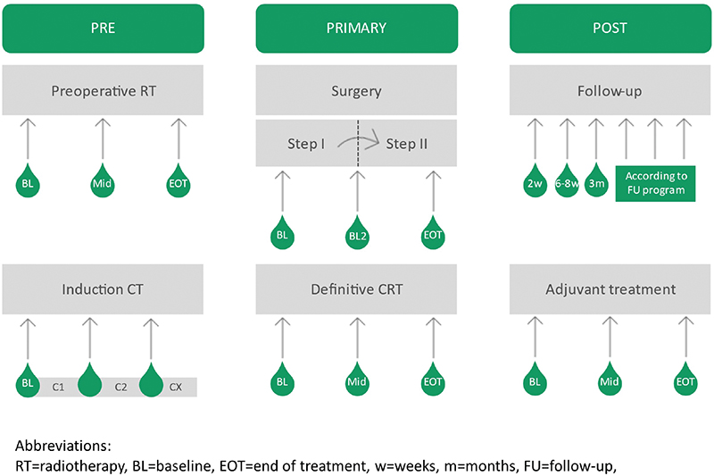
Figure 2. Suggested timepoints for sampling in future observational studies in primary surgery, chemoradiotherapy and during induction or adjuvant chemotherapy. Each sampling timepoint has a potential clinical relevance such as adding to the selection of primary surgery or CRT, treatment adaption based on sampling during CRT or CT, decisions on additional adjuvant chemo or radiotherapy, and detection of minimal residual disease after curative therapy.
HPVctDNA measurements during systemic treatment for advanced disease
A pre-treatment prognostic factor could add to clinical information and treatment decisions, particularly in the advanced setting with poor prognosis and limited efficacy from available systemic options. Recent findings suggest that ctDNA response might serve as a more reliable surrogate for OS compared to RECIST evaluations. Thus, assessing HPVctDNA response at the first evaluation of treatment response is essential. Conversely, early HPVctDNA progression may signal poor prognosis and a lack of benefit from systemic treatment, making sampling before the first three cycles relevant. Sampling at the time of progression could identify new targets for precision medicine, bringing value to future research in this area.
Clinically relevant designs for HPVctDNA guided studies
At current time there is strong data supporting the use of ctDNA as a marker of MRD across tumor types. ctDNA information is used to escalate or deescalate post-surgical adjuvant systemic therapy and for adding information to follow-up. Whereas escalation strategies in ctDNA-positive patients seem straightforward in most settings, the controversial points are feasibility of randomization, use of additional advanced imaging for example PET-CT scans, how to de-escalate and the primary endpoint. Some studies are designed with a strong endpoint such as recurrence or OS, whereas ctDNA clearance is increasingly used as primary endpoint. Studies that address ctDNA as replacement or substitute to imaging procedures are awaited.
Similar studies can be designed in SCCs, but to allow for ctDNA guided treatment decisions, it is essential that the method for HPVctDNA analysis has undergone pre-analytical and analytical validation, provides high sensitivity and specificity, and is feasible in low total DNA samples. In the MRD situations, binary reliable detection is needed. Finally, the assays must include both multiple relevant HPV subtypes and prove high feasibility in terms of fast laboratory results.
Less data allows for prospective studies of adaptation treatment, where reliable quantitative measures are needed. Validation of the observations from the current literature must be confirmed before entering prospective clinical intervention trials based on HPVctDNA results during treatment.
Conclusions
Strong data support the use of ctDNA as a marker of MRD across tumor types and potential as tool for adjuvant treatment guidance and follow-up. Similar studies can be designed in SCCs. Limited data warrants prospective studies of primary or palliative treatment adaption, and validation of the existing findings is essential before initiating clinical intervention trials based on HPVctDNA results during treatment. The HPVctDNA analysis method requires pre-analytical and analytical validation to ensure high sensitivity and specificity and feasibility in low DNA samples. It must include multiple relevant HPV subtypes and offer fast laboratory results while remaining feasible.
In conclusion, also in SCC data is emerging to confirm the major clinical potential of ctDNA measurement for risk assessment and treatment monitoring. Structured panSCC evaluations should be considered to allow for relevant sample sizes for statistical validation and to identify potential differences between the sub entities.
Data availability statement
No new data were created or analysed during this study. Data sharing is not applicable to this article.
Ethics declaration
This is a literature review including only already published manuscripts.
An ethics declaration is therefore not applicable to this manuscript.
References
[1] De Martel C, Georges D, Bray F, Ferlay J, Clifford GM. Global burden of cancer attributable to infections in 2018: a worldwide incidence analysis. Lancet Glob Health. 2020;8:e180–190. https://doi.org/10.1016/S2214-109X(19)30488-7
[2] Baumann M, Krause M, Overgaard J, Debus J, Bentzen SM, Daartz J, et al. Radiation oncology in the era of precision medicine. Nat Rev Cancer. 2016;16:234–249. https://doi.org/10.1038/nrc.2016.18
[3] Withers HR. The four R’s of radiotherapy. In: Lett JT, Adler H, editors. Advances in radiation biology. New York, NY: Academic Press; 1975. p. 241–247.
[4] Pajonk F, Vlashi E, McBride WH. Radiation resistance of cancer stem cells: the 4 R’s of radiobiology revisited. Stem Cells. 2010;28:639–648. https://doi.org/10.1002/stem.318
[5] Withers HR, Taylor JMG, Maciejewski B. The hazard of accelerated tumor clonogen repopulation during radiotherapy. Acta Oncol. 1988;27:131–146. https://doi.org/10.3109/02841868809090333
[6] Lacas B, Bourhis J, Overgaard J, Zhang Q, Grégoire V, Nankivell M, et al. Role of radiotherapy fractionation in head and neck cancers (MARCH): an updated meta-analysis. Lancet Oncol. 2017;18:1221–1237. https://doi.org/10.1016/S1470-2045(17)30458-8
[7] Overgaard J. Hypoxic Radiosensitization: adored and ignored. J Clin Oncol. 2007;25:4066–4074. https://doi.org/10.1200/JCO.2007.12.7878
[8] Eriksen JG, Maare C, Johansen J, Primdahl H, Evensen J, Kristensen CA, et al. OC-0271: 5-Y update of the randomized phase III trial DAHANCA19: primary (Chemo) RT +/- zalutumumab in HNSCC. Radiother Oncol. 2018;127:S137–S138. https://doi.org/10.1016/s0167-8140(18)30581-4
[9] Mehanna H, Robinson M, Hartley A, Kong A, Foran B, Fulton-Lieuw T, et al. Radiotherapy plus cisplatin or cetuximab in low-risk human papillomavirus-positive oropharyngeal cancer (De-ESCALaTE HPV): an open-label randomised controlled phase 3 trial. Lancet. 2019;393:51–60. https://doi.org/10.1016/S0140-6736(18)32752-1
[10] Gillison ML, Trotti AM, Harris J, Eisbruch A, Harari PM, Adelstein DJ, et al. Radiotherapy plus cetuximab or cisplatin in human papillomavirus-positive oropharyngeal cancer (NRG Oncology RTOG 1016): a randomised, multicentre, non-inferiority trial. Lancet. 2019;393:40–50. https://doi.org/10.1016/S0140-6736(18)32779-X
[11] Gebre-Medhin M, Brun E, Engström P, Haugen Cange H, Hammarstedt-Nordenvall L, Reizenstein J, et al. ARTSCAN III: a randomized phase III study comparing chemoradiotherapy with cisplatin versus cetuximab in patients with locoregionally advanced head and neck squamous cell cancer. J Clin Oncol. 2021;39:38–47. https://doi.org/10.1200/JCO.20
[12] Lee NY, Ferris RL, Psyrri A, Haddad RI, Tahara M, Bourhis J, et al. Avelumab plus standard-of-care chemoradiotherapy versus chemoradiotherapy alone in patients with locally advanced squamous cell carcinoma of the head and neck: a randomised, double-blind, placebo-controlled, multicentre, phase 3 trial. Lancet Oncol. 2021;22(4):450–462. https://doi.org/10.1016/S1470-2045(20)30737-3
[13] Martin D, Balermpas P, Gollrad J, Weiß C, Valentini C, Stuschke M, et al. RADIANCE – radiochemotherapy with or without Durvalumab in the treatment of anal squamous cell carcinoma: a randomized multicenter phase II trial. Clin Transl Radiat Oncol. 2020;23:43–49. https://doi.org/10.1016/j.ctro.2020.04.010
[14] Vendrely V, Ronchin P, Minsat M, Le Malicot K, Lemanski C, Mirabel X, et al. Panitumumab in combination with chemoradiotherapy for the treatment of locally-advanced anal canal carcinoma: results of the FFCD 0904 phase II trial. Radiother Oncol. 2023;186:109742. https://doi.org/10.1016/j.radonc.2023.109742
[15] Lassen P, Primdahl H, Johansen J, Kristensen CA, Andersen E, Andersen LJ, et al. Impact of HPV-associated p16-expression on radiotherapy outcome in advanced oropharynx and non-oropharynx cancer. Radiother Oncol. 2014;113:310–316. https://doi.org/10.1016/j.radonc.2014.11.032
[16] Serup-Hansen E, Linnemann D, Skovrider-Ruminski W, Hgødall E, Geertsen PF, Havsteen H. Human papillomavirus genotyping and p16 expression as prognostic factors for patients with American Joint Committee on Cancer stages I to III carcinoma of the anal canal. J Clin Oncol. 2014;32:1812–1817. https://doi.org/10.1200/JCO.2013.52.3464
[17] Lilja-Fischer JK, Eriksen JG, Georgsen JB, Vo TT, Larsen SR, Cheng J, et al. Prognostic impact of PD-L1 in oropharyngeal cancer after primary curative radiotherapy and relation to HPV and tobacco smoking. Acta Oncol. 2020;59:666–672. https://doi.org/10.1080/0284186X.2020.1729407
[18] Mehanna H, Taberna M, von Buchwald C, Tous S, Brooks J, Mena M, et al. Prognostic implications of p16 and HPV discordance in oropharyngeal cancer (HNCIG-EPIC-OPC): a multicentre, multinational, individual patient data analysis. Lancet Oncol. 2023;24(3):239–251. https://doi.org/10.1016/S1470-2045(23)00013-X
[19] Lefèvre AC, Pallisgaard N, Kronborg C, Wind KL, Krag SRP, Spindler KG. The clinical Alue of measuring circulating HPV DNA during chemo-radiotherapy in squamous cell carcinoma of the anus. Cancers (Basel). 2021;13(10):2451. https://doi.org/10.3390/cancers13102451
[20] Gameiro SR, Strauss J, Gulley JL, Schlom J. Preclinical and clinical studies of bintrafusp alfa, a novel bifunctional anti-PD-L1/TGFβRII agent: Current status. Exp Biol Med. 2022;247(13):1124–1134. https://doi.org/10.1177/15353702221089910
[21] Corcoran RB, Chabner BA. Application of cell-free DNA analysis to cancer treatment. N Engl J Med. 2018;379(18):1754–1765. https://doi.org/10.1056/nejmra1706174
[22] Mauri G, Vitiello PP, Sogari A, Crisafulli G, Sartore-Bianchi A, Marsoni S, et al. Liquid biopsies to monitor and direct cancer treatment in colorectal cancer. Br J Cancer. 2022;127(3):394–407. https://doi.org/10.1038/s41416-022-01769-8
[23] Boysen AK, Pallisgaard N, Andersen CSA, Spindler KLG. Circulating tumor DNA as a marker of minimal residual disease following local treatment of metastases from colorectal cancer. Acta Oncol. 2020;59:1424–1429. https://doi.org/10.1080/0284186X.2020.1806357
[24] Jeannot E, Becette V, Campitelli M, Calméjane MA, Lappartient E, Ruff E, et al. Circulating human papillomavirus DNA detected using droplet digital PCR in the serum of patients diagnosed with early stage human papillomavirus-associated invasive carcinoma. J Pathol Clin Res. 2016;2(4):201–209. https://doi.org/10.1002/cjp2.47
[25] Cabel L, Jeannot E, Bieche I, Vacher S, Callens C, Bazire L, et al. Prognostic impact of residual HPV ctDNA detection after chemoradiotherapy for anal squamous cell carcinoma. Clin Cancer Res. 2018;24:5767–5771. https://doi.org/10.1158/1078-0432.CCR-18-0922
[26] Bernard-Tessier A, Jeannot E, Guenat D, Debernardi A, Michel M, Proudhon C, et al. Clinical validity of HPV circulating tumor DNA in advanced anal carcinoma: an ancillary study to the EPITOPES-HPV02 trial. Clin Cancer Res. 2019;25:2109–2115. https://doi.org/10.1158/1078-0432.CCR-18-2984
[27] Mazurek AM, Małusecka E, Jabłońska I, Vydra N, Rutkowski TW, Giglok M, et al. Circulating HPV16 DNA in blood plasma as prognosticator and early indicator of cancer recurrence in radio-chemotherapy for anal cancer. Cancers (Basel). 2023;15(3):867. https://doi.org/10.3390/cancers15030867
[28] Tanaka H, Takemoto N, Horie M, Takai E, Fukusumi T, Suzuki M, et al. Circulating tumor HPV DNA complements PET-CT in guiding management after radiotherapy in HPV-related squamous cell carcinoma of the head and neck. Int J Cancer. 2021;148:995–1005. https://doi.org/10.1002/ijc.33287
[29] Lee JY, Garcia-Murillas I, Cutts RJ, De Castro DG, Grove L, Hurley T, et al. Predicting response to radical (chemo)radiotherapy with circulating HPV DNA in locally advanced head and neck squamous carcinoma. Br J Cancer. 2017;117:876–883. https://doi.org/10.1038/bjc.2017.258
[30] Dahlstrom KR, Li G, Hussey CS, Vo JT, Wei Q, Zhao CS, et al. Erratum: circulating human papillomavirus DNA as a marker for disease extent and recurrence among patients with oropharyngeal cancer. Cancer. 2015;121(19):3455–3464. https://doi.org/10.1002/cncr.29772
[31] Hanna GJ, Supplee JG, Kuang Y, Mahmood U, Lau CJ, Haddad RI, et al. Plasma HPV cell-free DNA monitoring in advanced HPV-associated oropharyngeal cancer. Ann Oncol. 2018;29:1980–1986. https://doi.org/10.1093/annonc/mdy251
[32] Hanna GJ, Lau CJ, Mahmood U, Supplee JG, Mogili AR, Haddad RI, et al. Salivary HPV DNA informs locoregional disease status in advanced HPV-associated oropharyngeal cancer. Oral Oncol. 2019;95:120–126. https://doi.org/10.1016/j.oraloncology.2019.06.019
[33] Chera BS, Kumar S, Beaty BT, Marron D, Jefferys S, Green R, et al. Rapid clearance profile of plasma circulating tumor HPV type 16 DNA during chemoradiotherapy correlates with disease control in HPV-associated oropharyngeal cancer. Clin Cancer Res. 2019;25:4682–4690. https://doi.org/10.1158/1078-0432.CCR-19-0211
[34] Cao Y, Haring CT, Brummel C, Bhambhani C, Aryal M, Lee C, et al. Early HPV ctDNA kinetics and imaging biomarkers predict therapeutic response in p16+ oropharyngeal squamous cell carcinoma. Clin Cancer Res. 2022;28:350–359. https://doi.org/10.1158/1078-0432.CCR-21-2338
[35] Adrian G, Forslund O, Pedersen L, Sjövall J, Gebre-Medhin M. Circulating tumour HPV16 DNA quantification – a prognostic tool for progression-free survival in patients with HPV-related oropharyngeal carcinoma receiving curative chemoradiotherapy. Radiother Oncol. 2023;186:109773. https://doi.org/10.1016/j.radonc.2023.109773
[36] Califano J, Yousef A, Mostafa H, Valsamakis A, Zhang X, Batis N, et al. Lead time to recurrence after posttreatment plasma and saliva HPV DNA testing in patients with low-risk HPV oropharynx cancer. JAMA Otolaryngol Head Neck Surg. 2023;149(9):812–813. https://doi.org/10.1001/jamaoto.2023.1730
[37] Cao H, Banh A, Kwok S, Shi X, Wu S, Krakow T, et al. Quantitation of human papillomavirus DNA in plasma of oropharyngeal carcinoma patients. Int J Radiat Oncol Biol Phys. 2012;82:351–358. https://doi.org/10.1016/j.ijrobp.2011.05.061
[38] Chera BS, Kumar S, Shen C, Amdur R, Dagan R, Green R, et al. Plasma circulating tumor HPV DNA for the surveillance of cancer recurrence in HPV-associated oropharyngeal cancer. J Clin Oncol. 2020;38:1050–1058. https://doi.org/10.1200/JCO.19
[39] Kedzia H, Gozdzicka-Józefiak A, Wolna M, Tomczak E. Distribution of human papillomavirus 16 in the blood of women with uterine cervix carcinoma. Eur J Gynaecol Oncol. 1992;13(6):522–526.
[40] Cabel L, Bonneau C, Bernard-Tessier A, Héquet D, Tran-Perennou C, Bataillon G, et al. HPV ctDNA detection of high-risk HPV types during chemoradiotherapy for locally advanced cervical cancer. ESMO Open. 2021;6:100154. https://doi.org/10.1016/j.esmoop.2021.100154
[41] Pornthanakasem W, Shotelersuk K, Termrungruanglert W, Voravud N, Niruthisard S, Mutirangura A. Human papillomavirus DNA in plasma of patients with cervical cancer. BMC Cancer. 2001;1:2. https://doi.org/10.1186/1471-2407-1-2
[42] Cheung TH, Yim SF, Yu MY, Worley MJ, Fiascone SJ, Chiu RWK, et al. Liquid biopsy of HPV DNA in cervical cancer. J Clin Virol 2019;114:32–36. https://doi.org/10.1016/j.jcv.2019.03.005
[43] Jeannot E, Latouche A, Bonneau C, Calmejane MA, Beaufort C, Ruigrok-Ritstier K, et al. Circulating HPV DNA as a marker for early detection of relapse in patients with cervical cancer. Clin Cancer Res. 2021;27:5869–5877. https://doi.org/10.1158/1078-0432.CCR-21-0625
[44] Campitelli M, Jeannot E, Peter M, Lappartient E, Saada S, De la Rochefordière A, et al. Human papillomavirus mutational insertion: specific marker of circulating tumor DNA in cervical cancer patients. PLoS One. 2012;7(8):e43393. https://doi.org/10.1371/journal.pone.0043393
[45] Parameshwaran K, Sharma P, Rajendra S, Stelzer-Braid S, Xuan W, Rawlinson WD. Circulating human papillomavirus DNA detection in Barrett’s dysplasia and esophageal adenocarcinoma. Dis Esophagus. 2019;32(12):doz064. https://doi.org/10.1093/dote/doz064
[46] Conesa-Zamora P, Doménech-Peris A, Orantes-Casado FJ, Ortiz-Reina S, Sahuquillo-Frías L, Acosta-Ortega J, et al. Effect of human papillomavirus on cell cycle-related proteins p16, Ki-67, Cyclin D1, p53, and ProEx C in precursor lesions of cervical carcinoma: a tissue microarray study. Am J Clin Pathol. 2009;132(3):378–390. https://doi.org/10.1309/AJCPO0WY1VIFCYDC
[47] Liu VWS, Tsang P, Yip A, Ng TY, Wong LC, Ngan HYS. Low incidence of HPV DNA in sera of pretreatment cervical cancer patients. Gynecol Oncol. 2001;82:269–272. https://doi.org/10.1006/gyno.2001.6289
[48] Dong SM, Pai SI, Rha SH, Hildesheim A, Kurman RJ, Schwartz PE, et al. Detection and quantitation of human papillomavirus DNA in the plasma of patients with cervical carcinoma. Cancer Epidemiol Biomarkers Prev. 2002;11(1):3–6.
[49] Sathish N, Abraham P, Peedicayil A, Sridharan G, John S, Shaji RV, et al. HPV DNA in plasma of patients with cervical carcinoma. J Clin Virol. 2004;31:204–209. https://doi.org/10.1016/j.jcv.2004.03.013
[50] Kay P, Allan B, Denny L, Hoffman M, Williamson AL. Detection of HPV 16 and HPV 18 DNA in the blood of patients with cervical cancer. J Med Virol. 2005;75:435–439. https://doi.org/10.1002/jmv.20294
[51] Shimada T, Yamaguchi N, Nishida N, Yamasaki K, Miura K, Katamine S, et al. Human papillomavirus DNA in plasma of patients with HPV16 DNA-positive uterine cervical cancer. Jpn J Clin Oncol. 2010;40:420–424. https://doi.org/10.1093/jjco/hyp193
[52] Kang Z, Stevanovic S, Hinrichs CS, Cao L. Circulating cell-free DNA for metastatic cervical cancer detection, genotyping, and monitoring. Clin Cancer Res. 2017;23:6856–6862. https://doi.org/10.1158/1078-0432.CCR-17-1553
[53] Bønløkke S, Stougaard M, Sorensen BS, Booth BB, Høgdall E, Nyvang GB, et al. The diagnostic value of circulating cell-free HPV DNA in plasma from cervical cancer patients. Cells. 2022;11(14):2170. https://doi.org/10.3390/cells11142170
[54] Ahn SM, Chan JYK, Zhang Z, Wang H, Khan Z, Bishop JA, et al. Saliva and plasma quantitative polymerase chain reaction-based detection and surveillance of human papillomavirus-related head and neck cancer. JAMA Otolaryngol Head Neck Surg. 2014;140:846–854. https://doi.org/10.1001/jamaoto.2014.1338
[55] Wang Y, Springer S, Mulvey CL, Silliman N, Schaefer J, Sausen M, et al. Detection of somatic mutations and HPV in the saliva and plasma of patients with head and neck squamous cell carcinomas. Sci Transl Med. 201;7(293):293ra104. https://doi.org/10.1126/scitranslmed.aaa8507
[56] Mazurek AM, Rutkowski T, Fiszer-Kierzkowska A, Małusecka E, Składowski K. Assessment of the total cfDNA and HPV16/18 detection in plasma samples of head and neck squamous cell carcinoma patients. Oral Oncol. 2016;54:36–41. https://doi.org/10.1016/j.oraloncology.2015.12.002
[57] Rutkowski TW, Mazurek AM, Śnietura M, Hejduk B, Jędrzejewska M, Bobek-Billewicz B, et al. Circulating HPV16 DNA may complement imaging assessment of early treatment efficacy in patients with HPV-positive oropharyngeal cancer. J Transl Med. 2020;18(1):167. https://doi.org/10.1186/s12967-020-02330-y
[58] Reder H, Taferner VF, Wittekindt C, Bräuninger A, Speel EJM, Gattenlöhner S, et al. Plasma cell-free human papillomavirus oncogene E6 and E7 DNA predicts outcome in oropharyngeal squamous cell carcinoma. J Mol Diagn. 2020;22:1333–1343. https://doi.org/10.1016/j.jmoldx.2020.08.002
[59] Haring CT, Bhambhani C, Brummel C, Jewell B, Bellile E, Heft Neal ME, et al. Human papilloma virus circulating tumor DNA assay predicts treatment response in recurrent/metastatic head and neck squamous cell carcinoma. Oncotarget. 2021;12(13):1214–1229. https://doi.org/10.18632/oncotarget.27992
[60] Siravegna G, O’Boyle CJ, Varmeh S, Queenan N, Michel A, Stein J, et al. Cell-free HPV DNA provides an accurate and rapid diagnosis of HPV-associated head and neck cancer. Clin Cancer Res. 2022;28(4):719–727. https://doi.org/10.1158/1078-0432.CCR-21-3151
[61] Tanaka H, Suzuki M, Takemoto N, Fukusumi T, Eguchi H, Takai E, et al. Performance of oral HPV DNA, oral HPV mRNA and circulating tumor HPV DNA in the detection of HPV-related oropharyngeal cancer and cancer of unknown primary. Int J Cancer. 2022;150:174–186. https://doi.org/10.1002/ijc.33798
[62] Routman DM, Kumar S, Chera BS, Jethwa KR, Van Abel KM, Frechette K, et al. Detectable postoperative circulating tumor human papillomavirus DNA and association with recurrence in patients with HPV-associated oropharyngeal squamous cell carcinoma. Int J Radiat Oncol Biol Phys. 2022;113:530–538. https://doi.org/10.1016/j.ijrobp.2022.02.012
[63] Cabel L, Bidard FC, Servois V, Cacheux W, Mariani P, Romano E, et al. HPV circulating tumor DNA to monitor the efficacy of anti-PD-1 therapy in metastatic squamous cell carcinoma of the anal canal: a case report. Int J Cancer. 2017;141:1667–1670. https://doi.org/10.1002/ijc.30863
[64] Veyer D, Pavie J, Pernot S, Mandavit M, Garrigou S, Lucas M-L, et al. HPV-circulating tumoural DNA by droplet-based digital polymerase chain reaction, a new molecular tool for early detection of HPV metastatic anal cancer? A case report. Eur J Cancer. 2019;112:34–37. https://doi.org/10.1016/j.ejca.2019.02.012
[65] Lee JY, Cutts RJ, White I, Augustin Y, Garcia-Murillas I, Fenwick K, et al. Next generation sequencing assay for detection of circulating HPV DNA (cHPV-DNA) in patients undergoing radical (chemo)radiotherapy in anal squamous cell carcinoma (ASCC). Front Oncol. 2020;10:505. https://doi.org/10.3389/fonc.2020.00505
[66] Damerla RR, Lee NY, You D, Soni R, Shah R, Reyngold M, et al. Detection of early human papillomavirus-associated cancers by liquid biopsy. JCO Precis Oncol. 2019;3:PO.18.00276. https://doi.org/10.1200/PO.18.00276
[67] Tie J, Wang Y, Tomasetti C, Li L, Springer S, Kinde I, et al. Circulating tumor DNA analysis detects minimal residual disease and predicts recurrence in patients with stage II colon cancer. Sci Transl Med. 2016;8(346):346ra92. https://doi.org/10.1126/scitranslmed.aaf6219
[68] Callesen LB, Hamfjord J, Boysen AK, Pallisgaard N, Guren TK, Kure EH, et al. Circulating tumour DNA and its clinical utility in predicting treatment response or survival in patients with metastatic colorectal cancer: a systematic review and meta-analysis. Br J Cancer. 2022;127:500–513. https://doi.org/10.1038/s41416-022-01816-4
[69] Spindler KG. Methodological, biological and clinical aspects of circulating free DNA in metastatic colorectal cancer. Acta Oncol. 2017;56:7–16. https://doi.org/10.1080/0284186X.2016.1253861
[70] Jakobsen A, Andersen RF, Hansen TF, Jensen LH, Faaborg L, Steffensen KD, et al. Early ctDNA response to chemotherapy. A potential surrogate marker for overall survival. Eur J Cancer. 2021;149:128–133. https://doi.org/10.1016/j.ejca.2021.03.006
[71] Reinert T, Schøler LV, Thomsen R, Tobiasen H, Vang S, Nordentoft I, et al. Analysis of circulating tumour DNA to monitor disease burden following colorectal cancer surgery. Gut. 2016;65:625–634. https://doi.org/10.1136/gutjnl-2014-308859
[72] Bønløkke S, Steiniche T, Sorensen BS, Nyvang GB, Lindegaard JC, Blaakær J, et al. Circulating cell-free HPV DNA is a strong marker for disease severity in cervical cancer. Mol Oncol. 2024;18(5):1231–1244. https://doi.org/10.1002/1878-0261.13538