ORIGINAL ARTICLE
Radiotherapy quality assurance in the PROTECT trial – a European randomised phase III-trial comparing proton and photon therapy in the treatment of patients with oesophageal cancer
Camilla S. Byskova  , Hanna R. Mortensenb, Marie-Claude Bistonc, Sara Broggid, Rebecca Bütofe,f, Richard Cantersg, Gilles Crehangeh, Gilles Defraenei, Jerome Doyenj, Mai L. Ehmsenb, Silvia Fabianok, Francesco Fracchiolal, Farid Goudjilh, Karin Haustermansi,m, Sarah E. Jensenb, Maria F. Jensenb, Marie Lecornun, Sebastian Makockie,f, Aurélia L. Manaj, Andrea Martignanol, Arturs Meijerso, Alfredo Mirandolap, Diana A. Mitreaj, Christina T. Muijsq, Ditte S. Møllera,r, Marianne Nordsmarka, Ester Orlandis,t, Panagiotis Balermpask, Pieter Populairei,m, Daniele Scartoniu, Jessica Serrandc, Muhammad Shamshadv, Najla Slimd, Valentina Vanoniw, Anthony Velan, Marie Vidalj, Gloria Vilches-Freixasg, Damien Webero, Lone Hoffmanna,r
aDepartment of Oncology, Aarhus University Hospital, Aarhus, Denmark; bDanish Centre for Particle Therapy, Aarhus University Hospital, Aarhus, Denmark; cCentre Léon Bérard, Lyon, France; dSan Raffaele Hospital, Milan, Italy; eDepartment of Radiotherapy and Radiation Oncology, Faculty of Medicine and University Hospital Carl Gustav Carus, TUD Dresden University of Technology, Dresden, Germany; fOncoRay – National Center for Radiation Research in Oncology, Faculty of Medicine and University Hospital Carl Gustav Carus, TUD Dresden University of Technology, Helmholtz-Zentrum Dresden-Rossendorf, Dresden, Germany; gDepartment of Radiation Oncology (Maastro), GROW School for Oncology and Reproduction, Maastricht University Medical Centre+, Maastricht, The Netherlands; hCurie Institute, Paris, France; iKU Leuven – University of Leuven – Department of Oncology – Laboratory of Experimental Radiotherapy, Leuven, Belgium; jCentre Antoine Lacassagne, Nice, France; kUniversity Hospital Zurich, Zurich, Switzerland; lDepartment of medical physics, APSS, Trento, Italy; mDepartment of Radiation Oncology, University Hospitals Leuven, Leuven, Belgium; nCentre François Baclesse, Caen, France; oPaul Scherrer Institute, Villigen, Switzerland; pNational Centre of Oncology Adrotherapy, Pavia, Italy; qDepartment of Radiation Oncology, University Medical Center Groningen, University of Groningen, Groningen, The Netherlands; rDepartment of Clinical Medicine, Faculty of Health Sciences, Aarhus University, Aarhus, Denmark; sDepartment of Clinical, Surgical, Diagnostic, and Pediatric Sciences, University of Pavia, Pavia, Italy; tClinical Department, National Center for Oncological Hadrontherapy (Fondazione CNAO), Pavia, Italy; uDepartment of Proton Therapy, APSS, Trento, Italy; vEBG MedAustron GmbH, Wiener Neustadt, Austria; wDepartment of Radiation Oncology, APSS, Trento, Italy
ABSTRACT
Purpose: To present results from the trial radiotherapy quality assurance (RTQA) programme of the centres involved in the randomised phase-III PROton versus photon Therapy for esophageal Cancer – a Trimodality strategy (PROTECT)-trial, investigating the clinical effect of proton therapy (PT) vs. photon therapy (XT) for patients with oesophageal cancer.
Materials and methods: The pre-trial RTQA programme consists of benchmark target and organ at risk (OAR) delineations as well as treatment planning cases, a facility questionnaire and beam output audits. Continuous on-trial RTQA with individual case review (ICR) of the first two patients and every fifth patient at each participating site is performed. Patient-specific QA is mandatory for all patients. On-site visits are scheduled after the inclusion of the first two patients at two associated PT and XT sites. Workshops are arranged annually for all PROTECT participants.
Results: Fifteen PT/XT sites are enrolled in the trial RTQA programme. Of these, eight PT/XT sites have completed the entire pre-trial RTQA programme. Three sites are actively including patients in the trial. On-trial ICR was performed for 22 patients. For the delineation of targets and OARs, six major and 11 minor variations were reported, and for six patients, there were no remarks. One major and four minor variations were reported for the treatment plans. Three site visits and two annual workshops were completed.
Interpretation: A comprehensive RTQA programme was implemented for the PROTECT phase III trial. All centres adhered to guidelines for pre-trial QA. For on-trial QA, major variations were primarily seen for target delineations (< 30%), and no treatment plans required re-optimisation.
KEYWORDS: Radiotherapy quality assurance; clinical trial; oesophageal cancer
Citation: ACTA ONCOLOGICA 2025, VOL. 64, 406–414. https://doi.org/10.2340/1651-226X.2025.42774.
Copyright: © 2025 The Author(s). Published by MJS Publishing on behalf of Acta Oncologica. This is an Open Access article distributed under the terms of the Creative Commons Attribution 4.0 International License (http://creativecommons.org/licenses/by/4.0/).
Received: 20 December 2024; Accepted: 20 February 2025; Published: 13 March 2025
CONTACT: Camilla S. Byskov cambys@rm.dk Department of Oncology, Aarhus University Hospital, Palle Juul-Jensens Boulevard 99, DK-8200 Aarhus N, Denmark
Supplemental data for this article can be accessed online at https://doi.org/10.2340/1651-226X.2025.42774
Competing interests and funding: The authors declare no conflicts of interest.
Introduction
Radiotherapy (RT) with photon therapy (XT) is standard of care for patients with locally advanced oesophageal cancer in combination with chemotherapy followed by surgery [1]. The oesophagus is in close proximity to vital organs at risk (OAR), e.g. heart and lungs, and the treatment will often result in radiation-induced side effects [2–7]. Proton therapy (PT) may reduce the dose to the surrounding OAR, whilst target dose remains similar, hence, can potentially decrease the radiation-induced side effects [6]. The European randomised phase III PROTECT-trial (PROton versus photon Therapy for Oesophageal Cancer – a Trimodality strategy; NCT05055648) will investigate the clinical effect of PT versus standard XT for oesophageal cancer patients treated with chemoradiotherapy followed by surgery [8].
It has previously been shown that a thorough quality assurance (QA) programme is important to reduce the number of major protocol deviations and to ensure high-quality results from clinical trials [9–14]. Protocol deviations are associated with increased risk of treatment failure and overall mortality [15] and are more frequent in centres where only few patients are recruited [12, 16]. In recent clinical trials involving RT of gastric and pancreatic cancer as well as for paediatric RT, resubmissions of target delineations were needed for approximately one-third of patients [16–18]. Also, regular completion of facility questionnaires (FQs), on-site visits and beam dosimetry audits are essential parts for the completion of international multicentre clinical trials and to prevent treatment heterogeneities from invalidating the significance of trial endpoints [19–21].
Therefore, in the PROTECT-trial, strict mandatory QA guidelines have been developed [8]. The completion of a pre-trial radiotherapy quality assurance (RTQA) programme consisting of benchmark delineation and treatment planning cases is mandatory, and prospective individual case reviews (ICRs) are performed for the first two patients, followed by every fifth patient at each treatment site. As part of the QA procedure, FQs have been distributed to gain insight in the patient treatment workflow across the participating centres and to ensure that all sites can deliver RT treatment according to the study protocol. A site visit is performed for associated PT and XT sites after the inclusion of the first two patients at each site, where all parts of the treatment procedure are discussed with members of the PROTECT RTQA group. Furthermore, all sites are required to perform patient-specific (PS) QA and to have a dosimetry audit performed.
The aim of this study was to present the first results from the QA programme under the PROTECT-trial, including FQs, pre-trial and on-trial RTQA, experiences from the first site visits completed as well as the two annual RTQA workshops.
Materials and methods
In short, the PROTECT trial aims to include 396 patients with locally advanced oesophageal cancer. Patients are randomised (1:1) to either neoadjuvant proton or photon chemoradiotherapy treatment [8]. Delineation guidelines have been published [22], and for all patients, both a PT and an XT treatment plan must be optimised using inverse planning, which should fulfil the requirements in the comprehensive RTQA document of the trial (Supplementary material S1).
The RTQA programme consists of five elements (Figure 1):
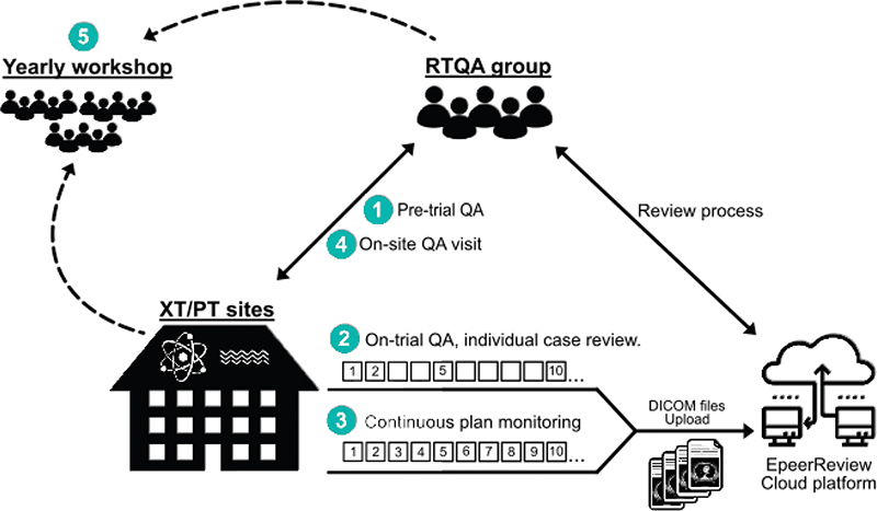
Figure 1. The PROTECT RTQA programme consists of (1) Pre-trial QA, (2) On-trial QA, (3) Continuous plan monitoring in ePeerReview, (4) On-site visits and (5) Annual workshops for everyone involved in the trial RTQA.
- Pre-trial QA:
- Five delineation benchmark cases
- Four treatment planning benchmark cases
- PT/XT FQ
- Dosimetry audit
- A plan for handling equipment breakdown
- On-trial QA:
- Prospective ICR of the first two patients enrolled in the trial at each centre
- ICR of every fifth patient
- Secondary dose calculation for all patients
- Patient-specific QA for all patients
- Continuous PT/XT treatment plan monitoring performed by the RTQA group.
- On-site QA visit:
- Discussion on computed tomography (CT) scans, delineation, treatment planning, treatment delivery, image guidance, treatment adaption strategy and plan QA. Criteria are set based on the trial RTQA document (Supplementary material S1).
- Annual workshop. All centres will discuss ICRs, technical solutions and scientific work.
RTQA group
The RTQA group consists of participants from six European centres, including four radiation oncologists, seven medical physicists and two RTQA managers. The RTQA group members arrange bimonthly meetings, where issues related to the approval of benchmark cases, patient inclusion, data acquisition, ICR, data analyses, etc. are discussed.
RTQA Programme, pre-trial QA
Five benchmark cases were prepared for pre-trial QA of both target and OAR (Body, Bowel Cavity, Heart, Liver, Lungs, Spinal Cord, planning organ at risk volume (PRV) Spinal Cord, Skin, Spleen, Kidneys and Stomach) delineations. The delineation benchmark cases were selected to represent different clinical stages with variations in location of the primary tumour and pathological lymph node(s). Target volumes and OARs were delineated according to published guidelines [22]. For target volumes, the mid-ventilation phase of the planning 4D-CT scan was used for delineation of the gross tumour volume of the primary tumour (GTVp), pathological lymph nodes (GTVn), the clinical target volume of the primary tumour (CTVp), the nodes (CTVn) and the total volume (CTVtotal). Generating an internal clinical target volume (iCTV) to account for respiratory motion was mandatory. All delineations had to be reviewed and approved by the RTQA group prior to inclusion of patients in the trial.
Four treatment planning benchmark cases were prepared and were chosen to represent different target sizes and locations for oesophageal cancer patients. The treatment planning benchmark cases are briefly described here and have been published previously [23]. When including patients in the PROTECT trial, each centre must choose between either a prescription dose of 41.4 Gy in 23 fractions or 50.4 Gy in 28 fractions. However, for the benchmark cases, prescription dose had to be 50.4 Gy in 28 fractions. Both a planning 4D-CT scan and a surveillance 4D-CT scan (recorded after 10 fractions) were included, and each centre had to calculate plan robustness towards setup and range uncertainties as well as respiratory motion. The impact of anatomical changes was evaluated using the surveillance scan. The plans were reviewed by the RTQA group, and feedback was given on primary and secondary dose constraints along with a report on mean doses to the heart and lungs compared to the results from the first eight European centres [23]. All benchmark plans had to be reviewed and approved by the RTQA group prior to inclusion of patients in the trial.
For both the delineation and the treatment planning benchmark cases, centres were asked to comply with the PROTECT RTQA guidelines (Supplementary file S1), and if these were not met, revisions had to be made, and delineations and plans re-submitted.
Facility questionnaires
The FQs contain questions related to all parts of the treatment procedure, including pre-treatment imaging, contouring, treatment planning and delivery, treatment interruptions and dose verification for both treatment modalities. The questionnaires are updated and collected yearly, and participants are asked to submit a new FQ if treatment procedures are changed during the study period.
RTQA programme, on-trial QA
For the first two patients included at each site, an accelerated QA programme must be performed. Target and OAR delineations as well as treatment plans must be reviewed within 24 hours from upload and a minimum of one working day before the patient will receive the first RT treatment. Delineations and treatment plans must adhere to the PROTECT RTQA guidelines (Supplementary material S1), which are also described by Thomas et al. [22] and Hoffmann et al. [23]. If either the delineations or the treatment plans do not adhere to the guideline requirements, corrections must be made as soon as possible, and treatment plans must be re-optimised within three fractions. A major protocol variation regarding target definition is defined as any case with the risk of violating target coverage throughout the treatment course. This includes but is not restricted to:
- Failure to generate an iCTVtotal
- Not including the entire oesophagus, where the CTV includes the GTV
- GTV not being within 10 mm of the propagation described on diagnostic CT, positron emission tomography (PET)-CT or endoscopic ultrasound will be discussed with the treating physician. If no substantial clinical reason, the GTV should be corrected. Smaller GTV will be considered a major variation and larger GTV a minor variation
- Less than 10 mm margin to metastatic lymph nodes unless shaping towards bone, vessels, etc.
- Not adding the described margins
- Scan range not sufficient according to section 1.1
- Not delineating all OARs included in the scan according to section 1.1 or not delineating OARs in the whole organ extent.
Violation of a first priority constraint (Table 1) for the nominal treatment plan is considered a major protocol variation except if this is deemed clinically necessary. Minor variations are defined as all other deviations from consensus guidelines due to clinical decision.
After the first two case reviews, every fifth patient must be reviewed within a week from the treatment start. A delineation and a treatment planning checklist have been developed to ensure consistency when the members of the RTQA group perform the reviews (Supplementary materials S2 and S3). For the patients who are not part of the on-trial QA, delineations, treatment plans and surveillance, CT scans as well as re-plans must be uploaded giving the RTQA group access to monitor on-trial data continuously. All on-trial data are uploaded to the ePeerReview Cloud platform (Varian medical Systems).
Image-guidance and adaptation
Image guidance based on volumetric imaging (enabling soft tissue visualisation and registration) should be performed daily before treatment and should be used for matching to ensure high precision of the target position during each treatment fraction. For XT, a soft tissue match on the target is preferred for daily setup. For PT, a bone match or soft tissue match on the target should be used [24]. Fiducial markers applicable for PT may be used but are not mandatory [25, 26].
The images acquired for daily image guidance must be used to verify the inter-fractional reproducibility of the target positioning. Each department must have a treatment adaptation strategy. The strategy and tolerance limits for target and OARs should be based on margins or uncertainties as determined by the individual centre. In case the tolerance limits are exceeded, action based on the adaptive strategy of the treating XT or PT centre must be performed. Adaptation should be performed within two working days.
Continuous PT/XT treatment plan monitoring
For all patients, both a PT and an XT treatment plan are inversely planned. Both PT and XT treatment plans for all patients must be uploaded in a pseudonymised form to the central dose plan bank, where the plans will be continuously monitored for adherence to the PT/XT guidelines. In case of adaptation, the recalculated plan and dose cube based on the adaptive plan for the modality used for treatment should also be exported. No strict time demand is set for continuous monitoring. Monitoring of plans will be used to check for variations of constraints and used for feedback to the centres. Furthermore, the continuous QA programme will monitor the difference in dose to OARs in the two arms.
Patient-specific QA
Patient-specific QA is mandatory, and all treatment plans should pass QA procedures with a γ (5%, 3 mm) above 95% or γ (3%, 3 mm) above 90%. The γ evaluation must be based on measurements of the treatment plans at the accelerator and may be based on the PSQA device used by the individual treatment site. All treatment plans must pass evaluation in a secondary, independent dose calculator with γ (5%, 3 mm) above 95% or γ (3%, 3 mm) above 90% [27].
Site visits
When two patients have been included for both PT and at least one associated XT site, a site visit must be performed. A site visit checklist is filled out prior to the visit for both the PT and the XT sites (Supplementary materials S4a b). At the site visit, a radiation oncologist, a physicist and a dosimetrist from the site to be audited and a minimum of two participants (a physician and a physicist) from the PROTECT RTQA group must be present. All parts of the treatment of oesophageal cancer patients are discussed. A site visit report is written, and the RTQA group will ask the site to change practice if needed (Supplementary material S5).
Dosimetry audit
It is mandatory for all sites included in the trial to have a dosimetry audit performed within the timeframe of the project. The audit should adhere to the European Organisation for Research and Treatment of Cancer (EORTC) guidelines [28]. Sites that do not have a dosimetry audit performed within 2 years of enrolment in the RTQA programme have the possibility to participate in an audit performed by IBA (IBA Dosimetry GmbH, Schwarzenbruck, Germany). The IBA audits are based on ion chamber measurements and measure absolute dose in the reference point for XT. For PT measurements, three monoenergetic fields (100, 170 and 220MeV) with a measurement depth of 2 cm and three modulated spread-out Bragg peaks (SOBP) measured in midpoint of SOBP were selected according to the IAEA TRS-398 Rev. 1 code of practice.
Annual workshop
An online workshop is arranged yearly by the RTQA group, where participants from all activated sites are invited. Here, the status of the trial is presented by the RTQA group, and topics on, e.g., adaptive strategies, challenging cases and translational studies can be discussed.
Results
Pre-trial QA: Eight PT sites and eight corresponding XT sites have completed the entire RTQA pre-trial programme and are approved for inclusion of patients (Table 2). Two Dutch centres also completed the pre-trial RTQA programme but will not include patients in the trial since in The Netherlands, a model-based approach is used for patient selection for PT. All sites received feedback upon completion of the benchmark cases and re-submitted delineations and plans if needed until everything was approved. Ten sites have started but not yet completed delineation and treatment planning benchmark cases.
FQs were distributed to 28 sites and returned from 20 sites. The questionnaires should be filled out, approved prior to inclusion of the first patient and must be updated in case of changes in the treatment procedure.
Selected results from the collected FQs are summarised in Table 3, and some are briefly presented here. Patients are imaged and treated in supine position with arms up at 17 sites, and in supine position with arms down at three sites. The treatment planning systems and dose calculation algorithms used are Eclipse and linear Boltzmann transport equation (seven sites) (Varian Medical Systems, Inc., Palo Alto, CA, USA), RayStation® and Monte Carlo (10 sites), Monaco® and Monte Carlo (one site) (Elekta AB, Stockholm, Sweden) and RayStation® and collapsed cone (two sites) (RaySearch Laboratories AB, Stockholm, Sweden). The prescribed dose is 41.4 Gy in 23 fractions at 10 sites and 50.4 Gy in 28 fractions at 10 sites. Three XT sites use intensity-modulated radiation therapy (IMRT), and seven XT sites use volumetric-modulated arc therapy (VMAT). For the PT sites, four use single-field uniform dose (SFUD), and six sites use intensity-modulated proton therapy (IMPT). Two XT sites use variable beam angles, and five sites use a number of full or partial arcs. One XT site has a class solution consisting of beam angles of 220°, 300°, 0°, 60° and 155° for Truebeam and 220°, 300°, 330°, 0°, 30°, 60° and 155° for Halcyon. Several different robust evaluation scenarios towards setup and range uncertainties are used at the different sites ranging from 6 to 14 scenarios at XT sites and from 12 to 70 scenarios at PT sites. Also, planning target volume (PTV) margins for XT differ from isotropic expansion of the iCTV of five, seven or eight mm to customised margins. All PT sites use the CTV robust concept, except one site that uses the PTV margin. Daily imaging is kV/x-ray based except for one site, where megavoltage (MV) cone-beam computed tomography (CBCT) is used, and setup is performed using either bone match, GTV/CTV/soft tissue match or both bones and soft tissue match for both XT and PT. Three XT sites and three PT sites use fiducial markers. All XT sites and four PT sites use CBCT for daily setup, whilst six PT sites use orthogonal or oblique kilovoltage imaging and markers.
On-trial QA: Sixty-four patients have been included in the study from three sites. A total of 22 ICRs have been performed. For the delineation of targets and OARs, six major and 11 minor variations were reported, and for six patients, there were no remarks. For major variations, the extension of CTV along the oesophagus and 2 cm into the stomach for distal tumours was the most frequent violation (three patients), followed by not generating the iCTV properly (two patients). For the last case, skin was delineated as 5 mm instead of 10 mm. Major variations were corrected and resubmitted before the third fraction of RT treatment.
The treatment planning ICRs were returned with minor variations in four cases and one major variation. For XT, the mean dose to iCTVTotal (Dmean) was >101% of the target dose for three patients (secondary constraint: 99% < Dmean < 101%). The maximum dose to 0.5 cm3 to stomach-iCTV was >103% of the target dose on the XT plan for one patient (secondary constraint). The XT plan of one patient violated the primary constraint of volume of the lungs receiving 5 Gy (V5Gy), which should be less than 70% (V5Gy = 96%). This was due to a large target size, iCTVTotal = 612 cm3. The treatment planning checklist used for the ICRs is shown in Supplementary material S3.
Three site visits have been performed: at Aarhus University Hospital, Department of Oncology and Danish Centre for Particle Therapy (Denmark), at the University Hospitals Leuven and Particle Therapy Interuniversity Centre Leuven (Belgium) and at the University Hospital Zürich and the Paul Scherrer Institute (Switzerland). Simulation CT scanning procedures, delineation, treatment planning, online treatment procedures, offline adaptive therapy, weekly surveillance 4D-CT scanning and patient-specific QA procedures were discussed at all site visits (Figure 2). The members from the RTQA group were present at the simulation CT-scan, dose planning and during treatment delivery of actual patients at each site. Topics like setup margins as well as robust evaluation were discussed at all site visits since both respiratory motion and anatomical changes during the course of RT may have a large impact on the treatment. All centres complied with the guidelines, and only minor comments were submitted to the sites after the visits.
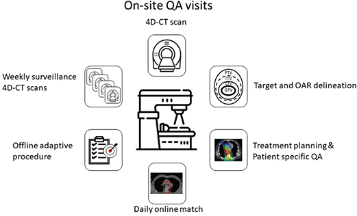
Figure 2. On-site visits are performed when a site/two corresponding sites has included two patients for both PT and XT. A physician and a physicist from the RTQA group will discuss all parts of the treatment process with a radiation oncologist and a physicist and dosimetrist from the site to be audited. XT: Photon therapy; PT: proton therapy; RTQA: radiotherapy quality assurance.
Eighteen sites have submitted beam output audits, which have all been approved. Results from the audit measurements will be presented separately and are briefly summarised here. Measurements were either based on ion chambers, thermoluminescent or optically stimulated luminescent dosimeters. Measurements were all within 3% agreement.
Secondary dose calculation will be installed at sites that presently do not have this opportunity.
Annual workshops: Two annual workshops were held online with 43 and 41 participants. The workshops included 15 and 11 presentations on, e.g., the QA review process, inter-fractional motion, online matching procedures, adaptation and treatment planning from the RTQA group and from external speakers.
Discussion
In this study, we have described the comprehensive RTQA programme implemented in the PROTECT trial, which encompasses both pre-trial and on-trial RTQA measures. These include benchmark cases, ICRs, FQs, on-site visits, patient-specific QA and beam output audits. The robust RTQA framework implemented in the PROTECT trial has been essential for ensuring consistent treatment delivery across participating centres, as deviations from protocol may compromise the reliability of study findings when patient inclusion ends [29]. Target delineation, in particular, has been shown to be susceptible to deviations, which can significantly affect clinical outcomes. For the target delineations in this study, six major deviations were found of 22 ICRs (27%), consistent with findings from previous studies [16–18, 30]. Importantly, no resubmissions of treatment plans were required, likely due to the stringent pretrial QA process, which included four treatment planning benchmark cases and detailed descriptions of the treatment planning and robustness evaluation provided in the RTQA guidelines. Whilst benchmark cases are important for training of staff at participating sites, deviations can still occur post-training, suggesting that on-trial QA should also be mandatory as recommended by the EORTC [31, 32] and in the paper by Wills et al. on QA of the SCOPE 1 (study of chemoradiotherapy in oesophageal cancer plus or minus erbitux) trial on oesophageal RT [33]. In SCOPE 1, a single benchmark case was utilised to train all participating centres in delineation and treatment planning prior to trial initiation. Despite this standardisation effort, protocol deviations were observed throughout the trial. Given resource limitations, continuous monitoring of delineations and treatment plans was conducted primarily in a retrospective manner. Additionally, the delineation reference standard was determined by two individuals rather than by consensus guidelines. FQs were returned by only 53% of participating centres, and submission was not a prerequisite for the RTQA approval. In the PROTECT trial, we have taken the RTQA process one step further by using several benchmark cases (five for delineation and four for treatment planning), consensus guidelines for delineation and treatment planning, mandatory FQs and beam output audits before patient inclusion, prospective on-trial RTQA of every fifth patient as well as site visits and annual workshops to exchange trial experience.
Deviations from clinical protocols can adversely impact patient outcomes, underscoring the critical importance of RTQA in clinical trials [12]. Despite its significance, there is currently considerable variability in the implementation of RTQA [34]. The RTQA framework used in the PROTECT trial aligns with best practices established in prior studies of radiation therapy in oncology trials [11, 12] and mirrors the approach taken in other multicentre trials, such as the EORTC and CONVERT (concurrent once-daily versus twice-daily chemoradiotherapy in patients with limited-stage small-cell lung cancer) lung cancer trials [9, 14]. Here, pre-trial QA improved compliance with the protocol and thereby improved the reliability of study outcomes. Such QA processes are especially vital in trials involving advanced radiation techniques, where even minor deviations in dose delivery or target delineation may significantly affect patient outcomes [16, 21].
Incorporating FQs into a comprehensive QA program is essential for maintaining protocol compliance and ensuring high-quality treatment delivery across multiple centres [19, 35]. Whilst on-site visits are resource- and time-intensive, they play a pivotal role in allowing RTQA personnel to thoroughly evaluate each stage of the treatment planning process [36]. These visits allow for direct interaction with the clinical staff responsible for patient treatment under the trial protocol, facilitating a comprehensive evaluation of the workflow. Moreover, the establishment of a personal contact during these visits fosters more effective communication and collaboration, which helps address any subsequent issues or deviations from the protocol.
A limitation of the project is that the on-trial ICR is very time-demanding for the clinical personnel involved. This may have caused some centres to decline from participation in the study and may even cause a limited number of patients to be included from the participating sites. Not all patients undergo on-trial RTQA, which is another limitation. In this paper, we have summarised the findings from the inclusion of the first 64 patients (16% of the total patient cohort to be included). The RTQA will continue during the entire trial period, and findings from all sites and all patients will be reported when the trial period ends.
In conclusion, the unique and strict RTQA programme within the PROTECT trial is essential for training and maintaining protocol adherence, minimising deviations and ensuring that the trial’s findings are both reliable and reproducible. Such a rigorous RTQA programme not only improves patient outcomes but also ensures that conclusions drawn from the trial can be applied in broader clinical practice.
Ethical approval
This study was approved by the Scientific Ethics Committees for the Central Denmark Region (Journal number 1-10-72-343-21).
Author contributions
CSB wrote the first draft of the manuscript and collected and presented all data; all other authors reviewed and edited the manuscript and contributed to the collection of data. HRM, KH, DSM, MN and LH contributed to the conceptualisation and study design. All authors read and approved the final manuscript.
Acknowledgements
This project has received funding from the Innovative Medicines Initiative 2 Joint Undertaking (JU) under grant agreement No. 101008134. JU receives support from the European Union’s Horizon 2020 research and innovation program and EFPIA and IBA and Varian - a Siemens Healthineer company. This project has received funding from the Novo Nordisk Foundation under grant No NNF19OC0057405, Denmark. This research project is also funded by Kom op tegen Kanker (Stand up to Cancer), the Flemish Cancer Society (Belgium). None of the above has any influence on study design, execution or interpretation of study results.
Data availability statement
The data that support the findings of this study are available from the corresponding author, CSB, upon reasonable request.
References
[1] Eyck BM, Van Lanschot JJB, Hulshof MCCM, van der Wilk BJ, Shapiro J, van Hagen P, et al. Ten-year outcome of neoadjuvant chemoradiotherapy plus surgery for esophageal cancer: the randomized controlled CROSS trial. J Clin Oncol. 2021;39:1995–2004. https://doi.org/10.1200/JCO.20.03614
[2] Bosch DJ, Muijs CT, Mul VEM, Beukema JC, Hospers GAP, Burgerhof JGM, et al. Impact of neoadjuvant chemoradiotherapy on postoperative course after curative-intent transthoracic esophagectomy in esophageal cancer patients. Ann Surg Oncol. 2014;21:605–11. https://doi.org/10.1245/S10434-013-3316-8
[3] Routman DM, Garant A, Lester SC, Day CN, Harmsen WS, Sanheuza CT, et al. A comparison of grade 4 lymphopenia with proton versus photon radiation therapy for esophageal cancer. Adv Radiat Oncol. 2019;4:63–9. https://doi.org/10.1016/J.ADRO.2018.09.004
[4] Shiraishi Y, Fang P, Xu C, Song J, Krishnan S, Koay EJ, et al. Severe lymphopenia during neoadjuvant chemoradiation for esophageal cancer: a propensity matched analysis of the relative risk of proton versus photon-based radiation therapy. Radiother Oncol. 2018;128:154–60. https://doi.org/10.1016/J.RADONC.2017.11.028
[5] Wang J, Wei C, Tucker SL, Myles B, Palmer M, Hofstetter WL, et al. Predictors of postoperative complications after trimodality therapy for esophageal cancer. Int J Radiat Oncol Biol Phys. 2013;86:885–91. https://doi.org/10.1016/J.IJROBP.2013.04.006
[6] Lin SH, Merrell KW, Shen J, Verma V, Correa AM, Wang L, et al. Multi-institutional analysis of radiation modality use and postoperative outcomes of neoadjuvant chemoradiation for esophageal cancer. Radiother Oncol. 2017;123:376–81. https://doi.org/10.1016/J.RADONC.2017.04.013
[7] Lin SH, Zhang N, Godby J, Wang J, Marsh GD, Liao Z, et al. Radiation modality use and cardiopulmonary mortality risk in elderly patients with esophageal cancer. Cancer. 2016;122:917–28. https://doi.org/10.1002/CNCR.29857
[8] Mortensen HR, Populaire P, Hoffmann L, Moeller DS, Appelt A, Nafteux P, et al. Proton versus photon therapy for esophageal cancer – a trimodality strategy (PROTECT) NCT050555648: a multicenter international randomized phase III study of neoadjuvant proton versus photon chemoradiotherapy in locally advanced esophageal cancer. Radiother Oncol. 2024;190:109980. https://doi.org/10.1016/J.RADONC.2023.109980
[9] Groom N, Wilson E, Lyn E, Faivre-Finn C. Is pre-trial quality assurance necessary? Experiences of the CONVERT Phase III randomized trial for good performance status patients with limited-stage small-cell lung cancer. Br J Radiol. 2014;87(1037):20130653. https://doi.org/10.1259/BJR.20130653
[10] Purdy JA. Quality assurance issues in conducting multi-institutional advanced technology clinical trials. Int J Radiat Oncol Biol Phys. 2008;71:S66–70. https://doi.org/10.1016/j.ijrobp.2007.07.2393
[11] Ibbott GS, Haworth A, Followill DS. Quality assurance for clinical trials. Front Oncol. 2013;3:311. https://doi.org/10.3389/fonc.2013.00311
[12] Weber DC, Tomsej M, Melidis C, Hurkmans CW. QA makes a clinical trial stronger: evidence-based medicine in radiation therapy. Radiother Oncol. 2012;105:4–8. https://doi.org/10.1016/j.radonc.2012.08.008
[13] Peters LJ, O’Sullivan B, Giralt J, Fitzgerald TJ, Trotti A, Bernier J, et al. Critical impact of radiotherapy protocol compliance and quality in the treatment of advanced head and neck cancer: results from TROG 02.02. J Clin Oncol. 2010;28:2996–3001. https://doi.org/10.1200/JCO.2009.27.4498
[14] Weber DC, Poortmans PMP, Hurkmans CW, Aird E, Gulyban A, Fairchild A. Quality assurance for prospective EORTC radiation oncology trials: the challenges of advanced technology in a multicenter international setting. Radiother Oncol. 2011;100:150–6. https://doi.org/10.1016/j.radonc.2011.05.073
[15] Ohri N, Shen X, Dicker AP, Doyle LA, Harrison AS, Showalter TN. Radiotherapy protocol deviations and clinical outcomes: a meta-analysis of cooperative group clinical trials. J Natl Cancer Inst. 2013;105:387–93. https://doi.org/10.1093/jnci/djt001
[16] Lukovic J, Moore AJ, Lee MT, Willis D, Ahmed S, Akra M, et al. The feasibility of quality assurance in the TOPGEAR international phase 3 clinical trial of neoadjuvant chemoradiation therapy for gastric cancer (an Intergroup Trial of the AGITG/TROG/NHMRC CTC/EORTC/CCTG). Int J Radiat Oncol Biol Phys. 2023;117:1096–106. https://doi.org/10.1016/j.ijrobp.2023.06.011
[17] Tchelebi LT, Winter KA, Abrams RA, Safran HP, Regine WF, McNulty S, et al. Analysis of radiation therapy quality assurance in NRG oncology RTOG 0848. Int J Radiat Oncol Biol Phys. 2024;118:107–14. https://doi.org/10.1016/j.ijrobp.2023.08.014
[18] Kelly SM, Turcas A, Corning C, Bailey S, Cañete A, Clementel E, et al. Radiotherapy quality assurance in paediatric clinical trials: first report from six QUARTET-affiliated trials. Radiother Oncol. 2023;182:109549. https://doi.org/10.1016/j.radonc.2023.109549
[19] Lambrecht M, Melidis C, Sonke JJ, Adebahr S, Boellaard R, Verheij M, et al. Lungtech, a phase II EORTC trial of SBRT for centrally located lung tumours – a clinical physics perspective. Radiat Oncol. 2016;11:7. https://doi.org/10.1186/S13014-015-0567-5
[20] Clark CH, Ga Aird E, Bolton S, Miles EA, Nisbet A, Snaith JA, et al. Radiotherapy dosimetry audit: three decades of improving standards and accuracy in UK clinical practice and trials. Br J Radiol. 2015;88(1055):20150251. https://doi.org/10.1259/bjr.20150251
[21] Clark CH, Hurkmans CW, Kry SF. The role of dosimetry audit in lung SBRT multi-centre clinical trials. Phys Med. 2017;44:171–6. https://doi.org/10.1016/J.EJMP.2017.04.003
[22] Thomas M, Mortensen HR, Hoffmann L, Møller DS, Troost EGC, Muijs CT, et al. Proposal for the delineation of neoadjuvant target volumes in oesophageal cancer. Radiother Oncol. 2021;156:102–12. https://doi.org/10.1016/j.radonc.2020.11.032
[23] Hoffmann L, Mortensen H, Shamshad M, Berbee M, Bizzocchi N, Bütof R, et al. Treatment planning comparison in the PROTECT-trial randomising proton versus photon beam therapy in oesophageal cancer: results from eight European centres. Radiother Oncol. 2022;172:32–41. https://doi.org/10.1016/j.radonc.2022.04.029
[24] Shamshad M, Møller DS, Mortensen HR, Ehmsen ML, Jensen MF, Hoffmann L. Bone versus soft-tissue setup in proton therapy for patients with oesophageal cancer. Acta Oncol. 2022;61:994–1003. https://doi.org/10.1080/0284186X.2022.2091949
[25] Apolle R, Brückner S, Frosch S, Rehm M, Thiele J, Valentini C, et al. Utility of fiducial markers for target positioning in proton radiotherapy of oesophageal carcinoma. Radiother Oncol. 2019;133:28–34. https://doi.org/10.1016/J.RADONC.2018.12.016
[26] Hoffmann L, Poulsen PR, Ravkilde T, Bertholet J, Kruhlikava I, Helbo BL, et al. Setup strategies and uncertainties in esophageal radiotherapy based on detailed intra- and interfractional tumor motion mapping. Radiother Oncol. 2019;136:161–8. https://doi.org/10.1016/J.RADONC.2019.04.014
[27] Miften M, Olch A, Mihailidis D, Moran J, Pawlicki T, Molineu A, et al. Tolerance limits and methodologies for IMRT measurement-based verification QA: recommendations of AAPM task group no. 218. Med Phys. 2018;45:e53–83. https://doi.org/10.1002/MP.12810
[28] EORTC Beam output audit guidelines [Internet]. n.d. [cited 2024 Dec 20]. Available from: https://www.eortc.org/quality-assurance/rtqa/beam-output-audit-boa/
[29] Pettersen MN, Aird E, Olsen DR. Quality assurance of dosimetry and the impact on sample size in randomized clinical trials. Radiother Oncol. 2008;86:195–9. https://doi.org/10.1016/j.radonc.2007.07.001
[30] Boustani J, Rivin Del Campo E, Blanc J, Peiffert D, Benezery K, Pereira R, et al. Quality assurance of dose-escalated radiation therapy in a randomized trial for locally advanced oesophageal cancer. Int J Radiat Oncol Biol Phys. 2019;105:329–37. https://doi.org/10.1016/J.IJROBP.2019.06.2542
[31] Budiharto T, Musat E, Poortmans P, Hurkmans C, Monti A, Bar-Deroma R, et al. Profile of European radiotherapy departments contributing to the EORTC Radiation Oncology Group (ROG) in the 21st century. Radiother Oncol. 2008;88:403–10. https://doi.org/10.1016/J.RADONC.2008.05.013
[32] Grant W, Hurkmans CW, Poortmans PM, Maingon P, Monti AF, Van Os MJH, et al. Quality assurance quality assurance standards drive improvements in the profile of radiation therapy departments participating in trials of the EORTC Radiation Oncology Group. Radiother Oncol. 2014;112:376–80. https://doi.org/10.1016/j.radonc.2014.09.003
[33] Wills L, Maggs R, Lewis G, Jones G, Nixon L, Staffurth J, et al. Quality assurance of the SCOPE 1 trial in oesophageal radiotherapy. Radiat Oncol. 2017;12:179. https://doi.org/10.1186/s13014-017-0916-7
[34] Brooks C, Miles E, Hoskin PJ. Radiotherapy trial quality assurance processes: a systematic review. Lancet Oncol. 2024;25:e104–13. https://doi.org/10.1016/S1470-2045(23)00625-3
[35] Gwynne S, Higgins E, Poon King A, Radhakrishna G, Wills L, Mukherjee S, et al. Driving developments in UK oesophageal radiotherapy through the SCOPE trials. Radiat Oncol. 2019;14:26. https://doi.org/10.1186/s13014-019-1225-0
[36] Bernier J, Horiot JC, Bartelink H, Johansson KA, Cionini L, Gonzalèz DG, et al. Profile of radiotherapy departments contributing to the Cooperative Group of Radiotherapy of the European Organization for Research and Treatment of Cancer. Int J Radiat Oncol Biol Phys. 1996;34:953–60. https://doi.org/10.1016/0360-3016(95)02177-9