ORIGINAL ARTICLE
Fixation method influences FLASH skin sparing in an in vivo leg model
Line Kristensena,b,c  , Cathrine Overgaardb,c , Jacob Graversen Johansena,c, Anna Holtz Hansenb,c, Niels Basslera,c , Per Rugaard Poulsena,c and Brita Singers Sørensena,b,c
aDanish Centre for Particle Therapy, Aarhus University Hospital, Aarhus, Denmark; bDepartment of Experimental Clinical Oncology, Aarhus University Hospital, Aarhus, Denmark; cDepartment of Clinical Medicine, Aarhus University, Aarhus, Denmark
ABSTRACT
Background and purpose: The FLASH effect, where ultra-high dose rate elicits a favourable normal tissue-sparing, has been shown in several preclinical studies. Study setup differences, for example fixation methods that affect blood flow, can influence radiation response but are unexplored for FLASH. This study compared FLASH’s acute skin-sparing effect with two fixation methods: a glued fixation (no blood flow restriction) and taped fixation (slight blood flow restriction).
Patient/material and methods: Female CDF1 mice were irradiated on their hind foot using a glue-fixation or tape-fixation method. Glue-fixated mice were only taped during the glueing procedure and had a 10-min unrestricted period afterwards before irradiation, while tape-fixated mice were taped shortly before and throughout irradiation. Mice received single-dose irradiation (19–58 Gy) with either conventional dose rate (CONV, protons 0.06 Gy/s, electrons 0.16 Gy/s) or FLASH (electrons, 223–233 Gy/s). Differences in skin toxicity were analysed.
Results: CONV-treated tape-fixated mice required a 16–17% higher dose to induce skin toxicity relative to glued mice for both protons and electrons. Meanwhile, the fixation method did not affect FLASH-treated mice. The resulting electron FLASH-sparing effect was reduced by 18% due to the shift in radiosensitivity for CONV-treated mice.
Interpretation: CONV-treated tape-fixated mice were more radioresistant than the glue-fixated mice, consistent with the expected response to mild hypoxia. FLASH-treated mice were unaffected. These findings demonstrate the impact of fixation and, in turn, oxygen level on the differential CONV versus FLASH skin response. The results highlight the importance of minimal systemic influence on animals during FLASH studies.
KEYWORDS: FLASH; ultra-high dose rate; normal tissue sparing; acute toxicity; murine model
Citation: ACTA ONCOLOGICA 2025, VOL. 64, 1029–1034. https://doi.org/10.2340/1651-226X.2025.43972.
Copyright: © 2025 The Author(s). Published by MJS Publishing on behalf of Acta Oncologica. This is an Open Access article distributed under the terms of the Creative Commons Attribution 4.0 International License (http://creativecommons.org/licenses/by/4.0/).
Received: 26 May 2025; Accepted: 26 June 2025; Published: 5 August 2025
CONTACT: Line Kristensen line.kristensen@clin.au.dk Aarhus University Hospital, Palle Juul-Jensens Boulevard 99, C106, DK-8200 Aarhus N, Denmark
Competing interests and funding: The authors declare the following financial interests/personal relationships, which may be considered potential competing interests: The study presented in this manuscript was supported by Varian, a Siemens Healthineers Company. BSS and PRP are co-inventors on a patent application (application no. 63257211 and EFS ID 44064136). The remaining authors declare that the research was conducted without commercial or financial relationships that could be construed as a potential conflict of interest.
Introduction
The FLASH effect, where ultra-high dose rate (UHDR) elicits a favourable normal tissue-sparing, has been shown in several preclinical studies, while the curative effect on cancer was maintained [1–8]. The sparing of FLASH has been investigated by comparing conventionally (CONV) low-dose-rate treated and UHDR-treated models, primarily using single fraction irradiations in several tissues. The murine skin model has been extensively used for FLASH studies [6, 9–12] and has recently been used to establish dose-response relationships after electron and proton FLASH irradiations [3, 10, 13, 14]. With the quick dose delivery, immobilisation of the animal model is crucial for target dose accuracy.
For unanaesthetised animals, the fixation of the target tissue needs to be secure enough that conscious animals cannot retract the tissue from the irradiation field. However, fixations should not induce changes in blood flow, as this can influence a tissue’s oxygen supply and, thereby, radiation response, making it more radioresistant [15]. Thus, fixations must balance the constraint of animals with minimal influence on blood flow. In murine irradiation studies by Sørensen et al. [3, 13] and Kristensen et al. [10, 14], the fixation of the target tissue was solved by a glue fixation of the target limb. The glue-fixation method is, however, primarily suitable for single-treatment use, as repeated glue-fixation procedures give a risk of skin damage.
With the recent movement towards fractionated experiments [16], an animal fixation method suitable for repeated use is needed. Tape-fixation of unanaesthetised mice has been used previously for fractionated experiments [17], and recently in a fractionated FLASH experiment (unpublished data). The tape fixation induces a risk of blood flow restriction, and the tissue might become locally hypoxic. As oxygen has been speculated to play a role in FLASH tissue sparing, as, for example seen with hyperoxia radio-sensitising skin to FLASH but not CONV irradiation [18], the hypoxia-induced radio-resistance may not be equal for CONV and FLASH-treated mice.
This study aimed to determine if fixation-induced changes in radiation response would have similar impact on a CONV-treated and FLASH-treated murine model. The study compared FLASH’s acute skin-sparing effect after single-fraction irradiation under two different fixation methods: a glued fixation (unimpeded blood flow) and a taped fixation (blood flow restriction). The study hypothesised that fixation would influence radio-sensitivity and that the tape-induced radio-resistance would not be equal for CONV and FLASH treatment.
Material and methods
Study overview
This study experimentally compared two fixation methods under conventional dose rate proton beam irradiation, followed by a retrospective analysis of preclinical animal studies that used either fixation method under electron irradiation [10] (unpublished data). The retrospective analysis in the electron beam study served to confirm findings from the proton beam study and to test the impact on FLASH treatment. All three studies used the same murine model and a consistent and reproducible experimental irradiation setup, allowing for comparative evaluation with a specific focus on the impact of fixation technique. The methodology is summarised next with an emphasis on fixation procedures. For full experimental details, see Kristensen et al. [10].
Murine model
All experiments used female C3D2F1 (CDF1) mice (11–19 weeks old) obtained from Janvier Labs (Le Genest-Saint-Isle, France). The mice were randomly assigned to treatment groups within each fixation method, with each animal considered an experimental unit. All experiments were approved by the Danish Animal Experiments Inspectorate (permits: 2017-15-0201-01218 and 2022-15-0201-01110) and conducted in compliance with the ARRIVE guidelines [19].
Fixation method
Unanaesthetised mice were immobilised in custom-designed jigs corresponding to their assigned fixation method. Each jig consisted of a ventilated hollow tube for the mouse body and PMMA plates to support the right hindlimb during fixation.
For glued fixation, the hindlimb was supported by a single plate running along the inner thigh, ending near the ankle (Figure 1). A small droplet of histoacrylic glue was applied at the contact point between the plate and the inner thigh near the ankle joint to immobilise the leg. The ankle joint was aligned just beyond the plate’s lower edge. During the 5-min glue setting period, the leg was temporarily held in place with tape, followed by a minimum 10-min restitution period with the tape loosened. The leg was released immediately post-irradiation. Target positioning was standardised and determined using template pictures from an excerpt of previous studies (Figure 1).
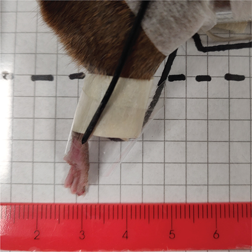
Figure 1. Target position example for a glued leg. The checkered template and ruler were used for water column depth determination.
For taped fixation, two supporting polymethyl methacrylate (PMMA) plates were used: one along the inner thigh extending to the toes and one perpendicular to support the sole. The hindlimb was fixated immediately before irradiation with a strip of tape loosely tied around the toes and the supporting plate, creating a triangular space around the foot (Figure 2B). The toes were aligned with the lower edge of the supporting plate. As with the glued fixation, the leg was released immediately post-irradiation. Likewise, the target position was determined through template pictures (Figure 2A).

Figure 2. Depictions of a tape-fixated mouse. (A) Target position example for a taped leg. The restrained mouse is in a jig with its right foot fixated. The checkered template was used for water column depth determination. (B) Plantar view of the fixated mouse. The loose tape around the foot forms a triangle with air between the tape and the toes.
To assess potential differences in limb position between the two fixation types, a subset of fixation images was analysed to compare foot placement.
Irradiations
For the proton beam study, two mouse cohorts were irradiated using either glued or taped fixation in the middle of a spread-out Bragg peak of a horizontal pencil beam scanning proton beamline (ProBeam, Varian, a Siemens Healthineers Company, Palo Alto, CA, USA) at the Danish Centre for Particle Therapy. Mice were treated using conventional low dose rates (CONV) of 0.06 Gy/s with five dose groups of 5–9 mice receiving single doses between 26 and 42 Gy. Treatment plans were made with the Eclipse treatment planning system (Varian Medical Systems, Palo Alto, CA, USA), and were verified dosimetrically with a plane-parallel and a thimble ionisation chamber. Dosimetric details are described in Overgaard et al. [20].
For the electron beam study, a retrospective analysis was conducted on preclinical studies with either a glued [10] or a taped (unpublished data) fixation. The mice were irradiated using a horizontal 16 MeV electron beam on a FLASH-enabled linac (TrueBeam accelerator, Varian, a Siemens Healthineers Company, Palo Alto, CA, USA). Mice received either CONV (0.16 Gy/s) or FLASH (taped: 223 Gy/s, glued: 233 Gy/s) dose rates, with groups of 6–10 mice receiving single doses between 19 and 58 Gy [10]. The absolute dose in the target position was measured with a diamond detector (flashDiamond, PTW Freiburg). CONV-doses were corrected for weekly output variations. FLASH doses were determined from Bergoz ACCT current transformer output and corrected for the dose at the target position. Dosimetric details of the electron beam setup are described in Kristensen et al. [10].
Data collection and analysis
Acute skin damage was assessed daily from days 9 to 28 post-irradiation. Toxicity was graded as 2.5, 3.0, and 3.5 based on hair loss, moist desquamation area, and toe visibility. Observers were blinded to treatment and previous grades. Grading data were binarised using an automated R script to ensure analysis blinding [21]. Dose-response relationships were modelled using logistic regression, from which TD50 values (dose to elicit a toxic response in 50% of mice) were derived. Statistical differences in TD50 values between fixation groups were analysed and visualised using GraphPad Prism with multiple comparison t-tests across the three toxicity grades [22].
Results
Target positioning in the water column was comparable between fixation groups (glue mean ± SD = 2.68 ± 0.15 cm (n = 82), tape mean ± SD = 2.81 ± 0.14 cm (n = 231)), reinforcing consistent setup conditions with the same doses delivered for both fixation types.
In the proton beam study of conventionally treated mice (n = 66), tape fixation shifted dose-response curves to higher doses, showing that higher doses were needed to induce skin damage with tape fixation. This shift was reflected in the TD50-values with 32.5 Gy for glued mice and 37.9 Gy for taped mice (Figure 3). The fixation method displayed a 17% difference under conventional proton irradiation, with reduced radiosensitivity for mice restrained with tape.

Figure 3. Dose–response relationship for acute skin toxicity (Grade 3) for mice treated with CONV protons during glued (red) or taped (orange) fixation. Black lines span the 95% confidence interval for 50% toxicity.
These findings prompted a comparison as to whether a similar fixation-induced difference existed in previously reported data for electron-irradiated mice, comparing glued mice [10] and taped mice (unpublished data). Similar to the proton beam study, the electron-irradiated mice with CONV dose rate showed tape fixation to produce a rightward shift in the dose-response curve (Figure 4), with a higher dose needed to induce skin damage. A significantly higher TD50 was found for tape-fixated mice compared to glued mice for CONV across all three toxicity grades (Figure 5A). The average difference in CONV TD50 was 16%, consistent with the proton beam study findings. In contrast, for FLASH-irradiated mice, dose-response curves and TD50 values did not significantly differ by fixation method (Figures 4 and 5B).

Figure 4. Dose–response relationship for mice treated with conventional (CONV, red nuances) and FLASH (blue nuances) electron irradiation. Black lines span the 95% confidence interval for 50% toxicity. (A) Moderate acute skin toxicity equalling grade 2.5, (B) Severe acute skin toxicity equal grade 3.0, (C) Severe acute skin toxicity equal grade 3.5. The separate curves for glued mice can be found in Kristensen et al. [10]

Figure 5. Dose to elicit a response in 50% of mice (TD50) in each acute skin grade for glued- and tape-fixated electron irradiated mice. Each dot represents the mean TD50, with error bars being standard error of the mean (SEM). * highlights a significant difference between means (p < 0.05) and highlights no significant difference. (A) For mice treated with a conventional dose rate. (B) For mice treated with FLASH.
The mean dose-modification factor (DMF) of electron beam FLASH was 1.51 (range 1.45–1.54) for glued mice [10] and 1.33 (1.25–1.41) for taped mice, indicating an 18% reduction in observed FLASH effect solely due to changes in the CONV baseline.
Discussion and conclusion
This study examined how two leg fixation methods, glue and tape, affected radiation-induced skin toxicity in both conventional (CONV) and ultra-high dose rate (FLASH) settings. The acute skin toxicity in response to irradiation was changed with different fixation methods for CONV-treated but not FLASH-treated mice. Despite only changing the fixation method from glue to tape, the electron FLASH skin sparing, the DMF, was reduced by 18% from 1.51 (DMF range 1.45–1.54) with glue fixation [10] to 1.33 (DMF range 1.25–1.41) with tape fixation. The difference in DMF was due solely to changes in radio-sensitivity under CONV conditions (Figure 4); FLASH responses were unaffected by fixation method. FLASH TD50 values were 1% different between fixation methods, with completely overlapping 95% confidence intervals (Table 1) and SEM (Figure 5B). Meanwhile, CONV TD50 had a 16% (range 10–19%) difference with non-overlapping 95% confidence intervals (Table 1) and SEM (Figure 5A).
| PROTON BEAM STUDY – CONV | |||
| Toxicity score | CONV TD50 Glue (Gy) | CONV TD50 Tape (Gy) | CONV TD50 Ratio |
| Grade 3.0 | 32.5 (30.9–34.0) | 37.9 (35.9–40.4) | 1.17 |
| ELECTRON BEAM STUDY – CONV | |||
| Toxicity score | CONV TD50 Glue (Gy) [10] | CONV TD50 Tape (Gy) | CONV TD50 Ratio |
| Grade 2.5 | 29.1 (28.1–30.2) | 32.1 (30.8–33.3) | 1.10 |
| Grade 3.0 | 30.1 (29.0–31.3) | 35.8 (34.1–37.6) | 1.19 |
| Grade 3.5 | 33.7 (32.1–35.2) | 39.9 (38.3–42.7) | 1.18 |
| Mean | 1.16 | ||
| ELECTRON BEAM STUDY – FLASH | |||
| Toxicity score | FLASH TD50 Glue (Gy) [10] | FLASH TD50 Tape (Gy) | FLASH TD50 Ratio |
| Grade 2.5 | 44.9 (43.2–46.7) | 45.2 (43.6–46.7) | 1.01 |
| Grade 3.0 | 47.0 (45.2–48.8) | 47.9 (46.7–49.1) | 1.02 |
| Grade 3.5 | 49.4 (47.5–51.4) | 49.9 (48.9–51.4) | 1.01 |
| Mean | 1.01 | ||
CONV-treated tape-fixated mice were more radioresistant than the glue-fixated mice. The CONV TD50 comparisons (Figures 3 and 5A) showed that taped mice needed significantly higher doses to achieve similar skin damage. The response after tape fixation relative to glue fixation was similar for the proton-irradiated (17%) and the electron-irradiated mice (16%). The similarity in fixation-induced radio-resistance across both modalities points towards a general tendency for similar blood flow restriction with the fixation method across the studies.
A major strength of this work is the consistency across the experimental conditions, including the same facility, water bath setup, mouse strain, mouse age, and the same observers, minimising confounding factors. Furthermore, the repeated testing of the fixation method across modalities with similar results highlights the fixation’s robustness and repeatability. The near-identical target placement for both fixations (mean 0.13 cm difference) further confirmed experimental consistency. However, the absence of direct physiological measurements, such as oxygen perfusion, limits the mechanistic interpretation. Future studies incorporating these measurements, or changing blood supply substantially, could clarify the role of blood supply and oxygen in fixation-induced radiosensitivity changes and FLASH sparing in general. The influence of substantial blood flow restriction on FLASH sparing is currently under investigation in a similar model, including tissue oxygen measures.
The similar radio-resistance observed in CONV-treated tape-fixed mice across both proton and electron modalities suggests a general mechanism, possibly involving reduced blood flow or local hypoxia due to the mechanical pressure of the tape. The effect must be due to a factor whose presence is influential during the irradiation. This hypothesis is seen in the light of Schwartz’s classic study [12] demonstrating reduced skin response when a radioactive source was tightly bound to the skin: an effect later linked to hypoxia and the oxygen enhancement ratio [23]. Although we did not directly measure blood flow or oxygen tension, the lack of a fixation effect under FLASH conditions supports the idea that oxygen somehow plays a role in FLASH sparing. If oxygen availability is already depleted or irrelevant during UHDR irradiation, mechanical changes in perfusion may not further influence the outcome. One hypothesis proposed on the mechanism of the FLASH sparing effect has been the influence of oxygen, for example, as an oxygen depletion [24–26] or oxygen-producing Reactive Oxygen Species (ROS) [27]. However, the hypotheses are highly debated [28].
As the mechanism behind FLASH tissue sparing is slowly unravelling, finding these steps for accurate study comparisons is crucial. Previous data have highlighted the influence of dose-sensitive endpoints on FLASH tissue sparing [12]. Another essential factor reported was the oxygen supply’s effect [29], where a complete blood supply restriction resulted in similar radiation response of CONV and FLASH treated mice, however within a limited dose range. This study adds observations that even small restrictions of blood flow and/or oxygen influence the CONV radiation response, but that an apparent change in FLASH dose-modification is not due to any change in response to FLASH. Furthermore, the study adds the fixation method to the list of key components to consider in across-study comparisons or highlights the difficulty in doing so.
In conclusion, the fixation method can substantially influence the perceived FLASH sparing effect, not by altering the response to UHDR itself, but by modifying the conventional baseline. Tape fixation increased the TD50 for CONV-treated mice by 16–17%, leading to an apparent 18% reduction in the FLASH dose modification factor. The fixation-induced shift in radio-sensitivity was consistent for CONV across protons and electrons, suggesting a shared underlying mechanism. These findings underscore the importance of standardised fixation protocols in preclinical radiation studies. Interestingly, the taped fixation did not induce any changed response in the FLASH-treated mice, suggesting that oxygen plays a role in the mechanism of FLASH, as even though there may be some degree of hypoxia induced, it does not alter the radiation response to FLASH.
Acknowledgements
The authors would like to thank Dorthe Grand, Maria Bech Arnoldus, and Marianne Kristiansen for their excellent help in animal care and handling. This study was supported by The Independent Research Fund Denmark (grant no. 1030-00125A), the Novo Nordisk Foundation (grant no. NNF195A0059372, NNF20OC0065282), DCCC Radiotherapy – The Danish National Research Center for Radiotherapy, Danish Cancer Society (grant no. R191-A11526, R353-A20702, R353-A20708, R374-A22587), and Varian, a Siemens Healthineers Company. BIGART 2025 was financially supported by the Acta Oncologica Foundation.
Data availability statement
Data are available for sharing upon request to the corresponding author.
Ethics declarations and trial registry information
All experiments followed the permit [2017-15-0201-01218] and [2022-15-0201-01110] and adhered to the ARRIVE guidelines.
Authors’ contributions
CO, NB and BSS designed and conducted the proton study. LK, JGJ, PRP and BSS designed and conducted the electron study. LK, CO and AHH collected the murine data. JGJ, NS and PRP performed dosimetric validations. LK and CO performed the data analysis and LK drafted the manuscript. All authors reviewed, edited and approved the final manuscript.
References
[1] Kim MM, Verginadis II, Goia D, Haertter A, Shoniyozov K, Zou W, et al. Comparison of FLASH proton entrance and the spread-out Bragg Peak dose regions in the sparing of mouse intestinal crypts and in a pancreatic tumor model. Cancers (Basel). 2021;13:4244. https://doi.org/10.3390/cancers13164244
[2] Wei S, Lin H, Choi JI, Simone CB, Kang M. A novel proton pencil beam scanning FLASH RT delivery method enables optimal OAR sparing and ultra-high dose rate delivery: a comprehensive dosimetry study for lung tumors. Cancers. 2021;13:5790. https://doi.org/10.3390/cancers13225790
[3] Sørensen BS, Sitarz MK, Ankjærgaard C, Johansen JG, Andersen CE, Kanouta E, et al. Pencil beam scanning proton FLASH maintains tumor control while normal tissue damage is reduced in a mouse model. Radiother Oncol. 2022;175:178–84. https://doi.org/10.1016/j.radonc.2022.05.014
[4] Montay-Gruel P, Acharya MM, Jorge PG, Petit B, Petridis IG, Fuchs P, et al. Hypofractionated FLASH-RT as an effective treatment against glioblastoma that reduces neurocognitive side effects in mice. Clin Cancer Res. 2021;27:775–84. https://doi.org/10.1158/1078-0432.CCR-20-0894
[5] Diffenderfer ES, Verginadis II, Kim MM, Shoniyozov K, Velalopoulou A, Goia D, et al. Design, implementation, and in vivo validation of a novel proton FLASH radiation therapy system. Int J Radiat Oncol Biol Phys. 2020;106:440–8. https://doi.org/10.1016/j.ijrobp.2019.10.049
[6] Cunningham S, McCauley S, Vairamani K, Speth J, Girdhani S, Abel E, et al. FLASH proton pencil beam scanning irradiation minimizes radiation-induced leg contracture and skin toxicity in mice. Cancers (Basel). 2021;13:1–15. https://doi.org/10.3390/cancers13051012
[7] Levy K, Natarajan S, Wang J, Chow S, Eggold JT, Loo PE, et al. Abdominal FLASH irradiation reduces radiation-induced gastrointestinal toxicity for the treatment of ovarian cancer in mice. Sci Rep. 2020;10:1–14. https://doi.org/10.1038/s41598-020-78017-7
[8] Velalopoulou A, Karagounis IV, Cramer GM, Kim MM, Skoufos G, Goia D, et al. FLASH proton radiotherapy spares normal epithelial and mesenchymal tissues while preserving sarcoma response. Cancer Res. 2021;81:4808. https://doi.org/10.1158/0008-5472.CAN-21-1500
[9] Verginadis II, Velalopoulou A, Kim MM, Kim K, Paraskevaidis I, Bell B, et al. FLASH proton reirradiation, with or without hypofractionation, reduces chronic toxicity in the normal murine intestine, skin, and bone. Radiother Oncol. 2025;205:110744. https://doi.org/10.1016/j.radonc.2025.110744
[10] Kristensen L, Rohrer S, Hoffmann L, Præstegaard LH, Ankjærgaard C, Andersen CE, et al. Electron vs proton FLASH radiation on murine skin toxicity. Radiother Oncol. 2025;206:110796. https://doi.org/10.1016/j.radonc.2025.110796
[11] Soto LA, Casey KM, Wang J, Blaney A, Manjappa R, Breitkreutz D, et al. FLASH irradiation results in reduced severe skin toxicity compared to conventional-dose-rate irradiation. Radiat Res. 2020:194:618–24. https://doi.org/10.1667/RADE-20-00090
[12] Sørensen BS, Kanouta E, Ankjærgaard C, Kristensen L, Johansen JG, Sitarz MK, et al. Proton FLASH: impact of dose rate and split dose on acute skin toxicity in a murine model. Int J Radiat Oncol Biol Phys. 2024;120(1):265–75. https://doi.org/10.1016/j.ijrobp.2024.04.071
[13] Singers Sørensen B, Krzysztof Sitarz M, Ankjærgaard C, Johansen J, Andersen CE, Kanouta E, et al. In vivo validation and tissue sparing factor for acute damage of pencil beam scanning proton FLASH. Radiother Oncol. 2022;167:109–15. https://doi.org/10.1016/j.radonc.2021.12.022
[14] Kristensen L, Poulsen PR, Kanouta E, Rohrer S, Ankjærgaard C, Andersen CE, et al. Spread-out Bragg Peak FLASH: quantifying normal tissue toxicity in a murine model. Front Oncol. 2024:14:1427667. https://doi.org/10.3389/fonc.2024.1427667
[15] Schwarz G. Über Desensibiliserung Gegen Röntgen-Und Radi-umstrahlen. Munchener Medizinische Wochenschrift. 1909;24:1–2.
[16] FRPT24 Abstract Book, FRPT24.; FLASH radiotherapy and particle therapy. 2024. Available from: https://2024.frpt-conference.org/
[17] Overgaard CB, Reaz F, Poulsen P, Spejlborg H, Overgaard J, Grau C, et al. The fractionation effect on proton RBE in a late normal tissue damage model in vivo. Radiother Oncol. 2025;206:110792. https://doi.org/10.1016/j.radonc.2025.110792
[18] Iturri L, Bertho A, Lamirault C, Brisebard E, Juchaux M, Gilbert C, et al. Oxygen supplementation in anesthesia can block FLASH effect and anti-tumor immunity in conventional proton therapy. Commun Med. 2023;3:1–13. https://doi.org/10.1038/s43856-023-00411-9
[19] Percie du Sert N, Hurst V, Ahluwalia A, Alam S, Avey MT, Baker M, et al. The ARRIVE guidelines 2.0: updated guidelines for reporting animal research. PLoS Biol. 2020;18:e3000410. https://doi.org/10.1371/journal.pbio.3000410
[20] Overgaard CB, Reaz F, Sitarz M, Poulsen P, Overgaard J, Bassler N, et al. An experimental setup for proton irradiation of a murine leg model for radiobiological studies. Acta Oncol (Madr). 2023;62:1566–73. https://doi.org/10.1080/0284186X.2023.2246641
[21] Posit team RStudio: Integrated Development for R 2025 [Internet]. Available from: https://posit.co/
[22] GraphPad Software GraphPad Prism for Windows 2024 [Internet]. Available from: www.graphpad.com
[23] Schwarz G. Merkwürdige Schwankungen Der Röntgenempfindlichkeit Bein Einem Und Demselben Patienten. Wiener Medizinische Wochenschrif. 1914;64:2597–8.
[24] Petersson K, Adrian G, Butterworth K, McMahon SJ. A quantitative analysis of the role of oxygen tension in FLASH radiation therapy. Int J Radiat Oncol Biol Phys. 2020;107:539–47. https://doi.org/10.1016/j.ijrobp.2020.02.634
[25] Poulsen PR, Johansen JG, Sitarz MK, Kanouta E, Kristensen L, Grau C, Sørensen BS. Oxygen enhancement ratio weighted dose quantitatively describes acute skin toxicity variations in mice after pencil beam scanning proton FLASH irradiation with changing doses and time structures. Int J Radiat Oncol Biol Phys. 2024;120(1):276–86. https://doi.org/10.1016/j.ijrobp.2024.02.050
[26] Pratx G, Kapp DS. Ultra-high-dose-rate FLASH irradiation may spare hypoxic stem cell niches in normal tissues. Int J Radiat Oncol Biol Phys. 2019;105:190–2. https://doi.org/10.1016/j.ijrobp.2019.05.030
[27] Montay-Gruel P, Acharya MM, Petersson K, Alikhani L, Yakkala C, Allen BD, et al. Long-term neurocognitive benefits of FLASH radiotherapy driven by reduced reactive oxygen species. Proc Natl Acad Sci U S A. 2019; 166:10943–51. https://doi.org/10.1073/pnas.1901777116
[28] Loo BW, Verginadis II, Sørensen BS, Mascia AE, Perentesis JP, Koong AC, et al. Navigating the critical translational questions for implementing FLASH in the clinic. Semin Radiat Oncol. 2024;34:351–64. https://doi.org/10.1016/j.semradonc.2024.04.008
[29] Zhang Q, Gerweck LE, Cascio E, Yang Q, Huang P, Niemierko A, et al. Proton FLASH effects on mouse skin at different oxygen tensions. Phys Med Biol. 2023;68(5):10. https://doi.org/10.1088/1361-6560/acb888