ORIGINAL ARTICLE
POT1 genetic testing in melanoma-prone families in Sweden: germline variant prevalence and tumor spectrum in identified carriers
Konstantinos Papadakisa*  , Francesca Portellia,b* , Karina Schultza,c , Hedvig Olsson Sterkya, Ismini Vassilakia,b, Jan Lapinsd,e , Michael R. Sargenf , Sofia Obolenskig,h , David J. Adamsg , Muyi Yanga, Veronica Höioma,c** and Hildur Helgadottir **a,c
aDepartment of Oncology and Pathology, Karolinska Institutet, Stockholm, Sweden; bDepartment of Clinical Pathology and Cancer Diagnostics, Karolinska University Hospital, Stockholm, Sweden; cTheme Cancer, Karolinska University Hospital, Stockholm, Sweden; dDermatology and Venereology Unit, Department of Medicine Huddinge, Karolinska Institutet, Stockholm, Sweden; eDepartment of Dermatology, Karolinska University Hospital, Stockholm, Sweden; fDivision of Cancer Epidemiology and Genetics, National Cancer Institute, National Institutes of Health, Rockville, Maryland, USA; gExperimental Cancer Genetics, Wellcome Sanger Institute, Wellcome Trust Genome Campus, Hinxton, Cambridge, UK; hDepartment of Dermatology, Leiden University Medical Centre, Leiden, The Netherlands
ABSTRACT
Background and purpose: Approximately 5–10% of cutaneous melanoma occurs in individuals with a family history of the disease. While known high-penetrance genes, such as CDKN2A, explain some cases, a substantial proportion of hereditary melanoma remains genetically undefined. Recently, germline variants in genes involved in telomere regulation, including POT1, TERT, ACD, and TERF2IP, have been identified in melanoma-prone families. This study investigated the prevalence and pathogenicity of POT1 variants in a Swedish familial melanoma cohort.
Patient/material and methods: A total of 168 familial melanoma cases were screened for CDKN2A, CDK4, BAP1, and POT1. The population frequency of pathogenic variants (PVs) was assessed using the SweGen and the gnomAD databases. Functional evaluation was performed using a saturation genome editing (SGE) assay. Telomere length analysis was performed using quantitative polymerase chain reaction (qPCR) on blood-derived DNA from melanoma patients and healthy controls. The melanomas of the carriers were reviewed by expert dermatopathologists.
Results: Among the 161 CDKN2A/CDK4/BAP1-negative melanoma families included in this cohort, only one likely PV in POT1 (c.676C > A, p.His226Asn) was identified (0.6%). Population data confirmed its rarity. The carrier family exhibited multiple early-onset melanomas, with two out of three invasive cases displaying spitzoid morphology, and several other tumors. No significant telomere length differences were observed between carriers and controls. Two additional POT1 variants of uncertain significance were detected; both were predicted to be benign.
Interpretation: POT1 PVs were rare in the studied Swedish familial melanoma cases, implying limited contribution to hereditary melanoma in this population. Nonetheless, the identification of a previously unknown likely PV further supports the need for continued genetic screening in selected cases. POT1 testing should be considered in families with multiple melanomas, early onset and spitzoid histopathology, and co-occurring with other syndromic tumors.
KEYWORDS: POT1; cutaneous melanoma; sarcoma; cancer; telomere; mutation; pathogenic variant
Citation: ACTA ONCOLOGICA 2025, VOL. 64, 1102–1108. https://doi.org/10.2340/1651-226X.2025.44048.
Copyright: © 2025 The Author(s). Published by MJS Publishing on behalf of Acta Oncologica. This is an Open Access article distributed under the terms of the Creative Commons Attribution 4.0 International License (http://creativecommons.org/licenses/by/4.0/).
Received: 3 June 2025; Accepted: 8 August 2025; Published: 25 August 2025
CONTACT: Konstantinos Papadakis konstantinos.papadakis@ki.se Department of Oncology and Pathology, Karolinska Institutet 171 64 Stockholm, Sweden
*These authors contributed equally (both first authors).
**These senior authors contributed equally (both last authors).
Competing interests and funding: The authors report there are no competing interests to declare.
Introduction
It is estimated that 5–10% of cutaneous malignant melanoma (in this article referred to as melanoma) occurs in individuals with a family history of the disease. Since only around 10–30% of familial melanoma cases are attributed to known germline aberrations in established high-penetrance genes, principally CDKN2A, a significant amount of the hereditary risk for melanoma is still largely undefined [1–4]. More recently, the focus has centered on germline variants in genes implicated in telomere dysregulation, including POT1, TERT, ACD, and TERF2IP, as a novel pathway underlying familial melanoma [5–9]. Telomeres are regions at the ends of linear chromosomes composed of repeating nucleotide sequences, shielding the chromosome termini from enzymatic degradation and aberrant DNA recombination during cell division. Telomeres progressively shorten with each round of cell division – an essential phenomenon in the replicative lifespan of cells and the aging process. To preserve telomere length and ensure genomic stability, the telomerase enzyme synthesizes new telomeric repeats with every cycle [10]. Among telomere regulating genes, germline pathogenic variants (PVs) in POT1 have been found at the highest frequencies in melanoma-prone families – up to 9% – though with considerable variation across different cohorts [5, 6, 9, 11, 12].
The POT1 (protection of telomeres 1) gene encodes a protein-key component of shelterin, a telomere-specific protein complex, consisting of six subunits, which is responsible for protecting telomeres and maintaining their length. POT1 subunit binds to the single-stranded 3’ telomeric DNA (ssDNA) overhang, enabling the whole shelterin complex to mediate and regulate the interaction of telomerase with telomeres [13, 14]. Therefore, protein-altering variants within this complex can dysregulate shelterin’s function in protecting telomeres from being inappropriately processed by the DNA-repair machinery. Germline genome sequencing studies have revealed several POT1 PVs that alter the protein’s capacity to bind to ssDNA, resulting in elongated and aberrant telomeres. These abnormalities ultimately promote carcinogenesis and predispose individuals to a familial cancer syndrome that, besides melanoma [5, 6, 15], includes glioma [16], chronic lymphoblastic leukemia [17], lymphoid and myeloid neoplasms [18], colorectal cancer [19], and angiosarcoma [20, 21]. In this article, we report the results of POT1 germline genetic testing that has been conducted in Stockholm, Sweden, as part of the routine gene panel testing for hereditary melanoma in families with melanoma and cancer susceptibility. We further report on the tumor spectrum and the histopathological features of the melanoma cases reviewed in identified carriers.
Patients/material and methods
Patients and families
Since 2021, all identified melanoma families in Stockholm have undergone POT1 testing, as it has been implemented in routine gene panel testing for familial melanoma (including also CDKN2A, CDK4, and BAP1). The Stockholm region is currently (2025) the only region in Sweden that has implemented POT1 in the routine gene panel for familial melanoma. For this testing, familial melanoma has been defined as three or more cases of melanoma (invasive or in situ) in a family, or two first-degree relatives (FDRs) with melanoma if one is diagnosed before the age of 55. At the Hereditary Melanoma Clinic, Karolinska University Hospital, genetic testing was offered to affected members of melanoma-prone families fulfilling the criteria. Melanoma diagnoses in families were verified by pathology or clinical records. This study was approved by the Swedish Ethical Review Authority (Dnr. 2012/1192-32 and 2018/803-31).
POT1 germline genetic testing in melanoma families
The POT1 germline genetic screening was performed as a clinical test at an accredited laboratory, Karolinska University Laboratory. The method used was accredited, with a library preparation performed using Ion AmpliSeq™ technology, followed by sequencing on Ion S5™ system, and data analysis using Ion Reporter™ software (Life Technologies). All POT1 exons and exon-intron boundaries were covered by the analysis. Only gene variants with a minor allele frequency (MAF) of less than 0.01 were considered. The clinical variant interpretation guidelines from the American College of Medical Genetics and Genomics (ACMG) and Association for Molecular Pathology (AMP) were used to assess whether a variant was (likely) pathogenic, of uncertain significance (VUS), or (likely) benign [22]. ClinVar and the VarSome databases were used for variant classification [23, 24]. In parallel, data from saturation genome editing (SGE) were used to functionally assess the impact of POT1 germline variants. This high-throughput approach introduces all possible single-nucleotide variants into a genomic region of interest and measures their functional consequences in a pooled assay. The method was performed as described by Obolenski et al. [25].
POT1 variant detection in normal population
To explore the frequency of POT1 PVs/likely PVs in the general population, we used the SweGen Variant Frequency Dataset [26] and non-Finnish Europeans (NFE) from the gnomAD v4.1.0 [27] as a control cohort. The SweGen dataset consists of genome variant frequencies for 1,000 Swedish individuals, representing a cross-section of the Swedish population. The gnomAD v4.1.0 dataset consists of exome data from over 800,000 individuals, whereof 590,031 are of NFE descent. MAFs were used to assess the population frequency of variants, with data obtained from SweGen, NFE, and gnomAD.
Telomere length analyses
Quantitative polymerase chain reaction (qPCR) was used to measure the relative telomere length using a protocol that was adapted from Joglekar et al. [28]. Briefly, the analyses were done using PowerUp™ SYBR™ Green Master Mix (Applied Biosystems), forward and reverse primers for the telomere gene and the house-keeping gene, human β-globin (HBG), respectively, and genomic DNA extracted from blood samples. The qPCR reactions were then conducted using the QuantStudio™ 7 Flex Real-Time PCR System (Applied Biosystems). All samples were run in triplicates. Relative telomere length was determined for 74 melanoma patients (53% from familial cases, including members of a family identified with a POT1 PV) and controls from the general population (blood donors). Relative quantification of gene expression was calculated using the double delta Ct (ΔΔCt) method, with the addition of between-plate control samples, as follows: 2^([ΔΔCt of Tel sample] – [average ΔΔCt HBG-samples]). Univariate analysis of variance in IBM SPSS statistics was used to assess differences in relative telomere length using a linear regression method (general linear model [GLM]). All measurements were adjusted for age and sex.
Melanoma pathological assessments
Histological slides of the available melanoma cases were reviewed by three pathologists with expertise in melanocytic tumors. Spitzoid morphology was defined, as previously described, by the presence of large spindled and/or epithelioid melanocytes in variable proportions, with absent to minimal pigment and prominent nucleoli [29–31]. In line with previous studies on melanomas in POT1 variant carriers [32, 33], tumors were classified as having spitzoid morphology if more than 25% of tumor cells exhibited these features. The histological subtype and the percentage of tumor with spitzoid morphology were reported. In some cases, BRAF V600E (VE1) immunohistochemistry (Ventana) was performed as part of the routine workup, using adequate controls on a Ventana BenchMark ULTRA automated staining system. In one case, NGS using Oncomine™ Focus Assay (Thermo Fisher Scientific, USA) was performed.
Results
Incidence of POT1 PVs in melanoma families and in the normal population
In total, 168 families fulfilling the definition of familial melanoma were tested for CDKN2A, CDK4, BAP1, and POT1. The median age at the earliest melanoma diagnosis in families was 40 years (range 12–78 years), 60% of families had ≥3 cases of melanoma, and 40% had multiple primaries. Seven families were identified with CDKN2A mutations, and none with CDK4 or BAP1 mutations. Only one family with a likely PV was identified; hence, the frequency of POT1 PVs in this familial melanoma cohort was 0.6%. The POT1 variant (NM_015450.3:c.676C > A, His226Asn), identified in the family, is a missense variant located in the region encoding the highly conserved oligonucleotide/oligosaccharide-binding (OB) 2 domain (Figure 1). It was reported as a VUS in Clinvar (variation ID: 1018835). The histidine 226 residue is, however, highly evolutionarily conserved, and individual predictors/meta scores used in the VarSome database asserted that this variant is likely to be disruptive. In the SGE assay, the variant c.676C > A was found not to affect cell depletion; however, the c.676_678delinsAAC:CaT→AaC, leading to the same His266Asn missense variant, showed a weak depletion effect in cells, suggesting a functional abnormality. In the SweGen dataset, the p.His226Asn variant was detected in 1/2,000 alleles (MAF = 0.0005) and in the gnomAD NFE dataset in 1,177,492 alleles (MAF = 6.2 × 10⁻⁷). It was not detected in any other population in gnomAD. No other POT1 PVs or likely PVs were described in the Swedish dataset. In the gnomAD NFE dataset, the combined MAF of predicted PV/likely PV was 0.00015.
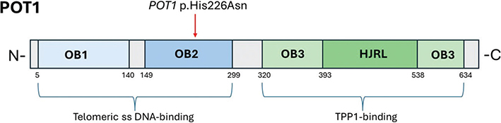
Figure 1. Schematic view of human POT1 protein functional domains. There are three oligonucleotide/oligosaccharide-binding (OB) fold domains, namely, OB1–3, with an embedded Holliday junction resolvase-like (HJRL) domain in the OB3 domain. The location of the likely pathogenic missense variant is indicated by the red arrow.
In our cohort, there were two other potential VUS detected, each in a different family. None of them have been reported in the general population (SweGen or gnomAD). One was a missense variant in exon 6 of the POT1 gene: c.56 G > T, p.(Gly19Val). This variant was detected in a patient from a melanoma-prone family, with a melanoma in situ diagnosed in the fifth decade of life. There was one additional histopathologically confirmed in situ melanoma, and two melanomas without histopathological confirmation, among other family members. The 19 amino-acid position is not well conserved, and the variant is predicted to be benign by individual predictors/meta scores used in the VarSome database. The other variant, –39 G > T, located in the –5’ UTR of POT1 was observed in a family with three affected members, where the earliest onset occurred in one patient’s fourth decade of life, and one member had multiple primaries. All had invasive melanoma, and the tumors were classified as superficial spreading (SSM) and nodular melanomas (NM). This variant is highly conserved in vertebrate species and may influence splicing; however, in the VarSome prediction databases, it was commonly predicted to be benign. The phenotype of families carrying these two variants, with no other cancer diagnoses in close relatives, was not considered typical of the POT1 predisposition syndrome either. Furthermore, both these two variants were considered unchanged/neutral in the SGE assay. Hence, neither the c.56 G > T nor the –39 G > T variant was considered pathogenic. The melanomas in these families were therefore not subjected to histopathological review.
Tumor spectrum in POT1 carrier family
The tumors identified in the family carrying the POT1 PV are shown in Table 1. To summarize, all three carriers were diagnosed with melanoma, with the earliest onset occurring in the third decade of life in one of the carriers, and multiple primaries were detected. Carrier no.1 was diagnosed with two invasive melanomas. One, located in the head and neck area, showed 100% spitzoid morphology and immunohistochemical reactivity for BRAF V600E, therefore being classified as a spitzoid BRAF-mutated melanoma (Figure 2A–B). Breslow score was reported as 0.6 mm, and the tumor was growing adjacent to an intradermal nevus (Figure 2A), suggesting that the melanoma had arisen from a precursor benign lesion. The other, located on the leg, was classified as an SSM with spitzoid features, with 90% of the tumor cells exhibiting spitzoid morphology. This melanoma had a Breslow thickness of 1.2 mm and stained positively for BRAF V600E (Figure 2C–D). Carrier no. 2 was diagnosed with a melanoma in situ on the leg, without spitzoid features, and stained positively for BRAF V600E. Carrier no. 3 was diagnosed, in the eighth decade of life, with a SSM on the leg, without spitzoid features, Breslow 0.6 mm, and an NRAS (p.Gln61Arg) mutation detected by NGS analysis. Interestingly, one FDR of a carrier, who tested negative for the POT1 PV, was diagnosed with an invasive melanoma in the third decade of life. This tumor was classified as an SSM and exhibited only partial (10%) spitzoid morphology. BRAF immunohistochemistry was not performed in this case. There were six additional FDRs from the same family branch that were not genotyped. All six were diagnosed with different types of tumors, including one case of melanoma. In the family, multiple other tumors were reported, including soft tissue tumors (n = 5), brain tumor (n = 1), and salivary gland tumors (n = 2).
| Family members | No. | Tumors, age of onset (decade) |
| Carriers | 1. | Melanoma (20s), Melanoma (20s) |
| 2. | Salivary gland tumor (50s), Melanoma in situ (50s), Basal cell carcinoma (50s), Lung carcinoid (60s), Renal angiomyolipoma (70s) | |
| 3. | Melanoma (70s), Tenosynovial giant-cell tumor (70s), Soft tissue sarcoma (70s) | |
| Non-carrier FDR of carrier | 1. | Melanoma (20s) |
| Non-genotyped FDRs of carriers | 1. | Melanoma (30s)† |
| 2. | Salivary gland tumor (50s) | |
| 3. | Soft tissue sarcoma (60s)† | |
| 4. | Malignant brain tumor (60s)† | |
| 5. | Lung cancer (60s)† | |
| 6. | Breast cancer (60s), Cerebral cavernous angioma (80s) | |
| FDR: First-degree relative, in the same family branch. †Deceased from the diagnosed tumor type. Note: Age of onset is reported by patient’s decade of life (e.g., ‘20s’ refers to the third decade of life, ages 20–29; ‘30s’ to the fourth decade of life, ages 30–39; and so on). |
||
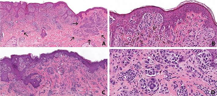
Figure 2. Histopathologic findings of cutaneous melanomas from an individual with a germline variant in POT1 (p.His226Asn). (A) Low magnification of spitzoid melanoma showing epidermal and dermal nests, growing adjacent to a nevus, marked by the arrows (hematoxylin and eosin, original magnification: X4). (B) Spitzoid morphology consisting of atypical enlarged epithelioid melanocytes with abundant eosinophilic cytoplasm. Some of the melanocytes are slightly pigmented (hematoxylin and eosin, original magnification: X20). (C) Superficial spreading melanoma with spitzoid features (hematoxylin and eosin, original magnification: X4). (D) Nests of enlarged spitzoid melanocytes including scattered multinucleated cells embedded in fibrosis (original magnification: X20).
Telomere length associated with POT1 PV
The adjusted mean telomere lengths in the white blood cells from two of the POT1 His226Asn variant carriers did not differ significantly from the values obtained from the blood donor control cohort (mean relative expression: 1.180 vs. 1.055, respectively, p = 0.645), nor when compared to other melanoma cases (mean relative expression: 1.180 vs 1.546, respectively, p = 0.492).
Discussion and conclusion
Discussion
Several genetic analysis studies of POT1 variants have implied their potentially significant role in familial melanoma and various familial cancer syndromes [5, 6, 9, 15–21]. By binding to telomeric ssDNA, as a protein-component of the shelterin complex, POT1 serves a crucial function in preserving telomere integrity [13, 14]. Genetic instability, due to aberrant telomere elongation, resulting in increased risk of carcinogenesis, may arise from dysfunctional variants of POT1 that disrupt telomerase regulation. For families with a history of melanoma or other related cancers, recent research supports the significance of including POT1 in genetic screening [5, 6, 15–21].
This study investigated the role of POT1 germline PVs in familial melanoma within a Swedish cohort. In Stockholm, Sweden, routine genetic testing for familial melanoma, initiated in 2021, revealed the rarity of POT1 PVs. Out of the 161 families in the familial melanoma cohort, meeting the criteria for melanoma susceptibility testing and being negative for CDKN2A, CDK4, and BAP1, only one family (1/161, 0.6%) was identified carrying a likely pathogenic POT1 variant. The c.676C > A (p.His226Asn) variant is located in the highly conserved OB2 domain – a region critical for POT1 function – suggesting its disruptive potential to protein’s activity. Given that the variant’s allele frequency in the SweGen dataset was 0.0005 and 1 in 1,610,494 alleles in the gnomAD database, the population-level analysis further confirmed its rarity.
Significant variation in both the prevalence and distribution of POT1 PVs is determined when comparing across different populations. Compared to our Swedish cohort, 1.8% of melanoma-prone families (4/228) in a Spanish cohort harbored POT1 PVs, all of which were negative for CDKN2A/CDK4 mutations [9]. In a multicenter study, POT1 PVs were also identified in Italian, French, and US cases – with frequencies reaching up to 9% in Italian families, where a founder mutation was identified, but considerably lower frequencies were observed in the other cohorts [5]. Moreover, it was reported that POT1 PVs/likely PVs were present in 3.8% of 105 high-risk melanoma families from the UK, Netherlands, and Australia, making POT1 the second most prevalent high-penetrance melanoma susceptibility gene in these populations after CDKN2A [6]. On the other hand, no potentially deleterious POT1 variants were discovered in a Dutch cohort of 451 familial melanoma families, indicating their extremely low frequency in the Dutch population [11]. Similarly, in an Austrian cohort, 53 POT1 variants were identified across a broader study population; however, only eight of those were exclusive to high-risk melanoma cases, further emphasizing the scarcity of POT1 PVs, even among individuals with a predisposition to melanoma [12]. These findings imply that POT1 may be a more prominent contributor to melanoma susceptibility in certain populations. However, the observed differences in prevalence could be attributed to variations in the study design, genetic background, and the inclusion criteria used for defining familial melanoma. The Spanish study inclusion criteria were less stringent, including families with at least two melanoma cases in first- or second-degree relatives [9], compared to our study requiring at least three melanomas or two FDRs with melanoma if one was diagnosed before the age of 55. The lower POT1 PVs frequency in the Swedish cohort, despite the more stringent selection criteria, could reflect a more complex etiology of melanoma in these families, potentially related to the pigmentation phenotype and sun exposure patterns among family members [34]. Our results are derived from one region, but similar to other capital cities, Stockholm – due to urbanization and a steady influx from various regions across Sweden – consists of a heterogeneous population and could, to an extent, represent the nationwide prevalence of POT1.
Consistent with previous studies linking POT1 aberrations to an increased tumor burden [5, 35], early-onset melanoma and multiple primary tumors were observed in our POT1-carrier family. One noteworthy feature in our cohort was the spitzoid morphology identified in the two of three invasive melanoma cases in the carriers. These tumors were therefore classified as BRAF-mutated melanomas with spitzoid features, rather than Spitz melanomas, which are molecularly defined by typical Spitz-associated gene fusions (ALK, ROS1, NTRK1-3, RET, MET, BRAF, and MAP3K8) or by HRAS activating point mutations [36, 37]. The presence of spitzoid features has also been reported in larger studies, in a high proportion of familial melanomas from individuals carrying germline variants in POT1 and other telomere maintenance genes, suggesting that dysfunctional telomere maintenance may contribute to spitzoid differentiation in melanocytic tumors [32, 33]. Therefore, our findings further support melanoma with spitzoid features as a defining trait within the POT1 familial cancer syndrome.
In the identified family with the p.His226Asn variant, a notable observation was the occurrence of an invasive melanoma with early onset in a confirmed non-carrier. This finding was verified through repeat genetic testing following a second blood sample, which again confirmed the absence of the variant. However, several other factors suggest that the identified POT1 variant in this family was disease-associated, including the in-silico analyses of the variant, the strong family history with multiple cases of melanoma (with early onset, spitzoid features, and multiple primaries), and several other tumors in the pedigree, including those frequently described in POT1 families (brain and connective tissue tumors). The non-complete segregation in this family was therefore interpreted as a likely phenocopy. Phenocopies have also been described in the setting of melanoma families with CDKN2A mutation and have been attributed to other risk-modifying genes and ultraviolet exposure patterns that can affect melanoma penetrance in carriers and result in a higher melanoma risk in non-carriers [38].
Previously published studies have described telomere elongation in patients carrying a POT1 PV, compared to those with wild-type (WT) POT1 [5], whereas in this study, no significant difference in telomere length was distinguished in the POT1-carriers compared to other melanoma cases or healthy controls. One possible explanation could be the low power of the telomere length analysis as it was performed on only two of the carriers (the third confirmed carrier was not available for this analysis). Of these two, one was in the seventh decade of life, and the other in the third decade at the time the blood for the analysis was collected, and it is possible that telomere length differences between POT1 carriers and non-carriers could become more prominent with age. Additionally, we only studied the average telomere length and not the telomere length heterogeneity, which has also been observed to be more pronounced in POT1 carriers, with some telomeres being excessively long while others remain short. Furthermore, the analyses were exclusively done from blood-derived DNA, which may not reflect the telomere status in the tumor. A more comprehensive analysis of telomere length in tumor-derived DNA might provide more information about the telomere-related pathogenicity of this variant.
Given the rarity of POT1 germline PVs and the limited data available, the lifetime cancer risk for individuals with POT1-carriers remains unclear. Moreover, evidence supporting the benefit of surveillance for early cancer detection in these patients is currently insufficient. Although a recent study proposed surveillance strategies similar to those for Li-Fraumeni Syndrome, including whole-body MRI, there is no firm evidence that POT1-carriers face comparable risks or would benefit similarly [39]. Until recently, no peer-reviewed national or international guidelines existed for cancer surveillance in individuals with POT1 PVs. More recently, a UK panel of experts from multiple specialties reviewed available evidence and reached a consensus on risk estimates and national clinical guidelines for managing individuals with POT1 PVs. The surveillance recommendations, in relation to cutaneous melanoma, include baseline full skin examination at diagnosis by a dermatologist for all individuals with POT1 PVs (age 18 or over), detailed discussion about symptom awareness and education on how to self-examine, decision on further surveillance to be made by a dermatologist based on individual’s phenotype and risk factor profile, and consideration of surveillance every 3–6 months for high-risk individuals tailored based on the individual and family. Regarding the other described POT1-associated tumors, surveillance is not, currently, routinely recommended, though in case of a strong family history, a cardiac or a brain MRI should be considered, for the early detection of angiosarcoma/cardiac angiosarcoma or glioma, respectively [40].
Conclusion
Our study reveals that POT1 aberrations are not a major contributor to hereditary melanoma in Sweden. However, the identification of a potentially POT1 PV in a family diagnosed with multiple early-onset melanomas underscores the need for continued genetic screening in hereditary melanoma cases, especially when CDKN2A, CDK4, and BAP1 aberrations are absent. The findings of this study contribute to the growing understanding of POT1 variants’ role in familial melanoma. Considering the reports on POT1 internationally and our results from a relatively small sample of melanoma families in this study, we recommend that POT1 testing should be considered on a case-by-case basis, specifically in families with a strong family history of melanoma with early-onset, multiple primaries, and spitzoid features, co-existing with other POT1-related tumors. Furthermore, the development of a patient information leaflet for individuals carrying POT1 PVs and their relatives is considered of essence, in order to raise patients’ awareness and explain their lifetime cancer risks compared with the general population. Such a resource can empower informed dialogue between patients and healthcare professionals about potential health concerns.
Acknowledgments
The authors thank the family members who participated in this study.
This work was supported by grants from the Swedish Cancer Society (20 0156 F, 21 1486 Pj, and 24 3826 Pj), ALF grant from Region Stockholm (grant numbers FoUI-975235 and FoUI-1023897), and the Cancer Research Funds of Radiumhemmet (grant numbers 194092 and 2240233 (HH) and 194103 (VH)). Dr. Michael Sargen is an employee of the United States Federal Government, and his work is supported by the Division of Cancer Epidemiology and Genetics, National Cancer Institute, National Institutes of Health.
Data availability statement
The data that support the findings of this study are available from the corresponding author upon reasonable request.
Ethics declarations & trial registry information
This study was approved by the Swedish Ethical Review Authority (Dnr. 2012/1192-32 and 2018/803-31).
Author contributions
HH and VH designed the study. All authors contributed to the data and results presented in the study. HH, VH, FP, and KP drafted the manuscript. All authors reviewed, edited, and approved the final manuscript.
References
[1] Goldstein AM, Chan M, Harland M, Hayward NK, Demenais F, Bishop DT, et al. Features associated with germline CDKN2A mutations: a GenoMEL study of melanoma-prone families from three continents. J Med Genet. 2007;44(2):99–106. https://doi.org/10.1136/jmg.2006.043802
[2] Pissa M, Helkkula T, Appelqvist F, Silander G, Borg Å, Pettersson J, et al. CDKN2A genetic testing in melanoma-prone families in Sweden in the years 2015–2020: implications for novel national recommendations. Acta Oncol. 2021;60(7):888–96. https://doi.org/10.1080/0284186X.2021.1914346
[3] Helgadottir H, Höiom V, Tuominen R, Nielsen K, Jönsson G, Olsson H, et al. Germline CDKN2A mutation status and survival in familial melanoma cases. J Natl Cancer Inst. 2016;108(11):djw135. https://doi.org/10.1093/jnci/djw135
[4] Helgadottir H, Schultz K, Lapins J, Vassilaki I, All-Eriksson C, Höiom V. BAP1 genetic testing among melanoma and cancer-prone families in Sweden. Acta Oncol. 2023;62(6):565–70. https://doi.org/10.1080/0284186X.2023.2216338
[5] Shi J, Yang XR, Ballew B, Rotunno M, Calista D, Fargnoli MC, et al. Rare missense variants in POT1 predispose to familial cutaneous malignant melanoma. Nat Genet. 2014;46(5):482–6. https://doi.org/10.1038/ng.2941
[6] Robles-Espinoza CD, Harland M, Ramsay AJ, Aoude LG, Quesada V, Ding Z, et al. POT1 loss-of-function variants predispose to familial melanoma. Nat Genet. 2014;46(5):478–81. https://doi.org/10.1038/ng.2947
[7] Aoude LG, Pritchard AL, Robles-Espinoza CD, Wadt K, Harland M, Choi J, et al. Nonsense mutations in the shelterin complex genes ACD and TERF2IP in familial melanoma. J Natl Cancer Inst. 2015;107(2):dju408. https://doi.org/10.1093/jnci/dju408
[8] Horn S, Figl A, Rachakonda PS, Fischer C, Sucker A, Gast A, et al. TERT promoter mutations in familial and sporadic melanoma. Science. 2013;339(6122):959–61. https://doi.org/10.1126/science.1230062
[9] Potrony M, Puig-Butille JA, Ribera-Sola M, Iyer V, Robles-Espinoza CD, Aguilera P, et al. POT1 germline mutations but not TERT promoter mutations are implicated in melanoma susceptibility in a large cohort of Spanish melanoma families. Br J Dermatol. 2019;181(1):105–13. https://doi.org/10.1111/bjd.17443
[10] Greider CW, Blackburn EH. Identification of a specific telomere terminal transferase activity in Tetrahymena extracts. Cell. 1985;43(2 Pt 1):405–13. https://doi.org/10.1016/0092-8674(85)90170-9
[11] Potjer TP, Bollen S, Grimbergen AJEM, van Doorn R, Gruis NA, van Asperen CJ, et al. Multigene panel sequencing of established and candidate melanoma susceptibility genes in a large cohort of Dutch non-CDKN2A/CDK4 melanoma families. Int J Cancer. 2019;144(10):2453–64. https://doi.org/10.1002/ijc.31984
[12] Müller C, Krunic M, Wendt J, von Haeseler A, Okamoto I. Germline variants in the POT1-gene in high-risk melanoma patients in Austria. G3 (Bethesda). 2018;8(5):1475–80. https://doi.org/10.1534/g3.117.300394
[13] Baur JA, Zou Y, Shay JW, Wright WE. Telomere position effect in human cells. Science. 2001;292(5524):2075–7. https://doi.org/10.1126/science.1062329
[14] de Lange T. Shelterin: the protein complex that shapes and safeguards human telomeres. Genes Dev. 2005;19(18):2100–10. https://doi.org/10.1101/gad.1346005
[15] Simonin-Wilmer I, Ossio R, Leddin EM, Harland M, Pooley KA, Martil de la Garza MG, et al. Population-based analysis of. J Med Genet. 2023;60(7):692–6. https://doi.org/10.1136/jmg-2022-108776
[16] Bainbridge MN, Armstrong GN, Gramatges MM, Bertuch AA, Jhangiani SN, Doddapaneni H, et al. Germline mutations in shelterin complex genes are associated with familial glioma. J Natl Cancer Inst. 2015;107(1):384. https://doi.org/10.1093/jnci/dju384
[17] Speedy HE, Kinnersley B, Chubb D, Broderick P, Law PJ, Litchfield K, et al. Germ line mutations in shelterin complex genes are associated with familial chronic lymphocytic leukemia. Blood. 2016;128(19):2319–26. https://doi.org/10.1182/blood-2016-01-695692
[18] Lim TL, Lieberman DB, Davis AR, Loren AW, Hausler R, Bigdeli A, et al. Germline POT1 variants can predispose to myeloid and lymphoid neoplasms. Leukemia. 2022;36(1):283–7. https://doi.org/10.1038/s41375-021-01335-w
[19] Chubb D, Broderick P, Dobbins SE, Frampton M, Kinnersley B, Penegar S, et al. Rare disruptive mutations and their contribution to the heritable risk of colorectal cancer. Nat Commun. 2016;7:11883. https://doi.org/10.1038/ncomms11883
[20] Calvete O, Garcia-Pavia P, Domínguez F, Bougeard G, Kunze K, Braeuninger A, et al. The wide spectrum of POT1 gene variants correlates with multiple cancer types. Eur J Hum Genet. 2017;25(11):1278–81. https://doi.org/10.1038/ejhg.2017.134
[21] Calvete O, Martinez P, Garcia-Pavia P, Benitez-Buelga C, Paumard-Hernández B, Fernandez V, et al. A mutation in the POT1 gene is responsible for cardiac angiosarcoma in TP53-negative Li-Fraumeni-like families. Nat Commun. 2015;6:8383. https://doi.org/10.1038/ncomms9383
[22] Richards S, Aziz N, Bale S, Bick D, Das S, Gastier-Foster J, et al. Standards and guidelines for the interpretation of sequence variants: a joint consensus recommendation of the American College of Medical Genetics and Genomics and the Association for Molecular Pathology. Genet Med. 2015;17(5):405–24. https://doi.org/10.1038/gim.2015.30
[23] Landrum MJ, Lee JM, Benson M, Brown GR, Chao C, Chitipiralla S, et al. ClinVar: improving access to variant interpretations and supporting evidence. Nucleic Acids Res. 2018;46(D1):D1062–D7. https://doi.org/10.1093/nar/gkx1153
[24] Kopanos C, Tsiolkas V, Kouris A, Chapple CE, Albarca Aguilera M, Meyer R, et al. VarSome: the human genomic variant search engine. Bioinformatics. 2019;35(11):1978–80. https://doi.org/10.1093/bioinformatics/bty897
[25] Obolenski S, Olvera-León R, Sun D, Adams DJ, Waters AJ. Protocol for the functional evaluation of genetic variants using saturation genome editing. STAR Protoc. 2025;6(2):103710. https://doi.org/10.1016/j.xpro.2025.103710
[26] Ameur A, Dahlberg J, Olason P, Vezzi F, Karlsson R, Martin M, et al. SweGen: a whole-genome data resource of genetic variability in a cross-section of the Swedish population. Eur J Hum Genet. 2017;25(11):1253–60. https://doi.org/10.1038/ejhg.2017.130
[27] Karczewski KJ, Francioli LC, Tiao G, Cummings BB, Alföldi J, Wang Q, et al. The mutational constraint spectrum quantified from variation in 141,456 humans. Nature. 2020;581(7809):434–43. https://doi.org/10.1530/ey.17.14.3
[28] Joglekar MV, Satoor SN, Wong WKM, Cheng F, Ma RCW, Hardikar AA. An optimised step-by-step protocol for measuring relative telomere length. Methods Protoc. 2020;3(2):27. https://doi.org/10.3390/mps3020027
[29] Yeh I. New and evolving concepts of melanocytic nevi and melanocytomas. Mod Pathol. 2020;33(Suppl 1):1–14. https://doi.org/10.1038/s41379-019-0390-x
[30] Barnhill RL. The spitzoid lesion: rethinking Spitz tumors, atypical variants, ‘Spitzoid melanoma’ and risk assessment. Mod Pathol. 2006;19(Suppl 2):S21–33. https://doi.org/10.1038/modpathol.3800519
[31] Sargen MR, Barnhill RL, Elder DE, Swetter SM, Prieto VG, Ko JS, et al. Evaluation and surgical management of pediatric cutaneous melanoma and atypical Spitz and non-Spitz melanocytic tumors (melanocytomas): a report from Children’s Oncology Group. J Clin Oncol. 2025;43(9):1157–67. https://doi.org/10.1200/JCO.24.01154
[32] Sargen MR, Calista D, Elder DE, Massi D, Chu EY, Potrony M, et al. Histologic features of melanoma associated with germline mutations of CDKN2A, CDK4, and POT1 in melanoma-prone families from the United States, Italy, and Spain. J Am Acad Dermatol. 2020;83(3):860–9. https://doi.org/10.1016/j.jaad.2020.03.100
[33] Goldstein AM, Qin R, Chu EY, Elder DE, Massi D, Adams DJ, et al. Association of germline variants in telomere maintenance genes. JAAD Int. 2023;11:43–51. https://doi.org/10.1016/j.jdin.2023.01.013
[34] Helgadottir H, Höiom V, Tuominen R, Jönsson G, Månsson-Brahme E, Olsson H, et al. CDKN2a mutation-negative melanoma families have increased risk exclusively for skin cancers but not for other malignancies. Int J Cancer. 2015;137(9):2220–6. https://doi.org/10.1002/ijc.29595
[35] Abu Shtaya A, Kedar I, Bazak L, Basel-Salmon L, Barhom SF, Naftali M, et al. A. Genes (Basel). 2024;15(3):365. https://doi.org/10.3390/genes15030355
[36] Gerami P, Chen A, Sharma N, Patel P, Hagstrom M, Kancherla P, et al. BRAF mutated and morphologically spitzoid tumors, a subgroup of melanocytic neoplasms difficult to distinguish from true Spitz neoplasms. Am J Surg Pathol. 2024;48(5):538–45. https://doi.org/10.1097/PAS.0000000000002194
[37] Raghavan SS, Peternel S, Mully TW, North JP, Pincus LB, LeBoit PE, et al. Spitz melanoma is a distinct subset of spitzoid melanoma. Mod Pathol. 2020;33(6):1122–34. https://doi.org/10.1038/s41379-019-0445-z
[38] Helgadottir H, Olsson H, Tucker MA, Yang XR, Höiom V, Goldstein AM. Phenocopies in melanoma-prone families with germ-line CDKN2A mutations. Genet Med. 2018;20(9):1087–90. https://doi.org/10.1038/gim.2017.216
[39] Baptista Freitas M, Desmyter L, Badoer C, Smits G, Vandernoot I, T Kint de Roodenbeke D. POT1 tumour predisposition: a broader spectrum of associated malignancies and proposal for additional screening program. Eur J Hum Genet. 2024;32(8):980–6. https://doi.org/10.1038/s41431-024-01611-0
[40] Tsoulaki O, Evans DG, Sinha K, Rajan N, Bakr F, Hatcher H, et al. UK clinical practice guidelines for the management of patients with constitutional. J Med Genet. 2025;62:559–65 https://doi.org/10.1136/jmg-2025-110638