ORIGINAL ARTICLE
Effect of artificial sweat, pigmentation and adhesives for fixing facial prostheses on physical and optical properties of two facial silicones
Gabriele Martins, DDSa, Daniela Micheline dos Santos, DDS, MS, PhDa, Bárbara Luise Medeiros dos Santos, DDSa, Fernanda Pereira de Caxias, DDS, MS, PhDa and Marcelo Coelho Goiato, DDS, MS, PhDa
aDepartment of Dental Materials and Prosthodontics, Araçatuba Dental School, São Paulo State University (UNESP), São Paulo, Brazil
ABSTRACT
The aim of this study was to evaluate the effect of sweat and adhesives on color stability, roughness (Ra) and Shore A hardness of two silicones for facial prostheses with different pigmentation. Samples of Silastic MDX4-4210 and A-2186 silicone were made for the proposed tests, distributed into 12 groups according to their pigmentation and adhesive used and were immersed in artificial sweat for 3 months, disinfected every 3 days with neutral soap. Measurements of color stability, roughness and hardness were performed according to ISO 21920 and CIEDE2000 using a spectrophotometer and ISO 868, respectively. There was a statistically significant color change in the two silicones used, with the groups that had the least change being those without adhesive applied to their surface (controls). There was also a reduction in roughness in all groups, with MDX4-4210 being the roughest silicone. In terms of Shore A hardness, all the groups became harder after the experimental period, with A-2186 achieving the highest results. Color change, Shore A hardness and roughness showed significant changes in the MDX4-4210 and A-2186 silicones for the proposed tests. Even so, all the results found were clinically acceptable, making both silicones excellent options for use in maxillofacial rehabilitation.
KEYWORDS facial silicone; adhesives; pigmentation
Citation: BIOMATERIAL INVESTIGATIONS IN DENTISTRY 2025, VOL. 12, 120–125. https://doi.org/10.2340/biid.v12.44660.
Copyright: © 2025 The Author(s). Published by MJS Publishing on behalf of Acta Odontologica Scandinavica Society. This is an Open Access article distributed under the terms of the Creative Commons Attribution 4.0 International License (http://creativecommons.org/licenses/by/4.0/), allowing third parties to copy and redistribute the material in any medium or format and to remix, transform, and build upon the material, with the condition of proper attribution to the original work.
Received: 4 August 2025; Accepted: 18 August 2025; Published: 25 September 2025
CONTACT Marcelo Coelho Goiato m.goiato@unesp.br Department of Dental Materials and Prosthodontics, Araçatuba Dental School, São Paulo State University (UNESP), José Bonifácio street, 1193, Vila Mendonça, 16015-050, Araçatuba, São Paulo, Brazil
Competing interests and funding: The author(s) declared no potential conflicts of interest.
Introduction
Oral and maxillofacial prosthetics is a humanitarian dental specialty of great importance for patients with orofacial defects, since the prosthetic restoration of facial deformities allows these patients to resume an active role in society [1].
Facial prostheses can be made using polymethyl methacrylate (PMMA) or silicone elastomers [2]. Studies in literature report that silicone elastomer is the most widely used option for making maxillofacial prostheses [3, 4], since in addition to being biocompatible [5–7], it is relatively inexpensive, can be characterized and pigmented [3], has a texture similar to the skin and physical [5, 7–9] and mechanical [9, 10] properties conducive to its use.
Typically, maxillofacial prostheses require replacement within a year due to fading of their color [3]. The success of the aesthetic result achieved, and the imperceptibility of the prosthetic reconstruction are determined by the nature and extent of the injury, the manual skill and anatomical knowledge of the specialist and the properties of the material used to make the facial prosthesis [11].
Literature shows that studies of the mechanical properties of silicone elastomers for facial prostheses have been the subject of several studies [12–19]. They have investigated hardness, roughness [13], tensile strength and modulus, elongation, tear strength, hardness, weight and color change [12], and properties of adhesives used for the prosthesis’s fixation [16–19]. These studies were performed in situ [13] or in simulated clinical conditions [12, 14, 15]. The hardness of silicone determines its flexibility, and simulating the flexibility of human skin as faithfully as possible promotes greater comfort, aesthetics and functionality of the prosthesis for the patient [20]. Silicones’ optical and mechanical properties can be influenced by exogenous and endogenous factors such as oil, sweat, use of skin products and cleaning protocol [13, 14].
A high surface roughness implies a greater accumulation of microorganisms, influenced by the length of time the prosthesis has been stored. Therefore, to avoid infections, it is necessary to use a chemical disinfection material when handling prostheses, such as washing with neutral pH soap and water [21].
Maxillofacial prostheses are subject to variations in humidity, such as contact with sweat, a factor that can degrade silicone over time [3, 4]. Studies show that humidity and precipitation have a greater effect on pigmented silicone elastomer than heat and sunlight [2]. In addition, changes in its physical and mechanical properties can occur with the application of pigment [9, 20, 21, 22, 23], where dimensional instability due to deterioration of the material leads to maladaptation of the prosthesis [10].
One of the biggest limitations of this material is the difficulty of adhesion [23], so the use of adhesive systems or primers is essential to improve the adhesion of silicone prostheses to human skin. These materials are applied to the inner surface of the prosthesis and inserted into the skin, keeping the bond more stable and secure [2, 23]. Literature shows that adhesive properties change along with the use and contact with skin [17].
The aim of this study was to evaluate the effect of sweat and adhesives on color stability, roughness (Ra) and Shore A hardness of two silicones for facial prostheses with different pigmentation. The null hypothesis was that there would be no statistical difference in color change, Shore A hardness and roughness between silicones MDX4-4210 and A-2186, under the influence of pigments, adhesives for fixation and artificial sweat.
Materials and methods
The following materials were used to make the samples: Silastic MDX4-4210 (Dow Corning Corporation Medical Products, Midland, MI, USA) and A-2186 Silicone Elastomer (Factor II, Inc.) for maxillofacial prostheses (Lakeside, AZ, USA), artificial sweat (0.5% sodium chloride, 0.5% disodium phosphate and 0.05% histidine L in 1,000 mL of distilled water), Drying Adhesive (Custom DME, LLC, North South Lake, USA) and Pros-Aide Adhesive (A.D.M. Tronics, Inc., Northvale, USA), suitable for maxillofacial prostheses and the Factor pigments Factor (Tan FI – 215, Factor II, Inc., Lakeside, USA) and ‘Flocking’ (Beige, Dimclay, São Paulo, Brazil), as described in Table 1.
Artificial ‘sweat’ for professional use refers to an aqueous solution that simulates the composition of human sweat. It has been obtained from pharmaceutical manipulation, prepared according to the International Standard Organization (ISO105-E04-2008E) [19], shown in Table 2. The pH was adjusted with NaOH or HCl [20].
The sequence of specimens was allocated into groups and predetermined using a randomization chart, possibly maintaining a degree of blinding of the samples.
One hundred and twenty samples (n = 120) were made in a prefabricated metal matrix with 10 circular compartments measuring 10 mm(Ø) × 3 mm each for the Shore A hardness, surface roughness (Ra and Rt) and color change tests. Of these, 60 samples (n = 60) were made with Silastic MDX4-4210 silicone (30 samples with Factor pigment and 30 with Flocking pigment) and the remaining 60 with A-2186 Silicone Elastomer (30 samples with Factor pigment and 30 with Flocking pigment). These samples were divided into 12 groups of 10 samples each according to their pigmentation and the adhesive that would later be applied to their surface. The flowchart is represented in Figure 1.
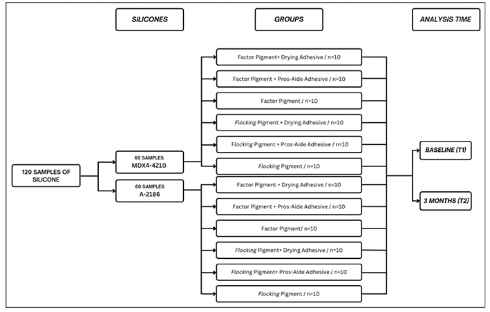
Figure 1. Distribution of samples according to group and proposed analysis time.
The silicones were handled according to the manufacturer’s instructions at a controlled temperature of 23º ± 2ºC and relative humidity of 50 ± 10%. The pigments were weighed using an analytical balance (BEL Analytical Equipment, Brazil) and incorporated into the silicone using a stainless-steel spatula until they formed a homogeneous mass, corresponding to 0.2% of the weight of the silicone [21]. After handling, the silicones were inserted into the metal matrix and subjected to pressure for 24 hours in a pneumatic polymerizing pan (Metal Vander, SP, Brazil) to avoid the appearance of bubbles in the material. They were then confined inside the matrix with the outer surface exposed to the environment for 48 hours for complete polymerization of the material [24].
After they were made, initial color, surface roughness and Shore A hardness readings were taken, and all the samples were stored in artificial sweat manipulated by pharmacists in a bacteriological oven at 37 ± 1°C (CIENLAB Equipamentos Científicos Ltda, Campinas, SP, Brazil) [22] for 3 months to simulate the effect of sweat on silicone facial prostheses influenced by different types of pigments and adhesive systems over time. During this storage and ageing process, the specimens were cleaned every 3 days with neutral pH soap (Karité, Delicious Care, Dove, Brazil) and water, and the samples that were in contact with the adhesive were renewed with a new layer. At the end of the 3 months, the final color, roughness and Shore A hardness readings of the samples were taken for statistical analysis.
Color was analyzed according to CIE/ISO new standard: CIEDE2000 using a spectrophotometer. The color changes (ΔE) were calculated using the CIELab system, established by the Commission Internationale de I’Eclairage [18]. This system can be calculated according to the formula: ΔE = [(ΔL)2 + (Δa)2 + (Δb)2]½ 15. The ‘L’ represents the brightness from 0 (black) to 100 (perfect white), the ‘a’ represents the amount of red (positive values) or green (negative values) and the ‘b’ represents the amount of yellow (positive values) or blue (negative values) [24]. For this process, the Ultraviolet-Visible Reflectance Spectrophotometer (UV-2450, Shimadzu, Japan) was used.
Surface roughness was analyzed according to ISO 21920, using contact profilometry, using an SJ-401 surface roughness profilometer (Mitutoyo, Kanagawa, Japan), which has a 2 mm diameter diamond. The settings defined were: λ = 0.08 mm cutting wavelength and 0.25 mm transverse length at a speed of 0.05 mm/s for the surface roughness characteristics Ra and Rt. Ra is the average roughness which is determined by the arithmetic mean of the absolute values of the ordinates of the roughness profile. Rt is the roughness depth that represents the maximum peak height in the measured profile. Three measurements were taken on each sample, with the average defined as the roughness value [23].
Hardness was analyzed according to ISO 868. The hardness test (Shore A) samples were read using a durometer model GDS 709 (Teclock, Osaka, Japan) under the American Society for Testing and Materials (ASTM) specifications D-2240 [20, 23, 25, 26]. This method is based on the penetration of a needle into the surface of the material with a constant load of 10 N [20]. The measurement was set between 0 and 100 Shore A with a 1% tolerance. Each sample was placed in the durometer at a distance of 2 mm from the device’s penetration needle [20]. The needle applied pressure to the samples for 15 seconds and three measurements were made on each one, so that the three values obtained could then be averaged.
Statistical analysis
The sample size calculation was performed retrospectively based on the observed effect size for the variable ‘ΔE’ (color change), which presented an F value of 10.590 in the analysis of variance (ANOVA) with 12 groups. The estimated effect size was f = 1.04 (a very large effect). Considering a significance level of 5% (α = 0.05) and statistical power of 80 and 90%, the minimum required sample size would be 3 specimens per group (total n = 36). As this study included 10 specimens per group (total n = 120), the sample size was more than sufficient to ensure statistical robustness.
Statistics were performed using Jamovi Software (Version 2.2.5.0, Jamovi Project, Australia). The tests performed were ANOVA with repeated measures, followed by the Tukey Honestly Significant Difference (HSD) test with a significance level of 5%. The evaluation of Color Chance was analyzed by Three-way ANOVA (Silicone × Pigment × Adhesive); Roughness tests were analyzed by four-way ANOVA (Time × Silicone × Pigment × Adhesive); Shore A hardness was analyzed by four-way ANOVA (Time × Silicone × Pigment × Adhesive).
Results
Concerning color change (ΔE), the three-way ANOVA showed a statistically significant change in the color change values (Table 3) only between the adhesives (*p < 0.001). As shown in Table 4, the control groups had the significantly least color change for both silicones.
Concerning roughness, the ANOVA analysis shown in Table 5 demonstrates that there was a statistically significant change (*p < 0.001) only to the time elapsed, but that the use of pigments and adhesives did not significantly alter the results. Furthermore, the statistical analysis showed a reduction in values after 3 months of immersion and levage (T2) for both silicones, regardless of the adhesive, with the MDX4-420 silicone having the highest roughness values, before and after the experimental period, compared to the A-2186 silicone (Table 6).
For the Shore A hardness tests, the four-way ANOVA shown in Table 7 highlights a statistically significant difference (*p < 0.001) in time, between time and silicone and between time and pigment used. Further statistical analysis showed that there was an increase in values after 3 months of immersion and washing (T2) for both silicones except for the MDX4-4210 silicone used with the Factor pigment. Indeed, the MDX4-4210 silicone groups with the Factor pigment stood out as having significantly lower Shore A hardness values at both times (T1 and T2) (Table 8).
Discussion
Based on the results of this study, the null hypothesis that there would be no statistical difference between color change, Shore A hardness and roughness between silicones MDX4-4210 and A-2186, under the influence of pigments, fixation adhesives and artificial sweat, was rejected.
The main challenge encountered in the performance of a maxillofacial prosthesis is the degradation of its appearance, whether due to changes in color or deterioration of physical properties. The average lifespan of facial prostheses is still only 1–1.5 years, mainly due to the fading of their color [27]. Studies point to the fact that humidity and precipitation have a greater effect on the pigmented silicone elastomer than heat and sun. In addition, human conditions such as sweat and sebum also contribute to the color change of prostheses [28]. It should be noted that there are perceptibility and acceptability thresholds for color differences and color changes in facial prostheses. The study by Lagouvardos et al. demonstrated that changes in luminosity were more noticeable and less accepted by observers than changes in chromatic coordinates (green/red and yellow/blue) [29]. Other authors found in their study that color changes are more perceivable in light skin colors regardless of gender and experience or the observer [30]. According to some authors, ΔE < 1 represents a visually imperceptible color change, values of ΔE ≥ 1.0 represent a visually perceptible color change, with ΔE < 3.3 being a clinically acceptable color change and a ΔE ≥ 3.3 being a clinically unacceptable color change, that is perceptible to untrained observers [31]. In this study, all ΔE values were between 1.43 and 1.9, below 3.3 and above 1, so all groups had a clinically acceptable color change that could be perceived by trained operators [31]. According to the results, the highest color change values were found in the groups that came into contact with adhesive, regardless of the pigmentation used, while the control groups (without adhesive application) had lower ΔE values. In addition, a statistically significant change was found only between the adhesives, which indicates that the presence of pigments in the composition of the adhesives used in this study and the difficulty in completely removing them after application was able to alter the color of the silicone [2]. There are few studies in the literature on adhesives for fixing maxillofacial prostheses and their relationship with color change, so more studies would be needed to corroborate this idea.
To avoid infections, it is necessary to use a chemical disinfection material when handling prostheses, such as washing with neutral pH soap and water. The material used for disinfection must not be aggressive to the tissues and must preserve the properties of silicone [21]. After 3 months of washing with neutral soap and immersion in artificial sweat, the tests carried out indicated a reduction in roughness values in all groups, regardless of the use of adhesives. This can be explained by the continuous polymerization process, which promotes a more complete polymer chain, making the silicone surface smoother over time [21]. Furthermore, when comparing the two silicones, it was observed that the MDX4-4120 silicone had a rougher surface compared to the A-2186 silicone at both times (T1 and T2). This is probably because MDX 4-4210 has a higher concentration of filler in its composition, which promotes greater surface roughness [21]. Despite the expected changes, all the results found in this study are clinically acceptable. According to Goiato et al., the typical clinically acceptable value for average roughness is Ra ≤ 0.8 μm [21].
According to Hatamleh et al. [32], the hardness of silicone elastomers is controlled by the surface characteristics of the polymer network and the density of the cross-linkers. The density of the cross-linkers could affect the length of the polymer chain [33] which results in degradation of the mechanical properties of silicon elastomers in time. It is possible that the increase in hardness of silicones added to pigments with nanoparticles maximizes the network of the silicone matrix, lengthens the polymer chain and leads to an increase in hardness values [34].
After the experimental period, the final Shore A hardness tests showed a statistically significant increase compared to the initial tests, with the exception of the groups with MDX4-4210 silicone and Factor pigment, which differed from the other groups in both tests (T1 and T2) and did not change statistically from the initial to the final time, having lower hardness values. In addition, the average Shore A hardness values were higher for the groups with silicone A-2186 at both times. These results may have been due to the continuous polymerization of the MDX4-4210 and A-2186 [35] silicones, which leads to an increase in their hardness over time, and probably the Factor pigment used in this study may have made it difficult for the MDX4-4210 silicone polymer chains to intertwine, reducing its hardness rate, polymerization [35] and leading to a lower hardness value than the others. This suggests that the MDX4-4210 silicone was influenced by the pigments more than the A-2186 silicone. The hardness values measured before and after the adhesives in our study are consistent with those reported by Sweneey et al. and according to Conroy, the ideal Shore A hardness should be between 25 and 55 [5], studies by Lewis and Castleberry stated that the ideal hardness should be 25–35 [36] units and is acceptable in clinical practice. Our results are between 35.3 and 40.6 shore A. The hardness values found in this study are clinically acceptable.
Despite numerous studies on the physical and mechanical properties of MDX4-4210 and A-2186 silicones, the present study brings a novel contribution by evaluating these silicones under combined clinical conditions, including artificial sweat, different adhesives and two types of pigmentation (including flocking) over a prolonged period of 3 months. The use of flocking fibers is an important innovation in the aesthetic characterization of facial prostheses. Flockings mimic the translucency and texture of human skin, contributing significantly to the natural appearance and patient satisfaction. Despite their clinical relevance, there is a lack of studies analyzing their influence on the physical and optical behavior of silicone prostheses, especially when associated with sweat exposure and adhesive application. Therefore, our findings offer clinically relevant insights that are not yet well-established in the literature. This integrative approach helps guide clinicians in choosing materials and protocols that ensure both durability and aesthetics, improving outcomes in maxillofacial rehabilitation. Furthermore, additional studies would be needed to evaluate the use of patches and artificial sweat using other methods in vitro and vivo.
Conclusion
The MDX4-4210 and A-2186 silicones, after pigmentation and application of facial adhesives, showed changes in color, Shore A hardness and roughness. Artificial sweat did not influence the results in the present study. Their results were considered clinically acceptable and an excellent option for use in maxillofacial rehabilitation.
Acknowledgments
The authors thank the Institutional Scientific Initiation Scholarship Program (PIBIC), CNPq [Proposal number: 146198/2024 – PIBIC].
References
[1] Goiato MC, Pesqueira AA, Ramos da Silva C, Gennari Filho H, Micheline Dos Santos D. Patient satisfaction with maxillofacial prosthesis. Literature review. J Plast Reconstr Aesthet Surg. 2009;62(2):175–80. https://doi.org/10.1016/j.bjps.2008.06.084
[2] Goiato MC, Santos Yanamoto NC, Melo Neto CLM, Freitas da Silva EV, Caxias FP, dos Santos DM. Analysis of the mechanical propertier of bond strength in adhesive systems bonded to HCR silicone facial. Int J Artif Organs. 2025;48(4):275–9. https://doi.org/10.1177/journals.sagepub
[3] Paulini MB, Micheline Dos Santos D, de Moraes Melo Neto CL, Bitencourt SB, da Silva EVF, de Caxias FP, et al. Analysis of physical properties of facial silicones with different pigmentations submitted to nonthermal plasma treatment and accelerated aging. J Prosthet Dent. 2020;124(6):815.e1–7. https://doi.org/10.1016/j.prosdent.2020.05.034
[4] Nóbrega AS, Neto CLMM, Dos Santos DM, Bertoz APM, MeloMoreno AL, Goiato MC. Effect of accelerated aging on the sorption and solubility percentages of silicone facial prostheses. Eur J Dent. 2022;16(1):223–6. https://doi.org/10.1055/s-0041-1731932
[5] Cevik P, Yildirim-Bicer AZ. Effect of different types of disinfection solution and aging on the hardness and colour stability of maxillofacial silicone elastomers. Int J Artif Organs. 2017;41(2) 108–14. https://doi.org/10.5301/ijao.5000659
[6] Cevik P, Eraslan O. Effects of the addition of titanium dioxide and silaned silica nanoparticles on the mechanical properties of maxillofacial silicones. J Prosthodont. 2017;26(7):611–15. https://doi.org/10.1111/jopr.12438
[7] Seçilmiş A, Öztürk AN. Nasal prosthesis rehabilitation after partial rhinectomy: a clinical report. Eur J Dent. 2007;1(2):115–18. https://doi.org/10.1055/s-0039-1698324
[8] Rai SY, Guttal SS. Effect of intrinsic pigmentation on the tear strength and water sorption of two commercially available silicone elastomers. J Indian Prosthodont Soc. 2013;13(1):30–5. https://doi.org/10.1007/s13191-012-0174-1
[9] Goiato MC, Haddad MF, Santos DM, Pesqueira AA, Moreno A. Hardness evaluation of prosthetic silicones containing opacifiers following chemical disinfection and accelerated aging. Braz Oral Res. 2010;24(3):303–8. https://doi.org/10.1590/S1806-83242010000300008
[10] Saboya ACL, Carvalho JCM, André M, Mattos BSC, Rossa R. Avaliação da dureza ‘shore a’ e da resistência ao rasgamento de alguns silicones acéticos modificados para uso em prótese facial. Rev Odontol Univ São Paulo. 1997;11(2):93–7. https://doi.org/10.1590/S0103-06631997000200005
[11] Nguyen CT, Chambers MS, Powers JM, Kiat-Amnuay S. Effect of opacifiers and UV absorbers on pigmented maxillofacial silicone elastomer, part 2: mechanical properties after artificial aging. J Prosthet Dent. 2013;109(6):402–10. https://doi.org/10.1016/S0022-3913(13)60328-5
[12] Polyzois GL, Tarantili PA, Frangou MJ, Andreopoulos AG. Physical properties of a silicone prosthetic elastomer stored in simulated skin secretions. J Prosthet Dent. 2000;83(5):572–7. https://doi.org/10.1016/S0022-3913(00)70017-5
[13] Quadros LCS, Silva-Lovato CH, Dotto MER, Ribeiro JS, Soto AF, Duque TM, et al. In situ study of the effect of endogenous and exogenous agents on color stability, hardness, and surface roughness of an elastomer for facial prostheses. J Prosthodont. 2024:1–10 https://doi.org/10.1111/jopr.13876
[14] Eleni PN, Krokida MK, Polyzois GL. Effects of storage in simulated skin secretions on mechanical behavior and color of polydimethylsiloxanes elastomers. J Craniofac Surg. 2011;22(3):830–6. https://doi.org/10.1097/SCS.0b013e31820f36e0
[15] Al-Dharrab AA, Tayel SB, Abodaya MH. The effect of different storage conditions on the physical properties of pigmented medical grade I silicone maxillofacial material. ISRN Dent. 2013;2013:582051. https://doi.org/10.1155/2013/582051
[16] Kiat-Amnuay S, Waters PJ, Roberts D, Gettleman L. Adhesive retention of silicone and chlorinated polyethylene for maxillofacial prostheses. J Prosthet Dent. 2008;99(6):483–8. https://doi.org/10.1016/S0022-3913(08)60113-4
[17] Liu L, Kuffel K, Scott D, Constantinescu G, Chung H-J, Rieger J. Silicone-based adhesives for long-term skin application: cleaning protocols and their effect on peel strength. Biomed Phys Eng Exp. 2018;4(1):1–11. https://doi.org/10.1088/2057-1976/aa91fb
[18] Polyzois GL. Bond strength of double-sided adhesive tapes used for facial prostheses. Spec Care Dentist. 1994;14(1):26–9. https://doi.org/10.1111/j.1754-4505.1994.tb01092.x
[19] Polyzois GL, Oilo G, Dahl JE. Tensile bond strength of maxillofacial adhesives. J Prosthet Dent. 1993;69(4):374–7. https://doi.org/10.1016/0022-3913(93)90183-O
[20] Polyzois GL, Eleni PN, Krokida MK. Effect of time passage on some physical properties of silicone maxillofacial elastomers. J Craniofac Surg. 2011;22(5):1617–21. https://doi.org/10.1097/SCS.0b013e31822e5e62
[21] Goiato MC, Pesqueira AA, dos Santos DM, Dekon SFC. Evaluation of hardness and surface roughness of two maxillofacial silicones following disinfection. Braz Oral Res. 2009;23(1):49–53. https://doi.org/10.1590/S1806-83242009000100009
[22] dos Santos DM, Goiato MC, Moreno A, Pesqueira AA, Haddad MF. Influence of pigments and opacifiers on color stability of an artificially aged facial silicone. J Prosthodont. 2011;20:205–8. https://doi.org/10.1111/j.1532-849X.2010.00657.x
[23] Haddad MF, Goiato MC, dos Santos DM, de Marchi Crepaldi N, Pesqueira AA, Bannwart LC. Bond strength between acrylic resin and maxillofacial silicone. J Appl Oral Sci. 2012;20(6):649–54. https://doi.org/10.1590/S1678-77572012000600010
[24] Nobrega AS, Malavazi EM, Melo Neto CLM, de Sousa Ervolino IC, Bannwart LC, de Magalhães Bertoz AP, et al. Influence of different pigment incorporation methods on color, dimensional stability, and detail reproduction of silicones. Eur J Dent. 2019;13(3):399–404. https://doi.org/10.1055/s-0039-1700768
[25] ISO 105-E04 [Internet]. 2008. Available from: https://standards.iteh.ai/catalog/standards/sist/bad3c3e2-4a94-4113-8be0-aa0ea33e8c0e/iso-105-e04-2008 [cited August 4th, 2025]
[26] Kulthong K, Srisung S, Boonpavanitchakul K, Kangwansupamonkon W, Maniratanachote R. Determination of silver nanoparticle release from antibacterial fabrics into artificial sweat. Part Fibre Toxicol. 2010;7:8. https://doi.org/10.1186/1743-8977-7-8
[27] Roberts AC. Facial reconstruction by prosthetic means. Br J Oral Surg. 1967;4:157‑82. https://doi.org/10.1016/S0007-117X(66)80032-X
[28] Gupta P, Deshpande S, Radke U, Ughade S, Sethuraman R. The color stability of maxillofacial silicones: a systematic review and meta-analysis. J Indian Prosthodon Soc. 2021;21:138. https://doi.org/10.4103/jips.jips_253_19
[29] Lagouvardos P, Spyropoulou N, Polyzois G. Perceptibility and acceptability thresholds of simulated facial skin color differences. J Prosthodont Res. 2018;62(4):503–8. https://doi.org/10.1016/j.jpor.2018.07.005
[30] Kurt M, Nemli SK, Güngör MB, Bal BT, Öztürk E. Perceptibility and acceptability thresholds for color differences of light and dark maxillofacial skin replications. Vis Res. 2024;223:108474. https://doi.org/10.1016/j.visres.2024.108474
[31] Dos Santos DM, Borgui Paulini M, Silva Faria TG, de Moraes Melo Neto CL, da Silva EVF, de Caxias FP, et al. Analysis of color and hardness of a medical silicone with extrinsic pigmentation after accelerated aging. Eur J Dent. 2020;14(4):634–8. https://doi.org/10.1055/s-0040-1715782
[32] Hatamleh MM, Polyzois GL, Silikas N, Watts DC. Effect of extraoral aging conditions on mechanical properties of maxillofacial silicone elastomer. J Prosthodont. 2011;20(6):439–46. https://doi.org/10.1111/j.1532-849x.2011.00736.x
[33] Mouzakis DE, Papadopoulos TD, Polyzois GL, Griniari PG. Dynamic mechanical properties of a maxillofacial silicone elastomer incorporating a ZnO additive: the effect of artificial aging. J Craniofac Surg. 2010;21:1867–71. https://doi.org/10.1097/SCS.0b013e3181f43f43
[34] Çevik P. Evaluation of Shore A hardness of maxillofacial silicones: the effect of dark storage and nanoparticles. Eur Oral Res. 2018;52(2):99–104. https://doi.org/10.26650/eor.2018.469
[35] Nobrega AS, Andreotti AM, Moreno A, Sinhoreti MAC, Dos Santos DM, Goiato MC. Influence of adding nanoparticles on the hardness, tear strength, and permanent deformation of facial silicone subjected to accelerated aging. J Prosthet Dent. 2016;116(4):623–9. https://doi.org/10.1016/j.prosdent.2016.03.004
[36] Lewis DH, Castleberry DJ. An assessment of recent advances in external maxillofacial materials. J Prosthet Dent. 1980;43(4):426–32. https://doi.org/10.1016/0022-3913(80)90215-2