ORIGINAL ARTICLE
Radio frequency sputtering of self-sanitizing material on NiTi archwires
Mahmood Nasera,b, Emad Al-Hassanib and Fatima Al-Hassanib
aDepartment of Materials Engineering, Faculty of Engineering, University of Kufa, Iraq; bCollege of Materials Engineering, University of Technology, Iraq
Abstract
Objective: The objective of this study was to coat orthodontic nickel titanium (NiTi) archwires with nano-particles (NP) of silver (Ag) combined with nano polytetrafluoroethylene (PTFE) to produce a smooth antimicrobial nanocomposite layer by using a radio frequency (RF) sputtering process and to evaluate the coated surfaces in terms of morphology, nano-roughness, adhesion strength, hardness, and antimicrobial activity.
Materials and methods: Super-elastic NiTi archwires (diameter = 0.4 mm, length = 160 mm) were surface cleaned and sterilized prior to the RF sputtering, using a mixture of nano Ag powder (20 nm; purity > 99.95%) and PTFE powder (25 nm; purity > 99.95%). X-ray diffraction apparatus (XRD), flex atomic force microscopy (AFM) and field emission scanning electron microscopy (FESEM) were used to characterize the morphology and nano roughness of the coated archwires. Lactobacillus acidophilus (L. acidophilus) and Streptococcus mutans (S. mutans) were selected to evaluate the antimicrobial activity.
Results: A uniform and homogeneous nanocomposite coating was obtained without agglomeration. Surface roughness values decreased with increasing sputtering time, while the coated samples exhibited excellent antibacterial activity against both bacterial strains. AFM analysis demonstrated that sputtering time strongly influenced adhesion resistance, hardness, and coating stability, and the antibacterial activity was highly effective against both L. acidophilus and S. mutans.
Conclusion: The sputtering time of 30 min gave a smooth coating layer on the surface of NiTi archwire with strong antimicrobial resistance that offers significant potential for dental applications.
KEYWORDS: Archwire; coating; radio frequency; roughness; nanoparticles
Citation: BIOMATERIAL INVESTIGATIONS IN DENTISTRY 2025, VOL. 12, 194–201. https://doi.org/10.2340/biid.v12.45035.
Copyright: © 2025 The Author(s). Published by MJS Publishing on behalf of Acta Odontologica Scandinavica Society. This is an Open Access article distributed under the terms of the Creative Commons Attribution 4.0 International License (http://creativecommons.org/licenses/by/4.0/), allowing third parties to copy and redistribute the material in any medium or format and to remix, transform, and build upon the material, with the condition of proper attribution to the original work.
Received: 20 July 2025; Accepted: 13 October 2025; Published: 11 December 2025
CONTACT Mahmood Shakir Naser Mahmoods.alammar@uokufa.edu.iq Department of Materials Engineering, University of Kufa, Iraq.
Competing interests and funding: The authors declare that they have no conflicts of interest.
Introduction
In orthodontics, the most important concerns are the microbial accumulation around the archwires which have an undesirable impact on the health of the surrounding tissues [1]. The human body presents a challenging environment for metallic biomaterials, with the oral cavity serving as a critical entry point. Here, the variations in intraoral conditions are both frequent and intricate, resulting in a distinctive medium that promotes corrosion [2]. The oral environment serves as a prime setting for the unavoidable growth of microorganisms. To date, more than 700 bacterial species have been documented, alongside a variety of fungi and viruses. The oral microbiota has developed alongside humans in a mutually beneficial or even symbiotic relationship: The host offers optimal physiochemical and nutritional environments, while microorganisms, particularly bacteria, fulfill crucial physiological functions such as digestion, differentiation of oral mucosa cells, and defense against external pathogens [3–5].
A metal archwire used in orthodontics, such as nickel-titanium (NiTi), possesses exceptional superelasticity, resistance to corrosion, thermal shape memory and biocompatibility. In addition, the increasing number of adult orthodontic patients has resulted in an increase in the need for more cosmetic orthodontic appliances. This demand has been observed in the development of esthetic wires made of polymer, metallic arch wires covered with polymer materials, and wires that have been gold-plated [6]. Altering the surface characteristics of orthodontic components presents a viable approach to enhance their functionalities, particularly in inhibiting the development of bacterial biofilms. These alterations can be achieved through the incorporation of supplementary layers that include substances potentially exhibiting antibacterial properties [7]. The large surface area of the antimicrobial nanoparticles facilitates their interaction with bacterial cells of negative charge to a significant extent leading to enhanced antimicrobial activity. Moreover, antimicrobial nanoparticles added to polymers or coated onto surfaces of biomaterials were found to exhibit superior antimicrobial characteristics in the oral cavity [8, 9].
The installation of a fixed orthodontic appliance in patients often leads to a notable deterioration in oral health and hygiene. This decline is generally linked to the discomfort experienced as teeth shift into their new positions, along with the challenges of maintaining proper oral cleanliness due to the presence of numerous irregular surfaces [10]. Consequently, the application of antimicrobial agents, such as particular nanoparticles, to orthodontic devices warrants exploration as a potential method for preventing adherence and microbial colonization [11]. Nanoparticles made of silver, often known as silver (Ag) NPs, are among the nanoparticles that are investigated most frequently, as a result of the high aspect ratio (surface-to-volume ratio) of these substances, along with their low cost, cytotoxicity, immunological response, and antibacterial action even at low concentrations. These nanoparticles have become a more desirable option for use in biomedical applications as a result of the extensive variety of potential that Ag-NPs possess [12, 13]. It is well known that polytetrafluoroethylene (PTFE) exhibits intrinsic non-adhesive characteristics as a result of its minimal surface energy. The incorporation of PTFE nanoparticles with the Ag NPs by radio frequency (RF) sputtering can produce a coating layer with favorable properties, like bacterial resistance, scaling resistance and smooth surface layer (affects surface roughness) [14, 15]. It was reported that the Ag/PTFE sputtered composite coatings demonstrated a high antibacterial efficacy, achieving removal rates of 99.99% for Staphylococcus aureus [16, 17]. Moreover, the Ag/PTFE sputtered composite coating was capable of releasing antibacterial Ag+ ions in a sustained manner, which effectively inhibited bacterial growth and reduced biofilm formation by approximately 50% after 7 days [18].
The motivation for this study is the clinical need to reduce microbial adhesion and biofilm formation on archwires, which contribute to enamel demineralization, gingival disease, and oral infections. The problem addressed is the lack of coatings that can simultaneously improve surface properties and enhance antibacterial performance. Previous studies have often investigated coatings based on either Ag, owing to its strong antimicrobial properties, or polymers such as PTFE, due to their low-friction characteristics, but rarely their combination. This has left a gap in understanding how Ag/PTFE nanocomposite coatings behave when applied to NiTi archwires. It was hypothesized that integrating Ag nanoparticles with PTFE in a nanocomposite coating, deposited by RF sputtering, would yield smoother surfaces with lower adhesion and enhanced antibacterial activity compared to single-component coatings. This assumption is supported by prior findings that Ag provides potent antibacterial effects, while PTFE reduces surface energy and friction when used as a coating material [19, 20]. In this study, Ag and PTFE were sputtered under different deposition times, and the resulting surfaces were systematically evaluated in terms of morphology, nano-roughness, adhesion strength, hardness, and antibacterial activity against Lactobacillus acidophilus and Streptococcus mutans.
Materials and methods
The primary materials used in this work and their properties are listed in Table 1.
Target preparation for RF sputtering process
The RF target was made from Ag/PTFE nano powders weighing 100 g. The powders were mixed together in four tanks mixer (MTI, USA) for 3 h at 100 rpm and then compressed by using a hydraulic press (Mega, PRDE (30T/50T), Spain). The mixed powder was compacted under a pressure of 500 MPa for 5 min, resulting in a disk specimen of 60 mm diameter and 5 mm thickness.
Archwire preparation for RF sputtering process
NiTi archwires (NTWs) were cleaned to eliminate any surface contamination by using an ultrasonic cleaner bath (MTI Corporation, USA) containing deionized water for 10 min. After drying, the NTWs were sterilized in an ultraviolet light cabinet (Analytic, Germany) for 30 min [21].
RF sputtering process
The RF target was affixed to the cathode, while the archwires were positioned on the anode, oriented towards the target holder, which was situated 60 mm away from the target within the RF sputtering apparatus (Barez Afarin industry, Iran). The sputtering chamber underwent initial evacuation to achieve a pressure of 6 × 10⁻⁴ Pa prior to the presenting of high-purity (99.9999%) working argon (Ar) gas with 20 mL min−1 flow rate. Throughout the sputtering process, the holder of the substrate was subjected to 20 RPM rotational speed. The Ag/PTFE layer was sputtered at 0.6 Pa gas pressure and utilizing a sputtering power of 60 W. Three sputtering durations were tested: 10, 20, and 30 min. All samples were prepared under ambient conditions.
Characterization techniques
X-ray diffraction was performed to define the presence of phases and crystallographic properties of the raw materials, the coated and the uncoated samples by using X-Ray diffractometer (Chongqing Drawell, China) with a nickel filter and copper generator. The speed of scanning was adjusted to 6 deg.min−1 and the diffraction angle (2θo) range was (10o–80o).
Surface topography, surface nanoroughness (Ra), adhesion strength and hardness, and particle size distribution of the coated and uncoated NTW were investigated by using a flex atomic force microscopy (AFM) with interchangeable cantilever holder (Nanosurf, Switzerland).
The surface morphology, size and distribution of nanoparticles and the coating layer thickness from the cross-section microstructure were investigated by using field emission scanning electron microscopy (FESEM) (TESCAN MIRA3, France). The elemental composition of the samples was examined using energy dispersive X-ray spectroscopy (EDX) in conjunction with the FESEM.
Antibacterial assay
Antibacterial activities of the surface-modified orthodontic wires were demonstrated against Lactobacillus acidophilus (L. acidophilus) and Streptococcus mutans (S. mutans) bacterial strains. The agar well diffusion technique was used to investigate the antimicrobial effect of the uncoated and coated NTWs. The prepared cultures were spread on petri dishes, and then the samples were placed in an incubator. After incubation for 24 h and at 37°C, the inhibition zone size was measured using a ruler.
Statistical analysis
Descriptive statistics was used in this study for the nano roughness and antimicrobial data which were organized, categorized and transferred into a computerized database structure using SPSS program and it includes Minimum (Min), Maximum (Max), Mean (M), standard deviation (SD), standard error (SE), and data were screened for normal distribution and homogeneity using Shapiro-Wilk and Levene’s tests respectively. These tests were employed to either accept or reject the statistical hypothesis, establishing a confidence interval of 95%. The significance level was deemed significant when the p-value fell below 0.05. One-way analysis of variance (ANOVA) was used to compare Ra among the four groups. When the ANOVA indicated significant differences (p < 0.05), pairwise comparisons were performed using Tukey’s honestly significant difference (HSD) post‑hoc test. Significance was set at p < 0.05. All analyses were conducted using IBM SPSS Statistics (Version 31.0.0.0, IBM Corp., USA).
Results
Surface characterization and morphology
The X-ray diffraction (XRD) patterns of the uncoated NTW (Figure 1A) showed two primary diffraction peaks corresponding to the NiTi alloy phase (110) and (211) respectively, while the Ag/PTFE coated NTW (Figure 1B) exhibited diffraction peaks of both Ag and PTFE from the thin coated layer. The diffraction peaks at 2θ value of 37.3o considered the primary peak of Ag which is correspond to the (111) plane that confirm the presence of Ag in addition to the two other weak peaks at 2θ value of 64.4o and 77.3o, which corresponds to (220) and (311) planes of metallic silver having face center cube (FCC) crystal symmetry and all the peaks corresponded to the standard Joint Committee on Powder Diffraction Standards (JCPDS) card No. 04-0783 of cubic AgNPs [22]. Finally, two diffraction peaks were observed at 2θ values of 16.8° and 18.3°, which correspond to the crystallinecarbon of PTFE phase [23]. From the XRD results, it can be seen that a Ag/PTFE coating layer was coated on NTW.
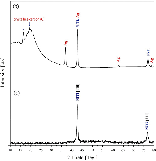
Figure 1. X-ray diffraction pattern of uncoated (A) and (B) silver/polytetrafluoroethylene-coated NiTi archwires.
Two and three-dimensional surface topographies of both coated and uncoated NTW at varying sputtering times are shown in Figure 2. The prominent microstructure on the surface varied in shape depending on the sputtering time. In Figure 2B, the brown area indicates the recessed part, while the area in white represent the raised part. The color transition in (Figure 2C and D) was slight, representing the protrusion height without any clear differences. The larger white parts in the Figures were assumed to the structure of the sputtered island. The results of the surface roughness measurements are shown in Table 2.
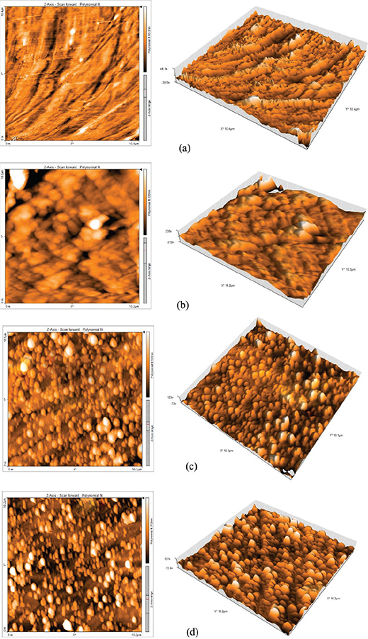
Figure 2. Surface morphology of uncoated NiTi archwires (NTW) (A) and silver/polytetrafluoroethylene coated NTW at different sputtering times (B) 10 min, (C) 20 min, and (D) 30 min.
The Shapiro-Wilk test found the data to be normally distributed, and Levene’s test found the variances to be homogeneous. One-way ANOVA showed a significant effect of sputtering time on surface roughness (Ra): F(3, 16) = 145.88, p = 7.8678 ×10⁻¹². The F statistic and corresponding p-value indicate a highly significant difference among the sputtering time groups (p < 0.05). Tukey’s HSD (Table 3) indicated that all pairwise comparisons between groups were statistically significant (p < 0.05). The uncoated wire (NT) exhibited moderate roughness, the 10 min sputtered group (1) showed the highest Ra, whereas increasing sputtering time to 20 min (2) and 30 min (3) significantly reduced roughness, with the smoothest surface observed at 30 min.
The particle size distribution of Ag/PTFE nanoparticles are shown in Figure 3. The average particle size of 48.11 nm is located within the nanoscale level (1~100 nm) [24].
AFM revealed distinct differences in adhesion among the sputtered Ag/PTFE nanocomposite coatings on NiTi wires. Figure 4 shows the adhesion forces/pull off obtained for the three sputtered coatings. Sample a (10 min sputtering) exhibited the lowest adhesion (−12.22 nN), indicating that shorter sputtering time produced a thinner, less consolidated surface with reduced interfacial forces. Sample b (20 min sputtering) presented moderate adhesion (−25.85 nN), while Sample c (30 min sputtering) showed an increased adhesion strength. The overall ranking of adhesion resistance was therefore: a (10 min) > b (20 min) > c (30 min).
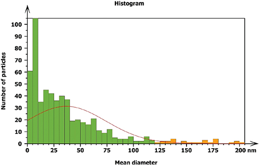
Figure 3. Histogram for the silver/polytetrafluoroethylene nanoparticles coated on the NiTi archwires at 30 min, explaining the percentage of nanoparticle size and its distribution.
Indentation results (Table 4) also demonstrated a strong dependence on sputtering time. Sample a (10 min) displayed the lowest modulus (~0.17 GPa), reflecting a relatively soft and less consolidated layer. Increasing sputtering to 20 min (Sample b) enhanced the modulus (~0.3 GPa), suggesting a thicker and more stable film. Sample c (30 min) provided intermediate values (~0.4 GPa) with good fitting, indicating that extended sputtering improved coating consolidation and durability, with an increase in adhesion.
The FESEM images of NTWs at various magnifications (35X, 250X and 10KX) are shown in Figure 5. The images demonstrate a distinct disparity between the surface topography of the uncoated archwire segment and that of the Ag/PTFE coated segment. The Ag/PTFE nanoparticle-coated NTW segments revealed a uniform distribution of spherical morphology.
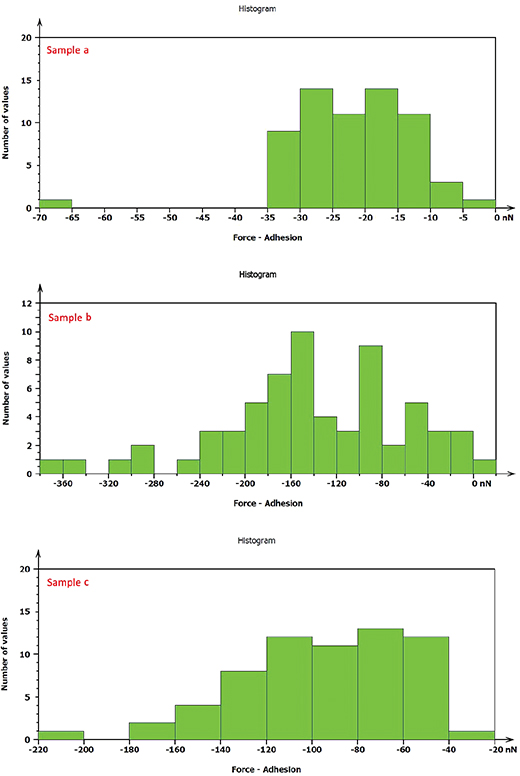
Figure 4. Adhesion strength/pull off of different coated NiTi archwires.
The EDX results (Figure 6) obtained during the FESEM analysis revealed the weight percentages of various elements, including Ag, fluorine (F), and carbon (C) from the coated layer, along with nickel (Ni) and titanium (Ti) from the substrate. The presence of carbon and fluorine served as the basis for identifying PTFE. This demonstrates that the RF sputtering occurs as a thin layer capable of being penetrated by the EDX electrons (Figure 7). The EDX mapping in Figure 8 illustrates the distributions of Ag, C, and F within the coating layer.

Figure 5. Field emission scanning electron microscopy images of the NiTi archwires (NTW) at different magnification powers for the (A, B, C) uncoated and (D, E, F) silver/polytetrafluoroethylene coated NTW at 30 min.
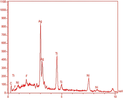
Figure 6. EDX spectroscopy charts for silver/polytetrafluoroethylene-coated NiTi archwires at 30 min.

Figure 7. Cross-section microstructure of the silver/polytetrafluoroethylene coating layer.
Antimicrobial assessment
The antimicrobial activity was illustrated by the formation of a clear inhibition zone around each NTW for both S. mutans and L. acidophilus while there was no microbial growth suppression of the uncoated NTW (Figure 8). The results of the inhibition zone measurements are shown in Table 5 and Figure 9.
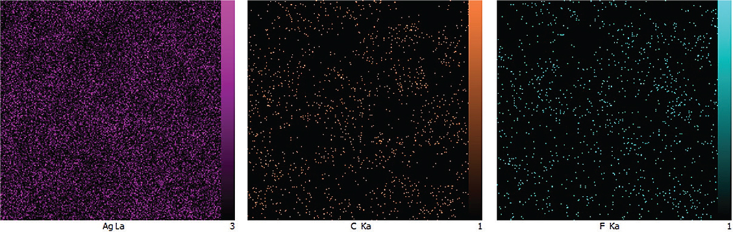
Figure 8. EDX mapping of the field emission scanning electron microscopy sample coated at 30 min.
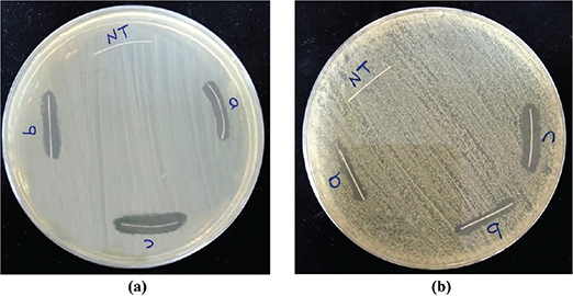
Figure 9. Antimicrobial assessment for the uncoated and coated NiTi archwires at different sputtering times for (A) S. mutans, (B) L. acidophilus.
All the coated NTW exhibited an antibacterial behavior but in different activity. The Shapiro-Wilk test found the data to be normally distributed, and Levene’s test found the variances to be homogeneous. The antibacterial analysis showed consistent trends for both S. mutans and L. acidophilus, ANOVA revealed highly significant differences in inhibition zone diameters among sputtering times (p < 0.0001) for both species. According to Tukey’s test (Table 6), all coated groups (a, b, and c) exhibited significantly larger inhibition zones compared to the uncoated control (NT). For both bacteria, the 30 min sputtered sample (c) demonstrated the largest inhibition zone, followed by 20 min (b) and 10 min (a), indicating enhanced antibacterial activity with prolonged sputtering time.
Discussion
The sputtered Ag/PTFE nanocomposite coatings produced on NiTi orthodontic wires exhibited significant improvements in surface morphology, adhesion, and antibacterial performance with increasing sputtering time. The standard peak intensities of the NTW XRD were lowered as a result of the incorporation and interaction between Ag and PTFE. AFM and FESEM analyses confirmed the formation of a smooth, dense, and uniform coating structure, particularly in the 30 min sputtered group. The reduction in surface roughness with extended sputtering time is attributed to improved film consolidation and uniform nanoparticle distribution reflecting the layer growth mechanism. This smoother morphology enhances coating adhesion to the NiTi substrate by reducing interfacial voids [25]. In addition, PTFE itself is known for its low surface energy and ability to form smooth, uniform layers. When deposited via RF sputtering, it can fill in micro-pores and irregularities on the substrate, resulting in a more compact and even surface morphology [26]. These findings agree with previous studies that reported improved nanocomposite film stability and durability under prolonged sputtering conditions [27]. While short deposition (10 min) resulted in a rougher, less consolidated layer and this can be attributed to the aggregation of the small tip structures into larger surface protrusions [28], these trends support the selection of 30 min sputtering when minimal surface roughness is required.
The mechanical characterization also revealed that higher sputtering durations enhanced the coating modulus and adhesion strength, indicating a more cohesive and stable film. Such structural uniformity plays a key role in the functional performance of coated orthodontic wires, as it prevents delamination during intraoral loading [29]. Moreover, the reduction in surface roughness was found to be inversely proportional to bacterial adhesion, suggesting that surface smoothness acts as a physical barrier against microbial colonization [30]. From a stability perspective, short sputtering (10 min) produced the lowest adhesion but also the weakest mechanical durability. Intermediate sputtering (20 min) improved both modulus and stability while maintaining low adhesion. Extended sputtering (30 min) yielded the most balanced performance, combining increased adhesion with reliable mechanical stiffness and long-term durability. The comparable results obtained for S. mutans and L. acidophilus indicate that the antibacterial mechanism of the Ag/PTFE nanocomposite coating is broadly effective against both Gram-positive bacterial species. The increasing inhibition with longer sputtering times is attributed to improved coating uniformity and Ag nanoparticle density, which enhances silver ion release and surface contact with bacterial membranes. The uncoated NTW showed no inhibition zones, confirming that antibacterial performance is directly related to the sputtered Ag/PTFE layer. Overall, the 30 min sputtered coating provided the most stable and efficient antibacterial response. These results are consistent with recent reports demonstrating that hybrid metallic–polymeric nanocomposites exhibit prolonged antimicrobial effectiveness and reduced cytotoxicity compared to single-phase coatings [31]. The enhanced surface quality and antibacterial stability achieved at 30 min sputtering suggest strong potential for clinical applications, particularly in maintaining long-term microbial resistance and minimizing frictional wear during orthodontic treatment.
Conclusion
In this study, it was concluded that extending the sputtering time during RF deposition of Ag/PTFE nanocomposite coatings on NiTi orthodontic wires significantly improves coating uniformity, adhesion, and antibacterial properties. The 30 min sputtering duration yielded the most balanced performance, with optimal surface smoothness, mechanical durability, and bacterial inhibition against S. mutans and L. acidophilus. These findings highlight the potential of Ag/PTFE nanocomposite coatings as a promising surface modification for orthodontic applications, combining mechanical stability with long-term antimicrobial protection.
References
[1] Zhang R, Han B, Liu X. Functional surface coatings on orthodontic appliances: reviews of friction reduction. Antibacterial Properties, and Corrosion Resistance. Int J Mol Sci. 2023;24:6919. https://doi.org/10.3390/ijms24086919
[2] Dawes C, Pedersen AML, Villa A, Ekstroe J, Proctor GB, Vissink A, et al. The functions of human saliva: a review sponsored by the World Workshop on Oral Medicine VI. Arch Oral Biol. 2015;60(6):863–74. https://doi.org/10.1016/j.archoralbio.2015.03.004
[3] He J, Li Y, Cao Y, Xue J, Zhou X. The oral microbiome diversity and its relation to human diseases. Folia Microbiol (Praha). 2015;60(1):69–80. https://doi.org/10.1007/s12223-014-0342-2
[4] Mystkowska J, Niemirowicz-Laskowska K, Lysik D, Tokajuk G, Dabrowski JR, Bucki R. The role of oral cavity biofilm on metallic biomaterial surface destruction–corrosion and friction aspects. Int J Mol Sci. 2018;19(3):743. https://doi.org/10.3390/ijms19030743
[5] Kilian M, Chapple IL, Hannig M, Marsh PD, Meuric V, Pedersenet AML, et al. The oral microbiome – an update for oral healthcare professionals. Br Dent J. 2016;221(10):657–66. https://doi.org/10.1038/sj.bdj.2016.865
[6] Kima IH, Parkb HS, Kimc YK, Kimd KH, Kwone TY. Comparative short-term in vitro analysis of mutans streptococci adhesion on esthetic, nickel-titanium, and stainless-steel arch wires. Angle Orthod. 2014;84(4):680–6. https://doi.org/10.2319/061713-456.1
[7] Kielan-Grabowska Z, Bacela J, Ziety A, Seremak W, Gawlik-Maj M, Kawala B, et al. Improvement of properties of stainless steel orthodontic Archwire using TiO2:Ag coating. Symmetry. 2021;13:1734. https://doi.org/10.3390/sym13091734
[8] Cao W, Zhang Y, Wang X, Li Q, Xiao Y, Liet P, et al. Novel resin-based dental material with anti-biofilm activity and improved mechanical property by incorporating hydrophilic cationic copolymer functionalized nanodiamond. J Mater Sci Mater Med. 2018;29:162. https://doi.org/10.1007/s10856-018-6172-z
[9] Hussain WS, Oleiwi JK, Hamad QA. Study of physical properties of biocomposite based on the polymer blends used for denture base applications. Eng Technol J. 2023;41(12):1474–87. http://doi.org/10.30684/etj.2023.141437.1496
[10] Al-Jewair TS, Suri S, Tompson BD. Predictors of adolescent compliance with oral hygiene instructions during two-arch multibracket fixed orthodontic treatment. Angle Orthod. 2011;81(3):525–31. http://doi.org/10.2319/092010-547.1525
[11] Borzabadi-Farahani A, Borzabadi E, Lynch E. Nanoparticles in orthodontics, a review of antimicrobial and anti-caries applications. Acta Odontol Scand. 2014;72(16):413–17. http://doi.org/10.3109/00016357.2013.859728
[12] Alaloosi HA, Noori FTM, Jidran AK. The fundamental of reduced graphene oxide with nanosilver composite films using the spin coating technique. Eng Technol J. 2022;40(08):1023–8. http://doi.org/10.30684/etj.v40i8.2205
[13] Abbas RH, Haleem AM, Judran AK. Fabrication of silver nanoparticles in aqueous solution by laser technique and study of their hemocompatibility and antibacterial effects against dental decay bacteria. Eng Technol J. 2023;41(04):543–52. http://doi.org/10.30684/etj.2023.136922.1329
[14] Marsh PD, Zaura E. Dental biofilm: ecological interactions in health and disease. J Clin Periodontol. 2017;44(18):12–22. http://doi.org/10.1111/jcpe.12679
[15] Xu W, Yuan X, Wei A, Feng Q, Wei Q. Characterisation of PET nonwoven deposited with Ag/FC nanocomposite films. Surf Eng. 2018;34(11):838–45. http://doi.org/10.1080/02670844.2017.1382063
[16] Li H, He Z, Wu W, Zheng L, Xu Q, Gao T, et al. Bacterial and corrosion resistance of polytetrafluoroethylene-silver composite coatings by magnetron sputtering. J Vac Sci Technol A. 2024;42(4):043409. https://doi.org/10.1116/6.0003545
[17] Zhang S, Wang L, Liang X, Vorstius J, Keatch R, Corner G, et al. Enhanced antibacterial and antiadhesive activities of silver-PTFE nanocomposite coating for urinary catheters. ACS Biomater Sci Eng. 2019;5(6):2804–14. https://doi.org/10.1021/acsbiomaterials.9b00071
[18] Zhang S, Liang X, Michael Gadd G, Zhao Q. A sol–gel based silver nanoparticle/polytetrafluorethylene (AgNP/PTFE) coating with enhanced antibacterial and anti-corrosive properties. Applied Surf Sci. 2021;535:147675. https://doi.org/10.1016/j.apsusc.2020.147675
[19] Slepička P, Slepičková Kasálková N, Fajstavr D, Frýdlová B, Sajdl P, Kolská Z, Švorčík V. Nanostructures on fluoropolymer nanotextile prepared using a high-energy excimer laser. Materials (Basel). 2023;16(12):4280. https://doi.org/10.3390/ma16124280
[20] Garipov IT, Khaydarov RR, Gapurova OU, Khaydarov RA, Firdaus ML, Efimova IL, et al. Silver nanoparticles as a new generation of antimicrobial prophylaxis. J Sib Fed Univ Biol. 2019;12(3):266–76. https://doi.org/10.17516/1997-1389-0301
[21] Oliveira DC, Thomson JJ, Alhabeil JA, Toma JM, Plecha SC, Pacheco RR, et al. In vitro Streptococcus mutans adhesion and biofilm formation on different esthetic orthodontic archwires. Angle Orthod. 2021;91(6):786–93. http://doi.org/10.2319/121220-998.1
[22] Oves M, Khan MS, Zaidi A, Ahmed AS, Ahmed F, Ahmad E, et al. Antibacterial and cytotoxic efficacy of extracellular silver nanoparticles biofabricated from chromium reducing novel OS4 strain of Stenotrophomonas maltophilia. PLoS One. 2013;8(3):e59140. https://doi.org/10.1371/journal.pone.0059140
[23] Shulga YM, Melezhik AV, Kabachkov EN, Milovich FO, Lyskov NV, Irzhak AV, et al. Characterisation and electrical conductivity of polytetrafluoroethylene/graphite nanoplatelets composite films. Appl Phys A. 2019;125:460. https://doi.org/10.1007/s00339-019-2747-x
[24] Abiodun‑Solanke IMF, Ajayi DM, Arigbede AO. Nanotechnology and its application in dentistry. Ann Med Health Sci Res. 2014;4(3):171–77. https://doi.org/10.4103/2141-9248.141951
[25] Wang L, Zhang S, Keatch R, Corner G, Nabi G, Murdoch S, et al. In-vitro antibacterial and anti-encrustation performance of silver-polytetrafluoroethylene nanocomposite coated urinary catheters. J Hosp Infect. 2019;103(1):55–63. https://doi.org/10.1016/j.jhin.2019.02.012
[26] Akram W, Rafique AF, Maqsood N, Khan A, Badshah S, Khan RU. Characterization of PTFE film on 316L stainless steel deposited through spin coating and its anticorrosion performance in multi acidic mediums. Materials. 2020;13(2):388. https://doi.org/10.3390/ma13020388
[27] Zhang S, Liang X, Michael Gadd G, Zhao Q. Advanced titanium dioxide-polytetrafluorethylene (TiO2-PTFE) nanocomposite coatings on stainless steel surfaces with antibacterial and anti-corrosion properties. Appl Sur Sci. 2019;490:231–41. https://doi.org/10.1016/j.apsusc.2019.06.070
[28] Kaune G, Ruderer MA, Metwalli E, Wang W, Couet S, Schlage K, et al. In situ GISAXS study of gold film growth on conducting polymer films. ACS Appl Mater Interf. 2009;1(2):353–60. https://doi.org/10.1021/am8000727
[29] Vidiš M, Truchlý M, Izai V, Fiantok T, Rajninec M, Roch T, et al. Mechanical and tribological properties of Ag/TiBx nanocomposite thin films with strong antibacterial effect prepared by magnetron co-sputtering. Coatings. 2023;13(6):989. https://doi.org/10.3390/coatings13060989
[30] Wu S, Zhang B, Liu Y, Suo X, Li H. Influence of surface topography on bacterial adhesion: a review (Review). Biointerphases. 2018;13(6):060801. https://doi.org/10.1116/1.5054057
[31] Gudkov SV, Li R, Serov DA, Burmistrov DE, Baimler IV, Baryshev AS, et al. Fluoroplast doped by Ag2O nanoparticles as new repairing non-cytotoxic antibacterial coating for meat industry. Int J Mol Sci. 2023;24(1):869. https://doi.org/10.3390/ijms24010869