ORIGINAL ARTICLE
Amphiroa fragilissima as a bioactive resource: exploring its antioxidant, anti-biofilm, anti-inflammatory, and antibacterial potential for dental applications
Dileepkumar Hemamalinia,b, S. Shantha Sundaria, K.M. Shahul Hameed Faizeeb and Sivakamavalli Jeyachandranc
aDepartment of Orthodontics and Dentofacial Orthopedics, Saveetha Dental College, Saveetha Institute of Medical and Technical Science (SIMATS), Saveetha University, Chennai, India; bDepartment of Orthodontics and Dentofacial Orthopedics, Sathayabama Dental College and Hospital, Sathyabama Institute of Science and Technology, Sathyabama University, Chennai, India; cLab in Biotechnology and Biosignal Transduction, Department of Orthodontics, Saveetha Dental College and Hospital, Saveetha Institute of Medical and Technical Sciences (SIMATS), Saveetha University, Chennai, India
ABSTRACT
Aim and objectives: To evaluate the antibacterial, antibiofilm, antioxidant, and anti-inflammatory properties of Amphiroa fragilissima and assess its potential for dental and orthodontic use.
Materials and methods: Methanolic extracts of A. fragilissima, collected from Rameshwaram, India, were tested against Streptococcus mutans, Enterococcus faecalis, Escherichia coli, and Shigella sonnei using the Kirby-Bauer method. Antibiofilm activity was analyzed via Crystal Violet staining. Antioxidant potential was assessed using 2,2-Diphenyl-1-picrylhydrazyl radical scavenging, and anti-inflammatory activity was measured via a bovine serum albumin assay.
Results: The extract showed dose-dependent antibacterial activity, with maximum inhibition observed at 100 µg/mL. Biofilm inhibition also increased with concentration. Antioxidant assays revealed significant radical scavenging activity, with results comparable to controls at higher concentrations. Anti-inflammatory testing showed reduced protein denaturation in treated samples, with effects similar to the positive control and significantly better than the blank.
Conclusion: Amphiroa fragilissima demonstrates strong antibacterial, antibiofilm, antioxidant, and anti-inflammatory activities, along with remineralization potential due to its calcium-rich composition. These properties support its potential as a natural, multifunctional agent for dental and orthodontic applications. Further in vivo studies are recommended to validate its clinical use.
KEYWORDS: Amphiroa Fragilisima; Bioactive polysaccharide; white spot lesion
Citation: BIOMATERIAL INVESTIGATIONS IN DENTISTRY 2025, VOL. 12, 184–193. https://doi.org/10.2340/biid.v12.45099.
Copyright: © 2025 The Author(s). Published by MJS Publishing on behalf of Acta Odontologica Scandinavica Society. This is an Open Access article distributed under the terms of the Creative Commons Attribution 4.0 International License (http://creativecommons.org/licenses/by/4.0/), allowing third parties to copy and redistribute the material in any medium or format and to remix, transform, and build upon the material, with the condition of proper attribution to the original work.
Received: 15 June 2025; Accepted: 20 November 2025; Published: 11 December 2025
CONTACT: Dileepkumar Hemamalini drhemaortho@gmail.com Department of Orthodontics and Dentofacial Orthopedics, Saveetha Dental College, Saveetha Institute of Medical and Technical Science (SIMATS), Saveetha University, Chennai, India
Introduction
Amphiroa fragilissima is a species of marine algae, commonly known as a red algae. It belongs to the family Amphiaroaceae and is typically found in marine environments, particularly in the intertidal zones. This species is notable for its delicate and fragile appearance, which is reflected in its name, ‘fragilissima’, which comes from the Latin word for fragile [1].
It is a member of the red algae group (Rhodophyta), which is characterized by pigments such as phycoerythrin, giving them their reddish color. Like many red algae, A. fragilissima can play an important role in the ecosystem by contributing to the structure of marine habitats, providing food, and supporting biodiversity [1].
The high mineral content and excellent biocompatibility of calcareous red algae such as A. fragilissima position them as promising candidates for applications in bone grafts and dental tissue repair. Amphiroa species, characterized by their rich deposits of calcium carbonate (mainly as high-magnesium calcite and aragonite), offer a natural source of minerals essential for hard tissue regeneration. Their composition suggests significant potential in promoting bone turnover and supporting the regeneration of bone and dental structures, particularly in the context of dental implants and periodontal therapies [2, 3].
In addition to their mineral richness, Amphiroa species contain bioactive molecules with antibacterial, anti-inflammatory, and antioxidant properties. Studies have reported that extracts from A. fragilissima demonstrate significant antimicrobial activity against oral pathogens such as Streptococcus mutans and Lactobacillus spp., which are primary contributors to dental caries and biofilm formation [4, 5].
The antioxidant activity of A. fragilissima has been attributed to its phenolic and sulfated polysaccharide content, which can scavenge free radicals and reduce oxidative stress – a key factor in inflammatory oral diseases. Anti-inflammatory effects have also been observed, with studies showing a downregulation of pro-inflammatory mediators when cells are treated with Amphiroa-derived compounds [5, 6].
The dissolution and demineralization of dental hard tissues such as enamel, dentin, and cementum are inevitable processes driven by environmental and genetic factors. Enamel, being the most mineralized and externally exposed tissue, is highly susceptible to demineralization. Partial enamel dissolution plays a role in the dynamic balance of mineral content – primarily calcium and phosphate ions – within oral fluids, maintaining a homeostatic state [7, 8].
Traditionally, fluoride-based therapies have been the cornerstone of efforts to regulate this mineral equilibrium and prevent dental caries. However, growing concerns over the systemic exposure to fluoride have led many regions, particularly in the European Union, the United States, and Canada, to reassess community water fluoridation policies [8, 9].
This contentious debate highlights the necessity of developing safe, sustainable, and effective alternatives to fluoride. A. fragilissima with its natural mineralization capacity and bioactive profile, offers a compelling, biocompatible substitute that could aid in enamel remineralization, reduce microbial colonization, and promote overall oral health.
This study aims to investigate the potential of calcareous red macroalgae A. fragilissima for dental applications, specifically focusing on its antioxidant, anti-inflammatory, antibiofilm, and antibacterial properties. Owing to its high calcium content and previously reported nutraceutical benefits related to bone health and mineral metabolism, A. fragilissima may offer a novel, sustainable strategy for enhancing enamel remineralization and oral health maintenance.
Materials and methods
Sample collection and preparation
The sample, which was collected from several locations in Rameshwaram, Tamil Nadu, India was determined to be A. fragilissima. The sample was cleaned in seawater to remove any debris (Figure 1) and allowed to dry for 2 days in the sun. It was then rinsed with tap water to remove any remaining sand and salt and allowed to dry in the sun for 7 days before being finely chopped with a scalpel to create a powder.
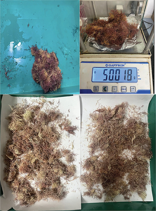
Figure 1. Processing, drying and fine powdering of Amphiroa fragilisima.
Solvent extract
The A. fragilissima powder (40 g) was combined with three volumes of methanol, following the method outlined by Terada et al. [10, 11]. The mixture was homogenized at 10,000 × g for 2 minutes using a homogenizer. Afterward, the homogenate was stirred continuously at room temperature for 30 minutes. The mixture was then centrifuged at 5,000 × g for 10 minutes at room temperature in a cooling centrifuge (REMI C-24 Plus, Goregaon [East], Mumbai, Maharashtra, India) to remove any undissolved debris.
Antibacterial activity
The antibacterial efficacy of the A. fragilisima extract was evaluated using the Kirby-Bauer method [12]. The bacteria selected for testing the antibacterial activity included S. mutans (Sdc_ortho5 16S Ribosomal RNA gene, partial sequence GenBank: OQ947767.1), Enterococcus faecalis (NCT34 16S Ribosomal RNA gene, partial sequence GenBank: OM283553.1), Shigella sonnei (NCT34 16S Ribosomal RNA gene, partial sequence GenBank: OM283552.1), and Escherichia coli (16S Ribosomal RNA gene, partial sequence, GenBank: U00096.3). The bacterial inoculum was cultured overnight in nutrient broth and a fixed volume was added to 10 mL aliquots of nutrient agar, mixed, and then poured over a nutrient agar base in sterile Petri dishes, forming the bacterial lawn. The surfaces of the plates were then inoculated with 200 μL of bacterial suspension. Using the well diffusion method, wells containing 25, 50, 75, and 100 μL of A. fragilisima extract were placed onto the agar plates. Each Petri dish was sealed individually to prevent contamination and media evaporation. The plates were incubated at 37°C for 24–48 hours. Antibacterial effects were indicated by the clear zone of inhibition surrounding the wells, and the diameter of each inhibition zone was measured in millimeters using a Vernier caliper (Baker Gauges India Pvt. Ltd. – Pune, Maharashtra).
Biofilm inhibition assay
Biofilm production and development were evaluated using light microscopy after Crystal Violet staining, following the protocol outlined by Zhou [13]. Biofilms were cultured in the wells of a 24-well microtiter plate and stained with Crystal Violet stain (Hi-media, India). Freshly cultured bacterial suspensions of S. mutans, E. coli, E. faecalis, and S. sonnei were added to each well of a 24-well polystyrene microtiter plate (Corning, Mumbai, India), which had previously been lined with sterile glass cover slips for microscopic examination. The plates were incubated from 48 to 72 hours, and the cover slips were then collected. Glass squares measuring 1 × 1 cm were used to assess the inhibitory effects of A. fragilisima extracts on in vitro biofilm formation. Biofilms were allowed to form on these glass squares, which were placed in 24-well polystyrene plates containing A. fragilisima extracts (25–100 µg/mL) and incubated for 24 hours at 30°C. A sterile medium without bacteria served as the control. After incubation, the glass squares were retrieved and rinsed twice with Phosphate Buffered Solution (PBS) before microscopic examination. The biofilms were stained with 6% Crystal Violet, and qualitative analysis was performed using an Olympus CX21i LED Microscope (Olympus Corporation, Tokyo, Japan) at 40x magnification.
DPPH radical scavenging activity
The DPPH (2,2-Diphenyl-1-picrylhydrazyl) radical scavenging activity was assessed following the method described by Shekar et al. [14]. To prepare the extract, 10 mg of A. fraglissima was dissolved in 10 mL of methanol. A stock solution of DPPH was prepared by dissolving 24 mg of DPPH (Sisco Research Laboratories Pvt. Ltd. [SRL], India) in 100 mL of methanol. This stock solution was stored under refrigeration until further use. Before analysis, it was diluted with methanol to obtain a working solution with an absorbance of approximately 0.98 ± 0.02 at 517 nm. For the assay, 3 mL of the DPPH working solution was mixed with 100 μL of either the standard solution (ascorbic acid as the positive control) or the A. fraglissima extract in a glass vial. The absorbance was then measured at 517 nm using a Shimadzu UV-1900i Spectrophotometer (Shimadzu Corporation, Kyoto, Japan) over a period of 30 minutes. PBS served as the negative control (blank). The percentage of radical scavenging activity was calculated for extract concentrations of 10, 20, 30, 40, and 50 µg/mL. The DPPH concentration at time t after reaction with the antioxidant sample is represented as DPPH] T = t. The standard utilized was ascorbic acid.
*% Radical Scavenging Activity (RSA) = (control-sample/control)*100
Bovine serum albumin assay
The anti-inflammatory potential of A. fraglissima was evaluated using a modified version of the bovine serum albumin (BSA) assay as described by Williams et al. [15]. A 0.4% (w/v) BSA solution was prepared using Tris-buffered saline by dissolving one tablet in 15 mL of deionized water, resulting in 0.05 M Tris and 0.15 M sodium chloride at pH 7.6 (25°C). The pH of this solution was then adjusted to 6.4 using diluted glacial acetic acid. Stock solutions of A. fraglissima were prepared in methanol at a concentration of 50 μg/mL (0.005%, w/v). From this stock, aliquots of 5.0, 10, and 20 µL – corresponding to final concentrations of 0.25, 0.50, and 1.00 µg/mL – were added to test tubes containing 1 mL of 0.4% BSA solution (prepared by dissolving 1 g of BSA in 80 mL of PBS, then adjusting the final volume to 100 mL with distilled water).
Both negative (dimethyl sulfoxide, DMSO) and positive (aspirin) controls were prepared and treated similarly. All tubes were heated in a water bath at 72°C for 10 minutes, then cooled to room temperature for 20 minutes. Turbidity, indicating the degree of protein denaturation, was measured at 660 nm using a Shimadzu UV-1900i Spectrophotometer (Shimadzu Corporation, Tokyo, Japan), with distilled water used as the blank. Each experiment was conducted in duplicate, and mean absorbance values were recorded. The percentage inhibition of protein denaturation – an indicator of anti-inflammatory activity – was calculated relative to the negative control using the following formula:
*%Inhibition = Abs of standard – Abs of sample/Abs of standard*100
All assays were conducted in triplicate using three independent biological replicates, each comprising separately prepared extracts. Within each biological replicate, technical triplicates were performed, and the mean ± SD was calculated from biological replicate means.
Statistical analysis
The results were analyzed by one way analysis of variance (ANOVA) and in case of significant differences by post hoc test (Tukey’s Honest Significant Difference [HSD]). The statistical analyses were performed using Statistical Package for the Social Sciences (SPSS) version 25 and the level of significance was set at p = 0.05.
Results
Antibacterial activity
The antibacterial activity of the tested samples was evaluated against E. faecalis, E. coli, S. sonnei, and S. mutans at different concentrations of A. fraglissima (25, 50, 75, and 100 µg/mL). The mean inhibition zone diameters (mm) and standard deviations as represented in Tables 1 and 2 and Figures 2 and 3. The results demonstrated a clear dose-dependent increase in the zone of inhibition for all tested organisms, indicating enhanced efficacy at higher concentrations.
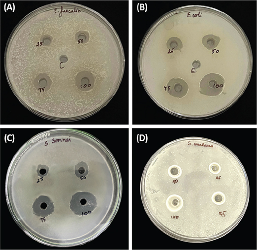
Figure 2. Antibacterial activity of Amphiroa fragilissima against (A) Enterococcus faecalis (B) Escherichia coli (C) Streptococcus mutans and (D) Shigella sonnei.
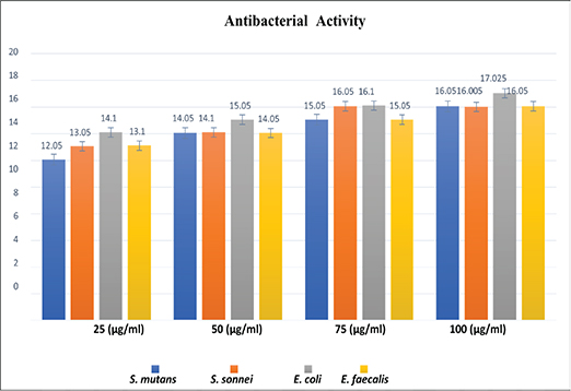
Figure 3. Antibacterial activity (Zone of inhibition) of Amphiroa Fragilisima at varied concentration.
At 25 µg, the mean inhibition zones ranged from 12.05 mm (S. mutans) to 14.10 mm (E. coli), while at 100 µg, they increased substantially, with E. coli showing the highest susceptibility (17.03 ± 0.035 mm), followed by S. mutans and E. faecalis (16.05 ± 0.07 mm) and S. sonnei (16.00 ± 0.007 mm). The overall differences in microbial inhibition across concentrations were statistically significant (p < 0.001).
Post hoc analysis (Tukey’s multiple comparisons) revealed that at 25 µg, E. coli exhibited significantly greater inhibition compared to S. mutans (mean difference: 2.05 mm, p < 0.001), S. sonnei (1.05 mm, p = 0.002), and E. faecalis (1.00 mm, p = 0.003). At 50 µg, E. coli continued to show significantly higher inhibition than S. mutans and S. sonnei (p < 0.001), while no significant differences were observed between S. mutans, S. sonnei, and E. faecalis.
At 75 µg and 100 µg, E. coli remained the most sensitive organism with statistically significant differences compared to all other species (p < 0.001). Notably, at the highest concentration (100 µg), the inhibition zones among S. mutans, S. sonnei, and E. faecalis were not significantly different from each other (p > 0.8), suggesting a saturation of antimicrobial response at this level.
Antibiofilm activity
The antibiofilm activity of A. fragilissima extract against S. mutans, E. coli, and E. faecalis demonstrated a clear concentration-dependent enhancement, with measurable inhibition observed from the lowest tested concentration of 25 µg/mL and a progressive, substantial increase in biofilm suppression at 50 µg/mL, 75 µg/mL, and reaching maximal efficacy at 100 µg/mL, as depicted in Figure 4.
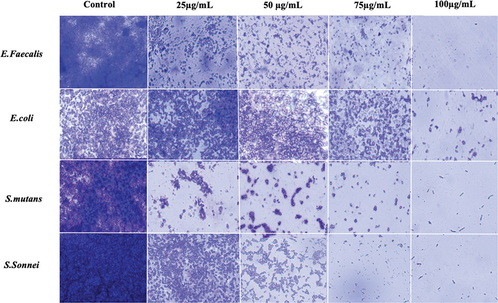
Figure 4. Light microscopic images showing antibiofilm activity at different concentrations of Amphiroa fragilisima against (A) Enterococcus faecalis (B) Escherichia coli (C) Streptococcus mutans (D) Shigella sonnei.
Antioxidant activity
The antioxidant response of A. fragilissima was evaluated at five different concentrations (10, 20, 30, 40, and 50 µg/mL) and compared to a blank and control group (Table 3 and Figure 5). At each concentration, there were significant differences among the blank, control, and A. fragilissima groups (p < 0.001). Although no significant differences were observed between the control and A. fragilissima groups, post hoc analysis showed that the absorbance values of the blank were consistently and significantly higher than those of both the control and sample groups (Table 4), indicating that both the control and A. fragilissima treatments exhibited notable radical scavenging activity. However, A. fragilissima did not significantly outperform the control.
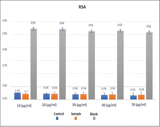
Figure 5. Antioxidant activity (% radical scavenging activity) of Amphiroa Fragilisima at varied concentration.
Anti-inflammatory
The anti-inflammatory response of A. fragilissima was tested at five different concentrations (10, 20, 30, 40, and 50 µg/mL) and compared to the blank and control groups (Table 5 and Figure 6). At all concentrations tested, ANOVA revealed significant differences between the blank, control, and A. fragilissima groups (p < 0.001). However, no significant differences were observed between the control and A. fragilissima groups at any concentration (p > 0.05). Post hoc analysis showed that the blank group consistently exhibited significantly higher absorbance values than both the control and sample groups across all concentrations (Table 6), indicating lower anti-inflammatory activity in the blank and confirming that both the control and A. fragilissima treatments exhibited a comparable and stronger anti-inflammatory effect.
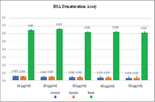
Figure 6. Anti-inflammatory activity (% bovine serum albumin denaturation) of Amphiroa Fragilisima at varied concentration. BSA: bovine serum albumin.
Discussion
Dental caries, periodontal disease, and orthodontic treatment–related complications such as enamel demineralization and soft tissue inflammation remain significant clinical challenges. The search for natural, multifunctional biomaterials that can address microbial, inflammatory, oxidative, and remineralization issues has gained momentum. Marine algae, particularly calcareous red algae, have garnered interest in this context due to their rich phytochemical content and high calcium carbonate composition. Amphiroa fragilissima, a calcareous red macroalga, demonstrates promising therapeutic potential owing to its antimicrobial, antibiofilm, antioxidant, and anti-inflammatory properties, as shown in this study. Given its inherent calcium-rich matrix, A. fragilissima is particularly relevant for dental and orthodontic applications, where biocompatibility, antimicrobial action, and enamel remineralization are critical [16–18].
The antimicrobial efficacy of A. fragilissima extract varied across tested concentrations and bacterial strains. At 25 µg/mL, E. faecalis exhibited significantly reduced susceptibility compared to E. coli, S. sonnei, and S. mutans. However, at higher concentrations (50–100 µg/mL), inhibition zones increased uniformly across all strains, suggesting a broad-spectrum bactericidal effect at sufficient dosage. This concentration-dependent activity aligns with previous findings for red algae containing secondary metabolites such as bromophenols, terpenoids, and alkaloids, which are known to disrupt bacterial membranes and interfere with protein synthesis and enzyme activity [19–21].
The antibiofilm activity was also evident and progressively increased with concentration. A. fragilissima effectively inhibited biofilm formation by S. mutans, E. coli, E. faecalis, and S. sonnei, with maximum inhibition observed at 100 µg/mL. This aligns with the findings of Prithviraj et al., who reported biofilm inhibition by using Amphiroa species by targeting bacterial quorum sensing. Their study on A. fragilissima demonstrated the potential of marine algal extracts as natural antivirulence agents. Such properties highlight the relevance of Amphiroa-based interventions in preventive orthodontic care. The ability to disrupt early biofilm formation is particularly valuable in orthodontics, where fixed appliances can promote plaque retention and white-spot lesions [22]. A qualitative approach was chosen to evaluate biofilm formation because the primary objective of this study was to observe biofilm architecture, coverage, and morphological characteristics, rather than to compare absolute biomass levels.
The antioxidant potential of A. fragilissima, assessed via radical scavenging assays, showed a concentration-dependent effect, with activity becoming comparable to the control at higher concentrations (40 and 50 µg/mL). These antioxidant effects are likely due to the presence of phenolic compounds and flavonoids, which help to neutralize reactive oxygen species (ROS) [5, 23]. The algal extract was evaluated against phosphate buffer solution as the negative control. Although its antioxidant activity were comparable to, but not higher than, the positive controls, this does not diminish its potential utility. Its main advantage lies in its ability to modulate biofilm formation on enamel surfaces while maintaining biocompatibility, which is critical for dental applications.
Özay and Pehlivan [24] emphasized that fluctuations in the composition and concentration of secondary metabolites – driven by environmental, physiological, and genetic determinants – substantially influence the biological efficacy of plant and algal extracts. This principle is pertinent to mitigating oxidative insult within inflamed gingival tissues and may consequently support improved longevity of orthodontic adhesive bonds.
The anti-inflammatory effect of A. fragilissima was evident, particularly at 50 µg/mL, where both the sample and control showed significantly reduced inflammatory responses compared to the blank group. No significant differences were observed between the sample and control groups across all concentrations, suggesting a consistent anti-inflammatory profile. Previous studies on red algae such as Halimeda opuntia and Halimeda tuna have demonstrated similar outcomes, with reductions in nitric oxide production and suppression of inflammatory cytokines such as TNF-α in macrophage models [25–27]. These findings suggest that A. fragilissima may exert its anti-inflammatory effects by modulating key inflammatory pathways, which could be useful for managing gingival inflammation during orthodontic therapy. In the anti-inflammatory assay, DMSO and aspirin were used as negative and positive controls, respectively. Although the activity of A. fragilissima was comparable to the positive control and as mentioned above this does not diminish its potential utility. The main advantage lies in its ability to modulate biofilm formation on enamel surfaces while maintaining safety and biocompatibility as key considerations for dental applications.
A defining characteristic of A. fragilissima is its high calcium carbonate content in the form of magnesium-substituted calcite and aragonite. A study of several seaweed species reported that A. fragilissima exhibited a calcium concentration of ~11720.4 ppm (i.e. ~1.172% w/w) in dry sample form. In another ecological study the calcified thallus of A. fragilissima was described as being deposited in the form of high‑Mg calcite, pointing to significant carbonate (~CaCO₃) content in its skeleton [28]. This feature of high Mg calcite not only supports its structural integrity but also offers potential remineralizing benefits. Calcium and carbonate ions released from algal particulates can aid in enamel repair, especially in demineralized regions adjacent to orthodontic brackets. Prior in vitro studies have demonstrated that marine algal powders can promote enamel microhardness recovery and reduce lesion depth, highlighting the potential of such materials as bioactive components in orthodontic adhesives, dental varnishes, and remineralizing toothpastes [18, 29, 30].
Collectively, the results of this study indicate that A. fragilissima possesses significant antimicrobial, antibiofilm, antioxidant, and anti-inflammatory properties. Coupled with its calcium-rich matrix, these bioactivities make it a compelling candidate for dental and orthodontic use. It may serve as a bioactive additive in orthodontic adhesives, preventive varnishes, and oral rinses to inhibit bacterial colonization, reduce gingival inflammation, mitigate oxidative stress, and support enamel remineralization. With further in vivo validation and formulation development, A. fragilissima holds promise as a natural, multifunctional agent in modern dental care and orthodontic therapeutics.
Limitation
Since this study was an in vitro assessment, its in vivo application in dentistry cannot be determined. Also, this study did not assess cytotoxicity, long-term stability, or directly compare A. fragilissima with standard remineralizing agents.
Future recommendation
Since all evaluated parameters yielded promising results, further investigation into the remineralizing potential, cytotoxity and long-term stability of this compound is warranted to substantiate its application in dentistry.
Conclusion
The study underscores the significant therapeutic potential of A. fragilissima, exhibiting strong antimicrobial, antibiofilm, antioxidant, and anti-inflammatory activities. Its abundant bioactive compounds, including terpenoids, phenolics, and calcium, contribute to its effective neutralization of free radicals and broad-spectrum antimicrobial effects. These properties highlight A. fragilissima as a promising natural agent for oral health applications, with potential benefits in managing biofilm-related infections and inflammatory conditions. Moreover, its calcium-rich composition suggests a valuable role in dental remineralization and orthodontic care. Overall, A. fragilissima represents a multifunctional bioactive resource with promising applications in pharmaceuticals, dentistry, and healthcare, meriting further in-depth investigation for clinical and therapeutic use.
References
[1] Harvey AS, Woelkerling WJ, Huisman JM. Amphiroa fragilissima (Linnaeus) J.V. Lamouroux. Ed. Patrick M. McCarthy and Anthony E. Orchard. In: Algae of Australia: marine benthic algae of North‑Western Australia, volume 2 (Red Algae). Canberra: CSIRO Publishing & Australian Biological Resources Study; 2018. p. 104–5.
[2] Oliveira JM, Grech J, Leonor IB, Mano JF, Reis RL. Calcium-phosphate derived from mineralized algae for bone tissue engineering applications. Mater Lett. 2007;61(15):3495–9. https://doi.org/10.1016/j.matlet.2006.11.099
[3] Basso D. Carbonate production by calcareous red algae and global change. Geodiversitas. 2012;34(1):13–33. https://doi.org/10.5252/g2012n1a2
[4] Behzadnia A, Moosavi-Nasab M, Oliyaei N. Anti-biofilm activity of marine algae-derived bioactive compounds. Front Microbiol. 2024;15:1270174. https://doi.org/10.3389/fmicb.2024.1270174
[5] Rathinasamy K, Dhamothrasamy K. Red sea weed Amphiroa fragilissima: ultrasonic extraction, phytochemical screening, in vitro anti-oxidant and anti-diabetic evaluations. Asian J Green Chem. 2025;9(5):587–604.
[6] Gopu M, Selvam K. Polysaccharides from marine red algae Amphiroa rigida and their biomedical potential: an in-vitro study. Biocatal Agric Biotechnol. 2020;29:101769. https://doi.org/10.1016/j.bcab.2020.101769
[7] Featherstone J. The science and practice of caries prevention. J Am Dent Assoc. 2000;131:887–99. https://doi.org/10.14219/jada.archive.2000.0307
[8] Ten Cate JM. Current concepts on the theories of the mechanism of action of fluoride. Acta Odontol Scand. 1999;57(6):325–9. https://doi.org/10.1080/000163599428562
[9] Peckham S, Awofeso N. Water fluoridation: a critical review of the physiological effects of ingested fluoride as a public health intervention. Sci World J. 2014;2014:293019. https://doi.org/10.1155/2014/293019
[10] European Commission, Scientific Committee on Health and Environmental Risks (SCHER). Critical review of any new evidence on the hazard profile, health effects, and human exposure to fluoride and the fluoridating agents of drinking water. Brussels: European Union; 2011.
[11] Terada R, Yamada H, Yoshida T. Methanol extraction of seaweed and its bioactive properties. J Phycol. 1987;23(4):345–52.
[12] Bauer AW, Kirby WMM, Sherris JC, Turck M. Antibiotic susceptibility testing by a standardized single disk method. Am J Cli Pathol. 1966;45(4):493–6. https://doi.org/10.1093/ajcp/45.4_ts.493
[13] Zhou P, Garcia BL, Kotsakis GA. Comparison of antibacterial and antibiofilm activity of bioactive glass compounds S53P4 and 45S5. BMC Microbiol. 2022;22(1):212. https://doi.org/10.1186/s12866-022-02617-8
[14] Shekar S, Reddy P, Srinivasan M. Evaluation of antioxidant activity by DPPH radical scavenging method. J Pharm Sci. 2015;104(7):2506–13.
[15] Williams LAD, O’Connor A, Ringer S, Whittaker JA, Conrad J, Vogler B, et al. The in vitro anti-denaturation effects induced by natural products and non-steroidal compounds in heattreated (immunogenic) bovine serum albumin is proposed as a screening assay for the detection of anti-inflammatory compounds, without the use of animals. West Indian Med J. 2008;57(4):327–31
[16] Shanmughapriya S, Manilal A, Sujith S, Selvin J, Kiran GS, Natarajaseenivasan K. Antimicrobial activity of seaweeds extracts against multiresistant pathogens. Ann Microbiol. 2008;58:535–41. https://doi.org/10.1007/BF03175554
[17] Bowen WH, Koo H. Biology of Streptococcus mutans-derived glucosyltransferases: role in extracellular matrix formation of cariogenic biofilms. Caries Res. 2011;45:69–86. https://doi.org/10.1159/000324598
[18] Carrilho MR, Bretz W. Red marine algae Lithothamnion calcareum supports dental enamel mineralization. Mar Drugs. 2023;21(2):109. https://doi.org/10.3390/md21020109
[19] Karthick M, Balachandar M, Raja M, Azhagu Raj R. Antibacterial activity of red alga Amphiroa anceps (Rhodophyceae: Lithophyllaceae) against selected human pathogens. Sci Acta Xaveriana. 2019;10(2): 15–19.
[20] Patra JK, Baek KH. Synergistic antibacterial potential of biosynthesised nanoparticles with algal metabolites. Front Microbiol. 2017;8:167.
[21] El-Bilawy EH, Al-Mansori ANA, Soliman SA, Alotibi FO, Al-Askar AA, Arishi AA, et al. Antifungal, antiviral, and HPLC analysis of phenolic and flavonoid compounds of Amphiroa anceps extract. Sustainability. 2022;14(19):12253. https://doi.org/10.3390/su141912253
[22] Piruthiviraj P, Maha Swetha BR, Balasubramanian C, Krishnamoorthy R, Gatasheh MK, Ahmad A, et al. Exploring the potential: inhibiting quorum sensing through marine red seaweed extracts – a study on Amphiroa fragilissima. J King Saud Univ Sci. 2024;36(4):103118. https://doi.org/10.1016/j.jksus.2024.103118
[23] Kelman D, Posner EK, McDermid KJ, Tabandera NK, Wright PR, Wright AD. Antioxidant activity of Hawaiian marine algae. Mar Drugs. 2012;10(2):403–16. https://doi.org/10.3390/md10020403
[24] Özay C, Pehlivan E. Factors affecting the biosynthesis and accumulation of plant secondary metabolites. J Fac Pharm Ankara. 2024;48(3):1248–63. https://doi.org/10.33483/jfpau.1488042
[25] Matin M, Koszarska M, Atanasov AG, Król-Szmajda K, Jóźwik A, Stelmasiak A, et al. Bioactive potential of algae and algae-derived compounds: focus on anti-inflammatory, antimicrobial, and antioxidant effects. Molecules. 2024;29(19):4695. https://doi.org/10.3390/molecules29194695
[26] Costa LS, Fidelis GP, Cordeiro SL, Oliveira RM, Sabry DA, Câmara RBG, et al. Biological activities of sulfated polysaccharides from tropical seaweeds. Biomed Pharmacother. 2010;64:21–8. https://doi.org/10.1016/j.biopha.2009.03.005
[27] Mhadhebi L, Mhadhebi A, Robert J, Bouraoui A. Antioxidant, anti-inflammatory and antiproliferative effects of aqueous extracts of three Mediterranean brown seaweeds of the genus Cystoseira. Iran J Pharm Res. 2014;13(1):207–20.
[28] Ambiye V, Untawale AG. Bioecology of an articulated coralline alga Amphiroa fragilissima from Anjuna, Goa, Central Western Coast of India. Mar Organisms. 1991;55:97–106. https://doi.org/10.5005/jp-journals-10015-2277
[29] Chakravarthy Y, Pallavi V, Santosh A, Revankar VD, Asmee MN, Manoharan S. Evaluation of quantum in human tooth remineralization and microhardness potential with two types of red algae: an in vitro comparative study. World J Dent. 2023;14(7):629–33. https://doi.org/10.5005/jp-journals-10015-2277
[30] Pangestuti R, Kim SK. Biological activities and health benefit effects of natural pigments derived from marine algae. J Funct Foods. 2011;3(4):255–66. https://doi.org/10.1016/j.jff.2011.07.001