ORIGINAL ARTICLE
Effect of carbonate apatite nanoparticles on the physicochemical properties and cytocompatibility of an experimental dental adhesive
Heba E. AbdelRazika,b  , Jon E. Dahlb and Simen E. Kopperudb,c
aDepartment of Orthopaedics and Traumatology, Faculty of Medicine, University of Turku, Turku, Finland; bNordic Institute of Dental Materials (NIOM), Oslo, Norway; cDepartment of Paediatric Dentistry and Oral Health Psychology, Faculty of Dentistry, University of Oslo, Oslo, Norway
ABSTRACT
Objective: The study aimed to evaluate the effect of incorporating silanized and non-silanized carbonate apatite nanoparticles (CA5n) on the physicochemical properties and cytotoxicity of a hydrophilic experimental adhesive.
Materials and methods: A dental adhesive composed of 70% bisphenol A diglycidyl ether dimethacrylate, 28.75% 2-hydroxyethyl methacrylate, 0.25% camphorquinone, and 1% ethyl N, N-dimethyl-4-aminobenzoate was used. CA5n, synthesized with a CO₃/PO₄ molar ratio of 5, was used in both silanized and non-silanized forms at concentrations of 0.5, 1, 2 or 4% (wt). The degree of conversion (DC) was measured using Fourier Transform Infrared spectroscopy (n = 5) immediately, 5 minutes, and 60 minutes after curing. Water sorption and solubility were evaluated over 28 days (n = 6). The cytotoxicity of 24-hour and 7-day extracts (n = 6) was assessed using 3-(4,5-dimethylthiazol-2-yl)-2,5-diphenyltetrazolium bromide (MTT) assay in triplicate. DC data were statistically analyzed using a mixed model. Independent-samples t-tests and Wilcoxon tests were used, depending on data normality, to analyze water sorption, solubility, and cell viability results. Statistical significance was set at p < 0.05.
Results: The addition of CA5n significantly increased the DC over time, regardless of the concentration or silanization status. All CA5n groups exhibited high water sorption and acceptable water solubility, unaffected by concentration or silanization. Water sorption increased significantly with higher CA5n concentrations compared to the control, except in the 0.5% non-silanized (NS) group. Water solubility remained below 7.5 μg/mm³ (ISO 4049 requirement), with no significant differences among groups. Cell viability decreased significantly across all experimental groups compared to the negative control, with similar effects observed in both 24-hour and 7-day extracts.
Conclusion: Adding CA5n to an experimental adhesive increased the DC and the water sorption. According to ISO 10993-5, the experimental adhesive, with and without CA5n, demonstrated low cytotoxicity potential.
KEYWORDS: Physicochemical properties; degree of conversion; water sorption/solubility; cytotoxicity; nanofillers; carbonate apatite; silanization; surface treatment; experimental adhesive
Citation: BIOMATERIAL INVESTIGATIONS IN DENTISTRY 2026, VOL. 13, 73–80. https://doi.org/10.2340/biid.v13.45433.
Copyright: © 2026 The Author(s). Published by MJS Publishing on behalf of Acta Odontologica Scandinavica Society. This is an Open Access article distributed under the terms of the Creative Commons Attribution 4.0 International License (http://creativecommons.org/licenses/by/4.0/), allowing third parties to copy and redistribute the material in any medium or format and to remix, transform, and build upon the material, with the condition of proper attribution to the original work.
Received: 21 September 2025; Accepted: 6 January 2026; Published: 11 February 2026
CONTACT: Heba E. AbdelRazik heba.e.abdelrazik@gmail.com; heba.e.abdelrazik@utu.fi MediCity Research Laboratory, Faculty of Medicine, University of Turku, 20520 Turku, Finland, and Nordic Institute of Dental Materials (NIOM), Oslo, Norway
Competing interests and funding: The authors report that there are no competing interests to declare.
KEY MESSAGES
- Adding CA5n to the experimental dental adhesive increased the degree of conversion and water sorption.
- Incorporation of CA5n maintained a low cytotoxicity.
- Silanization of CA5n did not significantly change the results compared to those of non-silanized CA5n.
Introduction
Dentin adhesion has long been challenging due to the complex composition of dentin, the size of the adhesion area, the depth of remaining dentin, enzymatic activity, and other factors [1]. Several factors influence the longevity of dental composite restorations, including the formulation of the adhesive materials and the bonding procedures employed [1]. Continuous advances in adhesive chemistry and application techniques have improved the simplicity and versatility of dental adhesives [2, 3]. However, achieving a durable and stable bond remains difficult, as the adhesive interface is continuously exposed to moisture, mechanical stress, and enzymatic degradation in the oral cavity [2, 4].
For long-lasting restorations, physicochemical properties such as the degree of conversion (DC), water sorption, and water solubility are relevant to achieving adequate bond strength [5]. Insufficient DC reduces polymer crosslinking, leading to weaker bond strength and increased residual monomer release, while excessive water sorption and solubility accelerate the hydrolytic degradation of the resin matrix [6, 7]. Hydrophobic adhesives typically have a limited ability to penetrate moist dentin, whereas hydrophilic formulations, although compatible with the wet substrate, tend to absorb more water on aging, accelerating plasticization and degradation [8]. To address these limitations, different strategies have been proposed, such as optimizing filler composition [8], modifying bonding protocols [9, 10], and altering curing methods [11].
A variety of inorganic nanofillers, including hydroxyapatite [12], bioactive glass [13], calcium phosphate derivatives [14], and zinc compounds [15], have been explored to enhance the performance of dental adhesives [16]. Among calcium phosphate-based materials (CaP), carbonate apatite has garnered particular interest due to its chemical and structural similarity to natural hard tissues. Carbonate apatite is a carbonate-substituted form of hydroxyapatite that more closely resembles the mineral component of enamel, dentin, and bone [17]. Previous studies have shown that bioactive carbonate apatite cements exhibit higher compressive strength than traditional calcium phosphate cements and can stimulate dentin-pulp regeneration through ion release [18], evolving signaling pathways such as Mitogen-Activated Protein Kinase (MAPK), Wingless-related integration site / Beta-catenin (Wnt/β-catenin), and Transforming Growth Factor-beta / Smad (TGF-β/Smad) [19–21].
Nanotechnology has made tremendous progress across many fields, which has promoted its use in medicine and dentistry [22]. The growing applications of nanostructured materials and their unique characteristics encourage researchers to deepen their exploration and improve their performance [23]. In the present study, nanoparticles made from carbonate apatite synthesized with a CO3/PO4 molar ratio of 5 (CA5n) were investigated as filler particles in an experimental dental adhesive. Due to biomimetic composition and nanoscale surface area, CA5n may enhance filler dispersion, improve polymer-filler interaction, and release calcium and phosphate ions that support mineral deposition and tissue compatibility [24]. Despite this potential, no data seem to exist in the literature on the effect of adding carbonate apatite nanoparticles to dental adhesives.
Evaluating the biological response to these materials is equally important to ensure their safety and compatibility with oral tissues. According to the ISO 10993-5:2009 standard for the biological evaluation of medical and dental materials, L-929 mouse fibroblast cells are the recommended in vitro model for assessing cytotoxicity [25]. The study aimed to evaluate the effect of incorporating silanized and non-silanized CA5n at concentrations of 0.5, 1, 2, and 4 (wt%) on the physicochemical properties and cytotoxicity of a hydrophilic experimental adhesive. The null hypothesis was that there was no difference in (1) DC, (2) water sorption and solubility, and (3) cytotoxicity between the experimental adhesive with and without silanized or non-silanized CA5n.
Materials and methods
Nanoparticles preparation and characterization
Carbonate apatite microparticle powder previously synthesized with a CO3/PO4 molar ratio of 5 was used to prepare carbonate apatite nanoparticles (CA5n) [26]. The microparticle powder was wet milled in a milling machine (Planetary Micro Mill, Pulverisette 7, Fritsch, Idar-Oberstein, Germany) at 300 rpm for 10 minutes, paused for 4 minutes, and the cycle was repeated 55 times. After that, the particles were collected in 15 mL centrifuge tubes, sonicated, and then centrifuged; the resulting pellet was collected. For characterization of CA5n, a Zetasizer Nano ZS (Malvern Instruments, Malvern, UK), an X-ray diffraction (XRD) instrument (Malvern Panalytical Empyrean, Malvern, UK), and a scanning electron microscope (SEM) (FEI/Thermo Scientific Inc., Hillsboro, Oregon, USA) were used (Figure 1).
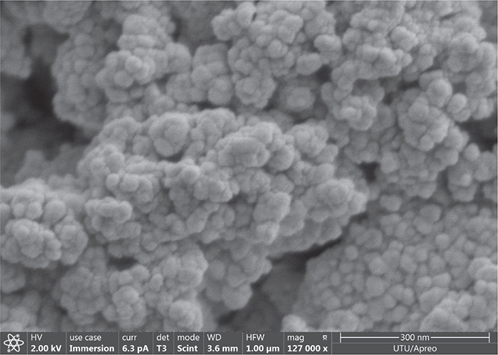
Figure 1. SEM image of synthesized carbonate apatite nanoparticles demonstrating sphere-shaped agglomerated nanoparticles of size less than < 100 nm. Magnification: 127,000X. SEM: scanning electron microscope.
Silanization of the nanoparticles was performed by preparing a silane mixture containing 4% by volume silane coupling agent (3-methacryloyloxypropyltrimethoxysilane) (Sigma-Aldrich, St. Louis, MO, USA) in 96% of 90% ethanol (≥ 99.45% EtOH, Sigma-Aldrich, St. Louis, MO, USA) [27, 28]. CA5n particles were added to the silane mixture and left for 90 minutes in the dark. The mixture was then washed twice with Elix water, centrifuged, and the silanized CA5n particles were collected. The collected nanoparticles were dried in an oven (Termaks Model TS8024, Bergen, Norway) preset to 50°C, then heated to 130°C for 15–20 minutes, and subsequently maintained at 130°C for an additional 15 minutes. The dried CA5n particles were cooled in a desiccator and then immediately added to the experimental adhesive in different concentrations.
Adding nanoparticles to the experimental adhesive
The experimental hydrophilic dental adhesive (Bisco Dental Products Co., Schaumburg, USA) was composed of 70% bis-GMA (bisphenol A diglycidyl ether dimethacrylate), 28.75% HEMA (2-hydroxyethyl methacrylate), 0.25% CQ (camphorquinone), and 1% EDMAB (ethyl N, N-dimethyl-4-aminobenzoate) [29]. Silanized and non-silanized CA5n were added at different percentages (0.5, 1, 2, 4% wt.) to the experimental adhesive. The CA5n containing adhesive was vortexed until the particles were well dispersed, then sonicated using a needle sonicator for 1 minutes (5 s on/2 s off, 30% amplitude) (Sonics Vibra-cell, Connecticut, USA) each time before use to prevent agglomeration and ensure homogeneity. The experimental dental adhesive without CA5n served as a control. The experimental setup is summarized in Table 1.
Degree of conversion
Test specimens (n = 5/group according to previous study [30]) were prepared using a circumferential stainless-steel mold with a thickness of 0.6 mm and a diameter of 6 mm. The adhesive was placed into the mold, immediately covered with a Mylar strip to prevent the formation of an oxygen-inhibition layer [31], and cured for 20 s using an LED light curing unit (Demi Ultra, Kerr, California, USA, irradiance 1767 mW/cm2 ± 5%) in continuous mode, resulting in a radiant exposure of 35.3 J/cm². Fourier Transform Infrared Spectroscopy (Varian model 670 FTIR, Agilent Technologies, California, USA), equipped with a universal attenuated total reflectance (ATR) accessory and baseline method, was utilized [32]. Post-curing measurements were taken at three time points: immediately, at 5 minutes, and at 60 minutes [29]. The ratio between the C = C aliphatic absorbance peaks at 1637 cm-1 and the C-C aromatic absorbance peaks at 1607 cm-1 was calculated to determine the DC according to the following equation [33]:

where ‘R’ is the ratio between the aliphatic and aromatic peaks at 1637 and 1607 cm-1, respectively.
Water sorption and solubility
The specimens were prepared following ISO 4049 specification [34], except for their dimensions [35]. Water sorption was monitored for 28 days, along with the water solubility of the material. Discs (n = 6/group) of experimental adhesive incorporated with 0.5, 1, 2, and 4% wt. nanoparticles of carbonate apatite (CA5n) with and without silanization were prepared with a diameter of 6 ± 0.1 mm and a thickness of 0.5 ± 0.02 mm, verified by a digital caliper (Mitutoyo Digimatic Caliper Europe GmbH, Neuss, Germany) [36]. Discs of experimental adhesive without CA5n served as controls. The discs were kept in a desiccator at 37°C and weighed daily using an analytical balance (Mettler Toledo Balance XS205DU, Greifensee, Switzerland) until a constant mass (m1) was reached, with a variation of less than 0.2 mg within 24 hours. Afterwards, the specimens were immersed in water and weighed daily for up to 7 days, and then again at 14 days and 28 days (m2). After 28 days in water, the specimens were transferred to new dry vials and stored in a 37°C incubator. They were weighed daily until they reached a stable weight (m3). Water sorption (Ws) and water solubility (Wsl) were calculated using the following equations [37]:

where ‘V’ is the sample volume in mm3.
Cell viability
The L-929 mouse fibroblast cell line and the 3-(4,5-dimethylthiazol-2-yl)-2,5-diphenyltetrazolium bromide assay (MTT assay), as specified in ISO 10993-5:2009 [25], were used to evaluate cytotoxicity following the incorporation of CA5n with and without silanization into the experimental dental adhesive. Discs (n = 6/group) were prepared as described for the water sorption and solubility test method. The prepared discs were stored individually in glass containers filled with 500 μL of cell medium. The surface area-to-volume ratio was 1.4 cm²/mL, within the range of 0.5–6 cm²/mL, as specified by ISO standard [25]. The sealed extract bottles were placed in a water bath (Julabo, Göteborg, Sweden) at 37°C for 24 hours and 7 days. L-929 cells, from the third passage, were used in triplicate, seeded at a cell density of 1 × 10⁴ cells/well in 96-well plates and incubated at 37°C, 5% CO2, and 95% humidity in an incubator (Sanyo Incubator, Tokyo, Japan) for 24 hours [38]. A positive control of 2-hydroxyethylmethacrylate (HEMA, Fluka Chemie AG, Buchs, Switzerland) (10 mM), a negative control with cell medium and cells, and blanks with only the cell medium were used in this study. The absorbance was measured at 570 nm using a spectrophotometer (Synergi H1, Biotek, Vermont, USA) [35, 38]. The L-929 cell line was purchased from the European Collection of Authenticated Cell Cultures (ECACC, Public Health England) and tested for mycoplasma using the MycoAlert Mycoplasma Detection kit (Lonza, Rockland, ME, USA).
Statistical analysis
A mixed model, a specific type of ANOVA (Stata version 17.0, College Station, TX, USA), was used to evaluate the DC results, with CA5n concentration and silanization as fixed factors and time as a repeated (within-specimen) factor. Concentration, silanization, and time were considered categorical variables. The Shapiro-Wilk test was used to assess the normality of the water sorption, solubility, and cell viability data. Based on the results of the normality test, the independent-samples t-test was applied to normally distributed data, whereas the Wilcoxon rank-sum test was used for data that did not meet the normality assumption (R version 4.4.1, Vienna, Austria). Statistical significance was set at p < 0.05.
Results
The SEM image (Figure 1) shows agglomerated CA5n nanoparticles with irregular to nearly spherical shapes. The particles’ size was below 100 nm, confirming the successful synthesis of nanoscale particles. The results of the DC measurements are listed in Table 2. Adding CA5n to the adhesive increased the DC, and the effect was independent of concentration and silanization of the nanoparticles. Time significantly increased the DC, with values at both 5 minutes and 60 minutes being significantly higher than immediately after curing in all groups (p < 0.001).
Water sorption and water solubility after 28 days are shown in Table 3. All CA5n groups showed significantly higher water sorption than the control group (p < 0.05), except for the 0.5% NS group (p = 0.064). All CA5n-containing groups exhibited acceptable water solubility, as defined by ISO 4049 specification [34] (<7.5 μg/mm³). There were no significant differences among the groups (p > 0.05).
MTT assay results are shown in Figure 2. Extracts after 24 hours and 7 days from all experimental groups and the control group showed significantly lower cell viability compared to the negative control (cell culture only) (p < 0.05). After 24 hours, cell viability was significantly higher in the 2% NS (p = 0.039) and 4% NS (p = 0.004) groups compared to the experimental adhesive without CA5n. In the silanized groups, particle concentration did not significantly influence cytotoxicity compared to the experimental adhesive without CA5n (p > 0.05). After 7 days, there were no significant differences in cytotoxicity among the adhesive groups (p > 0.05), except for the 2% Sil (p = 0.029) and 4% Sil (p = 0.009) groups, which exhibited significantly lower cytotoxicity than the other groups. Similar effects were observed in both 24 hours and 7-day extracts.
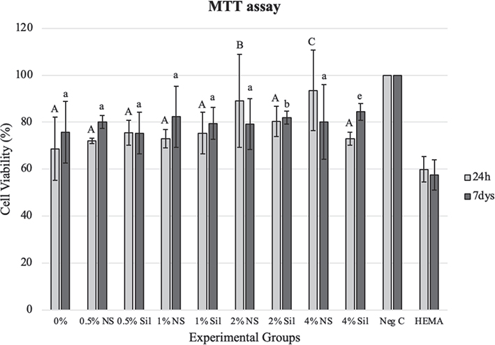
Figure 2. Effects of varying concentrations of CA5n added to the experimental adhesive on cell viability for 24-hour and 7-day extracts. Cell viability data are expressed as a percentage of the negative control cultures. NS: non-silanized particles; Sil: silanized particles. Different uppercase letters indicate statistical significance between different CA5n-containing groups and the control group (experimental adhesive without CA5n) at 24 hours (p < 0.05). Different lowercase letters indicate statistical significance between CA5n-containing groups and the control group (experimental adhesive without CA5n) at 7 days (p < 0.05).
Discussion
This study investigated the effect of incorporating carbonate apatite nanoparticles (CA5n) into a hydrophilic experimental dental adhesive on its physicochemical and biological properties. The results showed that adding CA5n increased the DC and the water sorption. Water solubility results met ISO 4049 standard [34] (<7.5 μg/mm3). Additionally, all formulations demonstrated acceptable cytotoxicity with L929 fibroblasts, as per ISO 10993-5 standard [25]. Silanizing the nanoparticles did not significantly change the results compared to those of non-silanized CA5n.
The DC reflects the efficiency of resin polymerization and is a critical determinant of mechanical strength, water resistance, and biocompatibility [3]. Typically, resin systems achieve conversion rates ranging from 52 to 75% [3]. Unreacted residual monomers may leach into the surrounding environment, eliciting toxic or irritating responses [3]. Previous studies have shown that adding inorganic fillers to dental adhesives can improve DC, likely by enhancing light scattering, reducing polymerization shrinkage, or modifying monomer-filler interactions [3]. However, other investigations reported no effect [39, 40] or even a detrimental impact [41, 42] of fillers on DC, possibly due to filler agglomeration, reduced monomer mobility [43, 44], mismatched refractive indices [45, 46], or increased viscosity [47]. It is therefore important to limit filler loading; in this study, the CA5n concentration was capped at 4% wt. Incorporating CA5n increased the DC to approximately 75%.
Silanization is often reported as an effective strategy to enhance filler-matrix adhesion and improve the mechanical and biological performance of resin composites by promoting covalent bonding between the inorganic filler surface and the organic resin matrix [27]. However, other studies have shown inconsistent or negligible effects, likely due to differences in formulation or incomplete silane coupling [48, 49]. Moreover, because silane molecules are relatively hydrophobic, excessive surface modification may compromise resistance to hydrolytic degradation [50]. In the current study, there was no significant difference in DC between the two surface treatments. This suggests that, under the highly hydrophilic conditions of the tested adhesive system, polymerization efficiency was predominantly controlled by monomer composition and water/solvent content, rather than by silane-mediated interfacial effects.
Two important parameters, water sorption and solubility, were assessed in this study as indicators of adhesive quality [51]. Water sorption reflects the diffusion of moisture into the polymer matrix, which can chemically degrade the material, weaken the bond between the organic matrix and inorganic fillers, and promote the leaching of residual monomers [5]. All tested groups, including the control, exhibited higher water sorption values than the ISO 4049 limit of 40 μg/mm³. However, silanization did not appear to significantly affect water sorption. The high water sorption and solubility may be attributed to solubility occurring primarily within the resin matrix [50], the high surface area of CA5n, which can increase the filler-matrix interfacial region and facilitate water diffusion, and the resin formulation’s intrinsic hydrophilicity [52, 53].
Cytotoxicity analysis was performed to assess the biological behavior of the modified adhesive [54]. The MTT assay was used as a verified colorimetric assay that measures the influence of the extract of the tested material on the metabolic activity of a cell culture [55]. The release of unreacted monomers into the extraction medium may influence the cytotoxic potential of a resin-based test material. Monomer elution typically peaks within the first 24 hours after curing, but can continue for several days [56]. Although most leaching is reportedly limited to this initial period, some studies have found increased cytotoxic effects after 72 hours of cell exposure, indicating ongoing monomer diffusion from the resin matrix [55, 57]. The extent of elution depends on factors such as monomer composition, solvent type, and the degree of polymerization achieved during curing [58, 59].
The observed cell viability was approximately 70% of that in the negative control group across all tested groups (experimental adhesive with and without CA5n). The results indicate that the adhesive’s modification did not adversely affect its cytotoxicity, which could be rated low according to ISO 10993-5:2009 [25]. Extracts from resin-based materials mainly contain monomers and other unreacted chemicals [60]. Filler particles are most likely absent from the extract, which explains why adding CA5n particles did not potentiate the cytotoxicity of the experimental dental adhesive. The lower cytotoxicity observed in some CA5n-containing groups may be attributed to reduced resin matrix volume, improved DC, or unreacted monomers being adsorbed by filler particles [61–63].
This study was limited to evaluating the physicochemical properties and cytotoxicity of experimental dental adhesives containing CA5n. Further research is needed to assess the remineralization potential of these materials at the resin-dentin interface and to investigate the cellular signaling pathways underlying their biological interactions. Such studies would offer a more comprehensive understanding of the bioactivity and long-term performance of these adhesive systems.
Conclusion
The addition of CA5n increased the DC of the experimental adhesive, indicating that the nanoparticles improved the polymerization process. The high water sorption and solubility found may be due to the adhesive’s inherent hydrophilicity rather than the CA5n filler itself. The low cytotoxic potential observed in the extracts was related to the experimental adhesive rather than adding CA5 nanoparticles. Silanization of CA5n did not significantly alter the evaluated properties compared to those of the non-silanized CA5n.
Acknowledgements
This study forms part of the corresponding author’s PhD research plan, approved by the University of Turku, and was funded through a visiting researcher grant to the corresponding author, conducted and supervised at the Nordic Institute of Dental Materials (NIOM). Carbonate apatite microparticles were kindly provided by the Nakamura Lab, MediCity Research Laboratory, Faculty of Medicine, University of Turku. CA5n preparation and characterization were supported by the Turku Collegium for Science, Medicine, and Technology (TCSMT), University of Turku, under Grant [number 2600517411]. We acknowledge the Materials Research Infrastructure (MARI) at the Department of Physics and Astronomy, University of Turku, for providing access to the milling lab and SEM facilities during the synthesis and characterization of CA5n nanoparticles. The NIOM staff’s technical support is acknowledged. We thank Naomi Azulay for the statistical analysis of the degree of conversion data, and Teferi Mekonnen Yitayew for the statistical analysis of the water sorption and solubility, and cell viability data.
Author contributions statement
Heba E. AbdelRazik contributed to the conception and design of the study, the preparation of nanoparticles, the conduct of experiments, data acquisition and interpretation, and writing of the manuscript. Jon E. Dahl contributed to the study’s conception and design and provided critical revisions to the manuscript. Simen E. Kopperud contributed to the conception and design of the study and to the critical revision of the manuscript.
Data availability statement
The data that support this study’s findings are available upon reasonable request.
References
[1] Bourgi R, Kharouf N, Cuevas-Suárez CE, Lukomska-Szymanska M, Haikel Y, Hardan L. A literature review of adhesive systems in dentistry: key components and their clinical applications. Appl Sci (Basel). 2024;14(18):8111. https://doi.org/10.3390/app14188111
[2] Cadenaro M, Josic U, Maravić T, Mazzitelli C, Marchesi G, Mancuso E, et al. Progress in dental adhesive materials. J Dent Res. 2023;102(3):254–62. https://doi.org/10.1177/00220345221145673
[3] Santos M, Fidalgo-Pereira R, Torres O, Carvalho O, Henriques B, Özcan M, et al. The impact of inorganic fillers, organic content, and polymerization mode on the degree of conversion of monomers in resin-matrix cements for restorative dentistry: a scoping review. Clin Oral Investig. 2024;28(8):454. https://doi.org/10.1007/s00784-024-05829-6
[4] Rubaie AJ, Schulze KA. Polymer chemistry of dental adhesives. Pac J Health. 2022;5(1):1. https://doi.org/10.56031/2576-215x.1014
[5] Al-Abbas MAFAA, Al-Badr RJ, Shamash MSA. Long-term water sorption/solubility of two dental bonding agents containing a colloidal dispersion of titanium dioxide. J Adv Pharm Technol Res. 2023;14(2):142–6. https://doi.org/10.4103/japtr.japtr_47_23
[6] Odabasi D, Guler C, Kucukaslan D. Evaluation of the amount of residual monomer released from different flowable composite resins. BMC Oral Health. 2024;24(1):244. https://doi.org/10.1186/s12903-024-04005-2
[7] Madhyastha PS, Naik DG, Natarajan S, Kumar Bhat MR, Vinodhini RS. Effects of water sorption and solubility on degradation of silorane and methacrylate-based dental composites. Indian J Dent Res. 2024;35(1):76–9. https://doi.org/10.4103/ijdr.ijdr_195_23
[8] Zhou W, Liu S, Zhou X, Hannig M, Rupf S, Feng J, et al. Modifying adhesive materials to improve the longevity of resinous restorations. Int J Mol Sci. 2019;20(3):723. https://doi.org/10.3390/ijms20030723
[9] Ahmed MH, De Munck J, Van Landuyt K, Peumans M, Yoshihara K, Van Meerbeek B. Do universal adhesives benefit from an extra bonding layer? J Adhes Dent. 2019;21(2):117–32. https://doi.org/10.3290/j.jad.a42304
[10] de Cardoso GC, Nakanishi L, Isolan CP, Jardim PDS, de Moraes RR. Bond stability of universal adhesives applied to dentin using etch-and-rinse or self-etch strategies. Braz Dent J. 2019;30(5):467–75. https://doi.org/10.1590/0103-6440201902578
[11] Xia Y, Zhang F, Xie H, Gu N. Nanoparticle reinforced resin-based dental composites. J Dent. 2008;36(6):450–5. https://doi.org/10.1016/j.jdent.2008.03.001
[12] Provenzi C, Cb Leitune V, Collares FM, Trommer R, Bergmann CP, Samuel SM. Interface evaluation of experimental dental adhesives with nanostructured hydroxyapatite incorporation. Appl Adhes Sci. 2014;2:2. https://doi.org/10.1186/2196-4351-2-2
[13] Par M, Spanovic N, Tauböck TT, Attin T, Tarle Z. Degree of conversion of experimental resin composites containing bioactive glass 45S5: the effect of post-cure heating. Sci Rep. 2019;9(1):1–9. https://doi.org/10.1038/s41598-019-54035-y
[14] Braga RR. Calcium phosphates as ion-releasing fillers in restorative resin-based materials. Dent Mater J. 2019;35(1):3–14. https://doi.org/10.1016/j.dental.2018.08.288
[15] Campos R, Oliveira C, Macedo J, França F, Basting R, Turssi C, et al. Effect of zinc chloride added to self-etching primer on bond strength to caries-affected dentin and chemical-physical-mechanical properties of adhesives. Int J Adhes Adhes. 2019;95:102412. https://doi.org/10.1016/j.ijadhadh.2019.102412
[16] Farooq I, Ali S, Al-Saleh S, Alhamdan E, Alrefeai M, Abduljabbar T, et al. Synergistic effect of bioactive inorganic fillers in enhancing properties of dentin adhesives – a review. Polymers (Basel). 2021;13(13):2169. https://doi.org/10.3390/polym13132169
[17] Ghiasi B, Sefidbakht Y, Mozaffari-Jovin S, Gharehcheloo B, Mehrarya M, Khodadadi A, et al. Hydroxyapatite as a biomaterial – a gift that keeps on giving. Drug Dev Ind Pharm. 2020;46(7):1035–62. https://doi.org/10.1080/03639045.2020.1776321
[18] Cahyanto A, Liemidia M, Karlina E, Zakaria M, Shariff K, Sukotjo C, et al. Bioactive carbonate apatite cement with enhanced compressive strength via incorporation of silica calcium phosphate composites and calcium hydroxide. Materials. 2023;16(5):2071. https://doi.org/10.3390/ma16052071
[19] Huang H, Luo L, Li L, Guan Y, Yan Y, Jiang Z, et al. Calcium phosphate cement promotes odontoblastic differentiation of dental pulp cells in vitro and in vivo. Coatings. 2022;12(4):543. https://doi.org/10.3390/coatings12040543
[20] Zhou L, Zhao S, Xing X. Effects of different signaling pathways on odontogenic differentiation of dental pulp stem cells: a review. Front Physiol. 2023;14:1272764. https://doi.org/10.3389/fphys.2023.1272764
[21] Amir M, Jeevithan L, Barkat M, Fatima S, Khan M, Israr S, et al. Advances in regenerative dentistry: a systematic review of harnessing Wnt/β-Catenin in dentin-pulp regeneration. Cells. 2024;13(13):1153. https://doi.org/10.3390/cells13131153
[22] Dipalma G, Inchingolo A, Guglielmo M, Morolla R, Palumbo I, Riccaldo L, et al. Nanotechnology and its application in dentistry: a systematic review of recent advances and innovations. J Clin Med. 2024;13(17):5268. https://doi.org/10.3390/jcm13175268
[23] Aktas OC, Puchert K, Vurucu EE, Ersöz B, Veziroglu S, Sen S. A review on nanocomposite coatings in dentistry. J Mater Sci. 2024;59(38):17991–8008. https://doi.org/10.1007/s10853-024-09915-8
[24] Yotsova R, Peev S. Biological properties and medical applications of carbonate apatite: a systematic review. Pharmaceutics. 2024;16(2):291. https://doi.org/10.3390/pharmaceutics16020291
[25] International Standard Organization. ISO 10993-5-2009-biological evaluation of medical devices: Geneva. Part 5: tests for in vitro cytotoxicity.
[26] Nakamura M, Hiratai R, Hentunen T, Salonen J, Yamashita K. Hydroxyapatite with high carbonate substitutions promotes osteoclast resorption through osteocyte-like cells. ACS Biomater Sci Eng. 2016;2(2):259–67. https://doi.org/10.1021/acsbiomaterials.5b00509
[27] Lung CYK, Sarfraz Z, Habib A, Khan AS, Matinlinna JP. Effect of silanization of hydroxyapatite fillers on physical and mechanical properties of a bis-GMA based resin composite. J Mech Behav Biomed Mater. 2016;54:283–94. https://doi.org/10.1016/j.jmbbm.2015.09.033
[28] Matinlinna JP, Dahl JE, Lassila LVJ, Vallittu PK. The effect of trialkoxysi lane coupling agent coatings on e-glass fibers on the flexural prop erties of fiber-reinforced composites. In Mittal, KL (Ed.), Silanes and Other Coupling Agents. 2007;4: 83–97.
[29] Cadenaro M, Antoniolli F, Codan B, Agee K, Tay F, Dorigo E, et al. Influence of different initiators on the degree of conversion of experimental adhesive blends in relation to their hydrophilicity and solvent content. Dent Mater J 2010;26(4):288–94. https://doi.org/10.1016/j.dental.2009.11.078
[30] de Souza Balbinot G, Collares FM, Herpich TL, Visioli F, Samuel SMW, Leitune VCB. Niobium containing bioactive glasses as remineralizing filler for adhesive resins. Dent Mater J. 2020;36(2):221–8. https://doi.org/10.1016/j.dental.2019.11.014
[31] Robertson L, Phaneuf M, Haimeur A, Pesun I, França R. Degree of conversion and oxygen-inhibited layer effect of three dental adhesives. Dent J (Basel). 2016;4(4):37. https://doi.org/10.3390/dj4040037
[32] Ling L, Chen Y, Malyala R. Assessment of degree of conversion and volumetric shrinkage of novel self-adhesive cement. Polymers (Basel). 2024;16(5):581. https://doi.org/10.3390/polym16050581
[33] Al-Qahtani A, Tulbah H, Binhasan M, Shabib S, Al-Aali K, Alhamdan M, et al. Influence of concentration levels of β-tricalcium phosphate on the physical properties of a dental adhesive. Nanomaterials. 2022;12(5):853. https://doi.org/10.3390/nano12050853
[34] International Standard Organization;ISO 4049:2019: Geneva. Dentistry – polymer-based restorative materials.
[35] Esteban Florez FL, Kraemer H, Hiers RD, Sacramento C, Rondinone A, Silvério K, et al. Sorption, solubility and cytotoxicity of novel antibacterial nanofilled dental adhesive resins. Sci Rep. 2020;10(1):1–10. https://doi.org/10.1038/s41598-020-70487-z
[36] Malacarne J, Carvalho RM, de Goes MF, et al. Water sorption/solubility of dental adhesive resins. Dent Mater J. 2006;22(10):973–80. https://doi.org/10.1016/j.dental.2005.11.020
[37] da Silva EM, Amaral CM, Jardim RN, Barbosa MP, Rabello TB. Influence of specimen dimension, water immersion protocol, and surface roughness on water sorption and solubility of resin-based restorative materials. Materials. 2024;17(5):984. https://doi.org/10.3390/ma17050984
[38] Morisbak E, Uvsløkk S, Samuelsen JT. In vitro effects of dental monomer exposure – dependence on the cell culture model. In Vitro Toxicol. 2020;67:104906. https://doi.org/10.1016/j.tiv.2020.104906
[39] Carneiro K, Araujo T, Carvalho E, Meier M, Tanaka A, Carvalho C, et al. Bioactivity and properties of an adhesive system functionalized with an experimental niobium-based glass. J Mech Behav Biomed Mater. 2018;78:188–95. https://doi.org/10.1016/j.jmbbm.2017.11.016
[40] Chladek G, Barszczewska-Rybarek I, Chrószcz-Porębska M, Mertas A. The effect of quaternary ammonium polyethylenimine nanoparticles on bacterial adherence, cytotoxicity, and physical and mechanical properties of experimental dental composites. Sci Rep. 2023;13(1):1–13. https://doi.org/10.1038/s41598-023-43851-y
[41] D'Acierno F, Liu L, Nguyen T, Michal C, Palma-Dibb R, Carvalho R, et al. Physical and mechanical properties of a dental resin adhesive containing hydrophobic chitin nanocrystals. Dent Mater J. 2022;38(12):1855–65. https://doi.org/10.1016/j.dental.2022.09.013
[42] Al-Hamdan R, Almutairi B, Kattan H, Alsuwailem N, Farooq I, Vohra F, et al. Influence of hydroxyapatite nanospheres in dentin adhesive on the dentin bond integrity and degree of conversion: a scanning electron microscopy (SEM), Raman, Fourier Transform-Infrared (FTIR), and microtensile study. Polymers (Basel). 2020;12(12):1–15. https://doi.org/10.3390/polym12122948
[43] Shah PK, Stansbury JW. Role of filler and functional group conversion in the evolution of properties in polymeric dental restoratives. Dent Mater J. 2014;30(5):586–93. https://doi.org/10.1016/j.dental.2014.02.015
[44] Halvorson RH, Erickson RL, Davidson CL. The effect of filler and silane content on conversion of resin-based composite. Dent Mater J. 2003;19(4):327–33. https://doi.org/10.1016/S0109-5641(02)00062-3
[45] Shortall AC, Palin WM, Burtscher P. Refractive index mismatch and monomer reactivity influence composite curing depth. J Dent Res. 2008;87(1):84–8. https://doi.org/10.1177/154405910808700115
[46] Bendary IM, Garcia IM, Collares FM, Takimi A, Samuel SMW, Leitune VCB. Wollastonite as filler of an experimental dental adhesive. J Dent. 2020;102:103472. https://doi.org/10.1016/j.jdent.2020.103472
[47] Li X, Liang S, Inokoshi M, Zhao S, Hong G, Yao C, et al. Different surface treatments and adhesive monomers for zirconia-resin bonds: a systematic review and network meta-analysis. Jpn Dent Sci Rev. 2024;60:175–89. https://doi.org/10.1016/j.jdsr.2024.05.004
[48] Rifane T, Cordeiro K, Silvestre F, Souza M, Zanotto E, Araújo-Neto V, et al. Impact of silanization of different bioactive glasses in simplified adhesives on degree of conversion, dentin bonding and collagen remineralization. Dent Mater J. 2023;39(2):217–26. https://doi.org/10.1016/j.dental.2023.01.005
[49] Michelotti G, Niedzwiecki M, Bidjan D, Dieckmann P, Deari S, Attin T, et al. Silane effect of universal adhesive on the composite-composite repair bond strength after different surface pretreatments. Polymers (Basel). 2020;12(4):950. https://doi.org/10.3390/POLYM12040950
[50] Araújo-Neto V, Moreira M, Ñaupari-Villasante R, De Paula D, Medeiros S, Loguercio A, et al. Nanofiller particles and bonding durability, water sorption, and solubility of universal adhesives. Oper Dent. 2021;46(6):690–7. https://doi.org/10.2341/20-239-L
[51] Esteves S, Barcellos D, Silva T, Silva M, Campos T, Rosetti E, et al. How Water Content Can Influence the Chemomechanical Properties and Physical Degradation under Aging of Experimental Adhesives. Int J Dent. 2022;2022:5771341. https://doi.org/10.1155/2022/5771341
[52] Robakowska M, Gierz Ł. Effect of nanosilica fillers on photopolymerisation behaviour and mechanical performance of dental resin composites. Coatings. 2025;15(11):1316. https://doi.org/10.3390/coatings15111316
[53] ElEmbaby AES, Nassar AE, Elawsya ME. Impact of silica nanoparticles incorporation on the properties of resin infiltration: an in vitro study. BMC Oral Health. 2024;24(1):1484. https://doi.org/10.1186/s12903-024-05107-7
[54] Kazak M, Sarialioglu Gungor A, Ozman Z, Donmez N. Comparative cell viability of dentin-bonding adhesive systems on human dental pulp stem cells: time-dependent analysis. BMC Oral Health. 2024;24(1):663. https://doi.org/10.1186/s12903-024-04438-9
[55] Ohlsson E, Bolay C, Arabulan S, Galler K, Buchalla W, Schmalz G, et al. In-vitro cytotoxicity of self-adhesive dental restorative materials. Dent Mater J. 2024;40(4):739–46. https://doi.org/10.1016/j.dental.2024.02.015
[56] Diemer F, Stark H, Helfgen E, Enkling N, Probstmeier R, Winter J, et al. In vitro cytotoxicity of different dental resin-cements on human cell lines. J Mater Sci Mater Med. 2021;32(1):4. https://doi.org/10.1007/s10856-020-06471-w
[57] Şişmanoğlu S, Demirci M, Schweikl H, Ozen-Eroglu G, Cetin-Aktas E, Kuruca S, et al. Cytotoxic effects of different self-adhesive resin cements: cell viability and induction of apoptosis. J Adv Prosthodont. 2020;12(2):89–99. https://doi.org/10.4047/jap.2020.12.2.89
[58] Maletin A, Ristic I, Veljovic T, Ramic B, Puskar T, Jeremic-Knezevic M, et al. Influence of dimethacrylate monomer on the polymerization efficacy of resin-based dental cements – FTIR analysis. Polymers (Basel). 2022;14(2):247. https://doi.org/10.3390/polym14020247
[59] Janani K, Teja K, Sandhya R, Alam M, Al-Qaisi R, Shrivastava D, et al. Monomer elution from three resin composites at two different time interval using high performance liquid chromatography – an in-vitro study. Polymers (Basel). 2021;13(24):4395. https://doi.org/10.3390/polym13244395
[60] Vervliet P, De Nys S, Duca R, Boonen I, Godderis L, Elskens M, et al. Identification of chemicals leaching from dental resin-based materials after in vitro chemical and salivary degradation. Dent Mater J. 2022;38(1):19–32. https://doi.org/10.1016/j.dental.2021.10.006
[61] Garcia IM, Assad-Loss TF, Schneider LFJ, Collares FM, Cavalcante LMA, Tostes MA. Cytotoxicity evaluation, antibacterial effect, and degree of conversion of QAM-containing adhesives. Braz Oral Res. 2024;38:e001. https://doi.org/10.1590/1807-3107bor-2024.vol38.0001
[62] Maimansomsuk S, Teangrom P, Teanchai C, Sinthuvanich C, Ruengket P, Attarataya J, et al. Methacrylic acid/tween 80 engineered amorphous calcium phosphate as an effective bioactive reinforcing nanofillers in dental adhesive. Dent Mater J. 2025;44(2):190–200. https://doi.org/10.4012/dmj.2024-285
[63] Wawrzynkiewicz A, Rozpedek-Kaminska W, Galita G, Lukomska-Szymanska M, Lapinska B, Sokolowski J, et al. The cytotoxicity and genotoxicity of three dental universal adhesives – an in vitro study. Int J Mol Sci. 2020;21(11):3950. https://doi.org/10.3390/ijms21113950