SYSTEMATIC REVIEW
Evaluating the remineralisation potential and clinical evidence of emerging biocompatible materials in restorative dentistry: a systematic review
Duraiarasan Isaiselvi, Kariannan Partheeban Indumathi, Siluvai Sibyl and Govindaraj Krishnaprakash
Department of Public Health Dentistry, Faculty of Medicine and Health Sciences, SRM Kattankulathur Dental College and Hospitals, SRM Institute of Science and Technology, Chengalpattu, India
ABSTRACT
Background: Biocompatible materials with remineralising and regenerative properties are now being increasingly used in restorative dentistry as possible materials in place of conventional agents. This systematic review aimed to evaluate the laboratory remineralisation potential and available clinical evidence of emerging biocompatible, non-fluoridated materials used in restorative dentistry. Patient-reported outcomes were explored where reported.
Materials and methods: A systematic review was performed on relevant databases as PubMed, Cochrane, EBSCOhost and Scopus.
Results: A total of 8 in vivo and 23 in vitro studies were included. Self-assembling peptide P11–4 (SAP P11–4) and chitosan-based materials demonstrated consistent remineralisation benefits, predominantly in laboratory studies, with limited supporting clinical evidence. Risk of bias was moderate for in vitro studies and low-to-some concerns for in vivo studies.
Conclusion: Self-assembling peptides and chitosan-based biomaterials show promising potential, though current evidence is largely laboratory-based and requires further clinical validation. Further well-designed clinical trials are warranted.
KEYWORDS: Aloe vera; chitosan; dental caries; grape seed; herbal; non-fluoridated biocompatible materials; remineralisation; restorative dentistry; self-assembling peptides
Citation: BIOMATERIAL INVESTIGATIONS IN DENTISTRY 2026, VOL. 13, 239–251. https://doi.org/10.2340/biid.v13.45679.
Copyright: © 2026 The Author(s). Published by MJS Publishing on behalf of Acta Odontologica Scandinavica Society. This is an Open Access article distributed under the terms of the Creative Commons Attribution 4.0 International License (http://creativecommons.org/licenses/by/4.0/), allowing third parties to copy and redistribute the material in any medium or format and to remix, transform, and build upon the material, with the condition of proper attribution to the original work.
Received: 6 December 2025; Accepted: 16 February 2026; Published: 15 April 2026
CONTACT: Dr. K. P. Indumathi indumatp@srmist.edu.in, indhuparthiban@yahoo.co.in Assistant Professor, Department of Public Health Dentistry, SRM Kattankulathur Dental College and Hospitals, Faculty of Medicine and Health Sciences, SRM Institute of Science and Technology, Kattankulathur, Chengalpattu District, Tamil Nadu-603203, India
Competing interests and funding: The authors assert that they possess no conflicting interests.
This study was self-funded
KEY MESSAGES:
- - Self-assembling peptide SAP P11–4 demonstrates strong clinical evidence for minimally invasive remineralisation of early carious lesions.
- - Chitosan-based restorative and preventive materials provide comparable outcomes to conventional materials with added biocompatibility benefits.
- - Although promising, most emerging biocompatible agents require further high-quality clinical trials to validate long-term effectiveness and patient outcomes.
Introduction
Dental caries is highly preventable, but remains one of the most prevalent chronic diseases in children and adults worldwide [1]. The WHO Global Oral Health Status Report estimated that globally 2 billion people suffer from caries of permanent teeth and 514 million children suffer from caries of primary teeth [2]. Shafer defined dental caries as an ‘irreversible microbial disease of the calcified tissues of the teeth, characterized by demineralization of the inorganic portion and destruction of the organic substance of the tooth, which often leads to cavitation’ [3]. The enamel and dentinal mineral loss are due to the acid attacks produced by the bacterial metabolism of carbohydrates and sugars [4]. Fortunately, dental caries is reversible during the initial phase of the disease, and the stagnation of enamel and dentin demineralisation can be prevented through the inhibition of biofilm formation and salivary protective factors [5]. Restoring a decayed tooth is essential for preventing further deterioration, preserving tooth structure, and maintaining oral function. Untreated decay can impair general health and quality of life by causing pain, infection, and tooth loss. According to the literature search [4, 6] numerous substances including fluorides, calcium glycerophosphate, and xylitol, have been employed to effectively allow demineralised dentin and enamel to remineralise.
Several researchers have used both traditional fluoridated and non-fluoridated dentifrices to examine the demineralisation and remineralisation of enamel lesions in permanent teeth. Though it has advantages, excessive levels of fluoride can be harmful leading to dental and skeletal fluorosis, hypersensitivity reactions, hypersalivation, dyspnoea, stomach irritation, muscular spasm, birth abnormalities, and so on [6, 7]. Therefore, alternative remineralising agents were explored, and consequently, a novel approach to managing enamel and dentin remineralisation has focused on identifying natural, effective, and non-fluoride-based treatment options. Biocompatible materials have been created to overcome these obstacles and enhance patient outcomes and treatment effectiveness. Despite the stability and aesthetic appeal of conventional restoratives like amalgam and composite resins, research into natural alternatives has been prompted by concerns about biocompatibility and durability. This systematic review was conducted to evaluate the laboratory remineralisation potential and available clinical evidence of emerging non-fluoridated biocompatible materials used in restorative dentistry.
Materials and methods
Research question: The research question was framed in PEO format.
In patients undergoing restorative dental procedures, how do emerging biocompatible materials perform in terms of clinical effectiveness, durability, and surrogate clinical outcomes, with patient-reported outcomes evaluated when reported?
P (Population): Patients receiving restorative dental treatments.
E (Exposure): Emerging biocompatible materials (e.g. herbal based materials like chitosan, cellulose, self-assembling peptides, grapeseed, non-fluoride-based materials).
O (Outcome): Surrogate clinical outcomes (lesion regression, surface integrity, remineralisation indices), laboratory outcomes (surface microhardness, SEM/EDX, Ca/P ratio), and patient- reported outcomes when reported.
Protocol and registration: The protocol for systematic review is registered with PROSPERO (International Prospective Register of Systematic Review) with registration number (CRD420251011817) and follows PRISMA 2020 guidelines. Figure 1 shows the PRISMA flowchart of study selection.
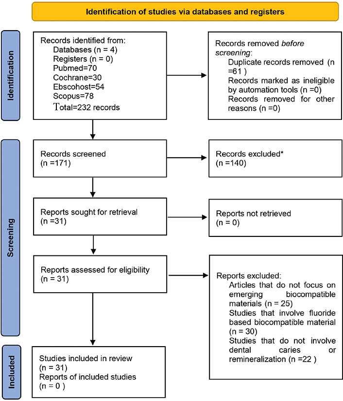
Figure 1. PRISMA 2020 flow diagram illustrating study selection and screening process.
Source: Authors’ analysis.
Inclusion criteria
Studies involving patients undergoing restorative dental procedures.
Use of emerging biocompatible materials (herbal and non-fluoride based) such as plant-derived biomaterials, self-assembling peptides, chitosan, cellulose, Aloe vera, and grape seed extract in restorative dentistry.
Articles available in English.
Studies published from 2015 to 2025 to focus on emerging materials.
Original studies, including experimental, observational, and clinical trials, together with case–control studies, were selected for review.
Exclusion criteria
Studies evaluating interventions not related to restorative dentistry.
Articles published in languages other than English.
Studies that focus exclusively on fluoride-based materials.
Studies not addressing dental caries or remineralisation.
Search strategy
A broad search was conducted in databases like PUBMED, COCHRANE, EBSCOhost and SCOPUS using the keywords like plant derived materials, grapeseed, chitosan, Aloe vera, self-assembling peptides, herbal, non-fluoridated materials, remineralising agent, dental caries, remineralisation, and restorative-dentistry. BOOLEAN expression like AND and OR were used. Duplicates were removed. The search was last updated on 15 March 2025. Two reviewers independently extracted data from included studies; any disagreements were resolved by discussion.
Data synthesis
A quantitative meta-analysis was not feasible due to heterogeneity in study designs, interventions, and outcome measures among the included studies. Therefore, a narrative synthesis of the results was conducted in accordance with PRISMA 2020 guidelines.
Risk of bias assessment (Methods section)
Risk of bias for included case series and cohort studies was evaluated using the Quality in Non- Randomised Studies (QUIN) tool. The overall risk judgments (Low, Moderate, or High) across seven bias domains are presented in Figure 2.
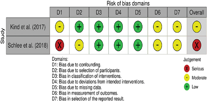
Figure 2. Risk of bias assessment for in vitro studies using Quality in Non- Randomised Studies (QUIN) tool.
Source: Authors’ analysis.
For in vivo research (clinical trials), methodological quality assessment was performed according to the Cochrane Risk of Bias 2.0 (RoB 2.0) tool, as shown in Figure 3. This assessment covered five domains: the randomising process, deviations from intended interventions, missing outcome data, outcome assessment, and selection of reported outcomes. Each study was categorised as having low risk, some concerns, or high risk of bias, depending on the transparency and rigour of its reported methods.
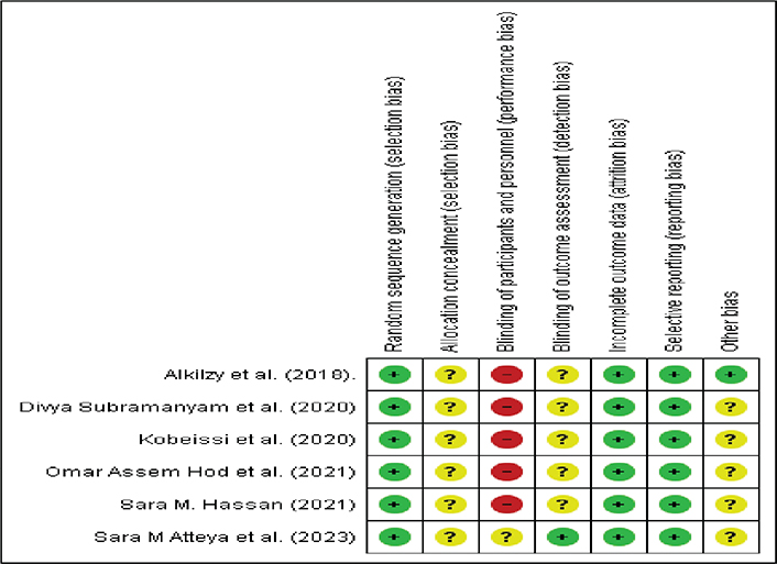
Figure 3. Risk of bias assessment for in vivo studies using Cochrane Risk of Bias 2.0 (RoB 2.0) tool.
Source: Authors’ analysis.
For in vitro studies, a structured and modified ROB-based assessment tool was used to evaluate seven criteria: sample size determination, randomisation, standardisation of procedures, blinding of outcome assessment, repetition of experiments, appropriate statistical analysis, and use of control groups. Each study was assessed and classified into low, moderate, or high-risk categories as illustrated in Figure 4. The detailed search strategy is provided in Appendix A.
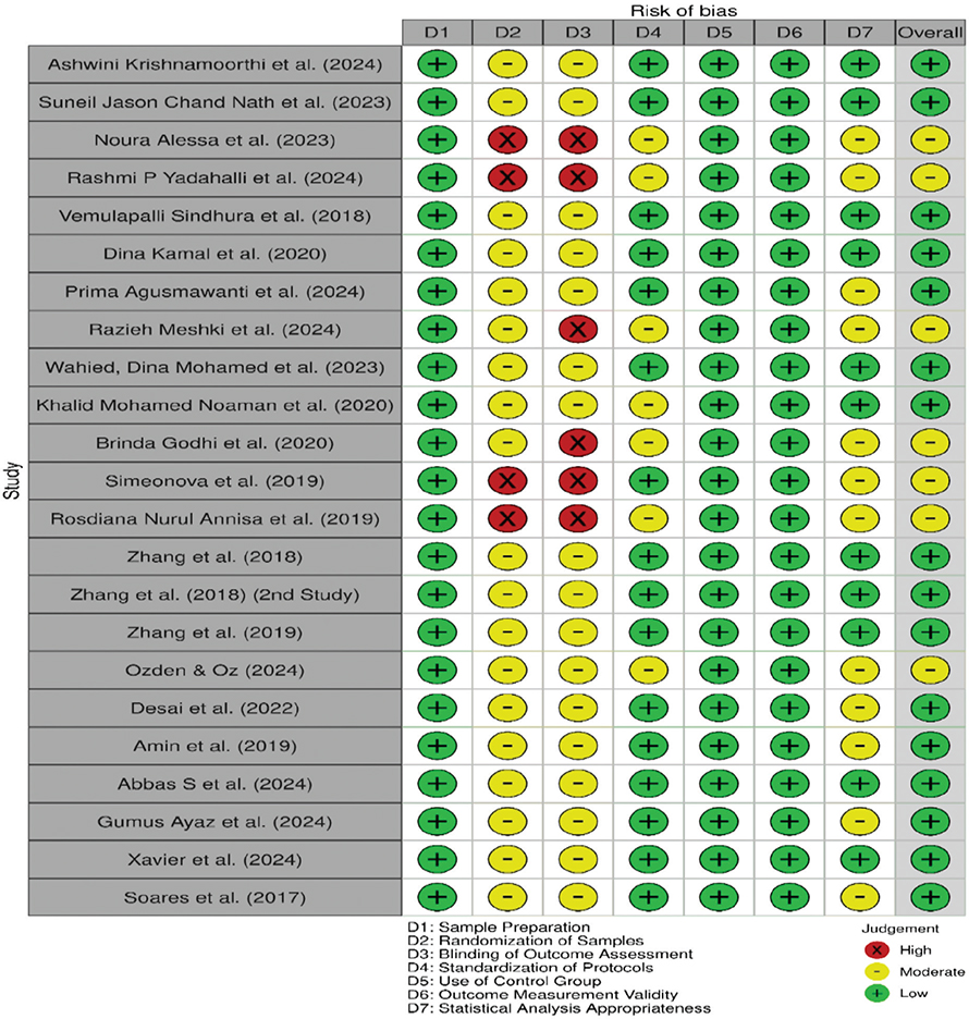
Figure 4. Risk of bias assessment for in vitro studies using a modified ROB-based assessment tool.
Source: Authors’ analysis.
Results
Findings are presented descriptively, with relative improvements reported where available. A total of 31 studies were included, consisting of 8 in vivo clinical studies and 23 in vitro studies. The majority of included studies reported favourable remineralisation outcomes with emerging biocompatible materials compared to controls. A total of 77 full-text articles were excluded for the following reasons: lack of focus on emerging biocompatible materials (n = 25), use of fluoride-based biocompatible agents (n = 30), and absence of outcomes related to dental caries or remineralisation (n = 22), as shown in the PRISMA flow diagram (Figure 1). Detailed characteristics of the included studies are summarised in Table 1 (in vivo studies) and Table 2 (in vitro studies).
| S. No | Author (year) | Study design | Specimens used | Follow-up duration | Materials used | Mode of application | Outcome measures used | Clinical effectiveness | Patient-reported outcomes |
| 1 | Sara M Atteya et al. (2023) [8] | RCT (split-mouth) | Permanent teeth, non-cavitated carious lesions | 6 months | SAP P11–4 + Nano Silver Fluoride vs SAP alone | Topical application | DIAGNOdent, ICDAS, morphometric | SAP + NSF showed higher remineralisation than SAP alone | Not reported |
| 2 | Divya Subramanyam et al. (2020) [9] | RCT | Primary molars | 6 months | Aloe vera gel vs Formocresol | Direct pulp therapy (pulpotomy) | Clinical signs, radiographic evaluation | Comparable clinical success rates between Aloe vera and FC | Not directly assessed |
| 3 | Omar Assem Hod et al. (2021) [10] | RCT | Primary molars | 6 months | Chitosan-modified GIC vs conventional GIC | Restorative filling | USPHS criteria | No significant difference between groups | Not reported |
| 4 | Schlee et al. (2018) [11] | Case series | Permanent teeth (proximal lesions) | 12 months | SAP P11–4 | Topical, direct application into lesion | Radiographic regression, subtraction analysis | Lesion depth reduced, early arrest observed | Not reported |
| 5 | Alkilzy et al. (2018) [12] | RCT | Permanent anterior teeth | 6 months | SAP P11–4 + Fluoride varnish | Topical application | ICDAS scoring, DIAGNOdent | Significant remineralisation in SAP group | Not reported |
| 6 | Kobeissi et al. (2020) [13] | RCT | Children with WSLs | 6 months | SAP P11–4 vs Tricalcium phosphate fluoride varnish | Topical application | DIAGNOdent, WSL index | SAP had comparable or slightly better outcomes | Not reported |
| 7 | Kind et al. (2017) [14] | Cohort Study | Permanent teeth with early caries | 12 months | Self-assembling peptide | Topical application into lesion | Visual scoring, QLF | Significant improvement in enamel appearance and lesion regression | Not reported |
| 8 | Sara M. Hassan (2021) [15] | RCT | Permanent teeth with WSLs | 6 months | Ginger & Rosemary Extract vs Fluoride Varnish | Topical gel | DIAGNOdent, WSL index | Herbal group showed comparable remineralisation to fluoride | Not reported |
| SAP: Self-assembling peptide; GIC: glass ionomer cement; RCT: Randomized Controlled Trial; NSF: Nano Silver Fluoride; USPHS: United States Public Health Service; FC: Formocresol; ICDAS: International Caries Detection and Assessment System; WSL: White Spot Lesion; QLF: Quantitative Light-Induced Fluorescence; CSP: Calcium Sucrose Phosphate; CPP: Casein Phosphopeptide; ACP: Amorphous Calcium Phosphate; ACPF: Amorphous Calcium Phosphate Fluoride; VHN: Vickers Hardness Number; SEM: Scanning Electron Microscopy; LRAP: Leucine-Rich Amelogenin Peptide; NaF: Sodium Fluoride; CT: Computed Tomography; CS: Chitosan; EDX: Energy Dispersive X-ray Analysis; VMT: Vickers Microhardness Test; SDF: Silver Diamine Fluoride; NHA: Nano Hydroxyapatite; TCP: Tricalcium Phosphate; CMC: Carboxymethyl Chitosan; TEM: Transmission Electron Microscopy; PAA: Polyacrylic Acid; BG: Bioactive Glass; APF: Acidulated Phosphate Fluoride; GSE: Grape Seed Extract; CESP: Chicken Eggshell Powder; SMH: Surface Microhardness; BAG: Bioactive Glass; PEO: Population Exposure Outcome; ROB: Risk of Bias; PRISMA: Preferred Reporting Items for Systematic Reviews and Meta-Analyses | |||||||||
| S. No | Author (year) | Study design | Specimens used | Follow-up duration | Materials used | Mode of application | Outcome measures used | Remineralisation outcome | Results/conclusion |
| 1 | Ashwini Krishnamoorthi et al. (2024) [16] | In vitro | Primary molars | 21 days | P11–4, CSP, Bioactive glass, CPP-ACP, CPP-ACPF | Topical application (micropipette) | Surface Vickers microhardness (VHN), SEM | All P11–4 combinations showed increased surface microhardness and improved enamel morphology under SEM; highest effectiveness observed in P11–4 + CPP-ACPF. | P11–4 combined with calcium phosphate-based agents led to significant remineralisation; P11–4 + CPP-ACPF group showed the highest efficacy. |
| 2 | Suneil Jason Chand Nath et al. (2023) [17] | In vitro | Premolars | 8 days | P11–4, P26, LRAP, 5% NaF, DI water | Topical application (P11–4 via sponge; others in solution) | Micro-CT (MD), Nano indentation (H, EM) | P11–4 and P26 significantly increased mineral density, hardness, and elasticity modulus compared to NaF and LRAP, especially at deeper enamel layers (60 µm). | P11–4 & P26 outperformed NaF & LRAP at 60 µm depth; SAPs show strong remineralisation potential. |
| 3 | Noura Alessa et al. (2023) [18] | In vitro | Premolars | 12 days | Bioactive glass, P11–4, Ozone remineralising agents, Deionised water (Control group) | Topical application (ozone via ozone unit & others in solution) | Vickers microhardness (VHN) | Increased microhardness post-remineralisation. | Bioactive glass and self-assembling peptides showed higher remineralising capacity. |
| 4 | Rashmi P Yadahalli et al. (2024) [19] | In vitro | Premolars | 14 days | Bioactive glass, P11 4, Arginine bicarbonate remineralising agents, deionised water(control) | Topical application (in solution) | Vickers microhardness (VHN) | Increased microhardness post-remineralisation. | Bioactive glass and self-assembling peptides showed higher remineralising capacity. |
| 5 | Vemulapalli Sindhura et al. (2018) [20] | In vitro | Premolars | 1 week, 1 month, 3 months | SAP P11 and CPP ACP | Topical (SAP via brush after etching; CPP-ACP via gloved finger) | SEM, EDX, Ca/P Ratio | P11–4 showed gradual increase in Ca/P ratio over time; CPP-ACP showed early spike then decline. | At 3 months, SAP P11–4 showed higher Ca/P ratio and better mineral deposition than CPP-ACP. |
| 6 | Dina Kamal et al. (2020) [21] | In vitro | Molars | 1 week, 4 weeks | Fluoride varnish, CPP-ACPF varnish, SAP P11–4, SAP + Fluoride, SAP + CPP-ACPF | Topical application | Surface Vickers microhardness (VHN), SEM | All groups showed remineralisation; combination groups (SAP + Fluoride and SAP + CPP-ACPF) showed higher microhardness values. | Combination of SAP with CPP-ACPF or Fluoride showed superior and faster remineralisation; synergistic effect evident from 1 to 4 weeks. |
| 7 | Prima Agusmawanti et al. (2024) [22] | In vitro | Mandibular incisors | 7 days | QP3VH, NaF, QP3VH + NaF, QP3VH + CS, and saline distilled water | Topical application (in solution) | SEM, EDX, Ca/P Ratio | QP3VH-based groups showed notable enamel surface improvement; combination with chitosan and/or NaF further enhanced remineralisation. | QP3VH peptide significantly improved remineralisation; synergistic effect with NaF or chitosan shown by smoother enamel and higher Ca/P ratio. |
| 8 | Razieh Meshki et al. (2024) [23] | In vitro | Primary canine | 7 days | Bioglass-Chitosan solution, Chitosan, Bioglass solution, SDF, Remineralisation solution, Distilled water | Topical application (except SDF, applied once) | Vickers microhardness (VHN) Test (VMT) | Increased microhardness post-remineralisation. | Bioglass-chitosan solution showed the highest improvement in Vickers microhardness (VHN); chitosan and bioglass also effective. |
| 9 | Wahied, Dina Mohamed et al. (2023) [24] | In vitro | Permanent molars | 42 hours | Nano β-TCP, Nβ-TCP + Chitosan, NHA, NHA + Chitosan | Topical application (Gel applied with micro-brush) | Vickers microhardness (VHN) Number (VHN), SEM imaging | All test groups showed increased microhardness; NHA + Chitosan most effective. | NHA + Chitosan group exhibited 55.10% Vickers microhardness (VHN) increase; chitosan enhanced both NHA and Nβ-TCP efficacy. |
| 10 | Khalid Mohamed Noaman et al. (2020) [25] | In vitro | Premolars with subclinical lesions | 3 and 6 months | Phosphorylated Chitosan + ACP, CPP-ACP, Fluoride | Topical brushing | DIAGNOdent, EDAX (Ca/P ratio) | Chitosan group showed highest remineralisation; time-dependent increase. | Chitosan significantly more effective at 6 months; fluoride lowest remineralisation. |
| 11 | Brinda Godhi et al. (2020) [26] | In vitro | Premolars | 21 days | Chitosan (2.5 mg/ml), Ginger-Manuka honey mix | Topical application | QLF parameters: ΔF, ΔF Max, ΔQ | Chitosan showed significant remineralisation; Ginger-Honey moderate. | Chitosan highest potential; Ginger-Honey moderate from Day 14 onward. |
| 12 | Simeonova et al. (2019) [27] | In vitro | Permanent molars and premolars | 42 hours (6 h × 7 sessions) | Hybrid chitosan/calcium phosphates microgels | Immersion for 6 h per cycle | SEM, ATR-IR spectroscopy | Formation of enamel-like nanocrystalline structures. | Chi_CaP MGs effectively promoted remineralisation, forming stable enamel-like phases. |
| 13 | Rosdiana Nurul Annisa et al. (2019) [28] | In vitro | Dentin collagen samples from human teeth | Not explicitly stated | Carboxymethyl Chitosan + Amorphous Calcium Phosphate (CMC/ACP) | Applied as cavity base; stored in PBS at 37°C | EDX, TEM | Increased Ca and P levels; intra- and extrafibrillar mineral deposition. | CMC/ACP showed superior dentin remineralisation and mineral content restoration. |
| 14 | Zhang et al. (2018) [29] | In vitro | Molars | 7 days | Bioactive glass (BG), BG + PAA, Chitosan pre-treatment (CS-BG, CS-BG + PAA), remineralising solution (RS), deionised water | Topical application | Raman spectroscopy, Knoop surface & cross-sectional Vickers microhardness (VHN), SEM | CS-BG group showed greatest surface and subsurface remineralisation; CS-BG + PAA had larger enamel-like crystals. | Chitosan enhanced subsurface remineralisation even in presence of pellicle; potential for lesion consolidation. |
| 15 | Zhang et al. (2018) [30] | In vitro | Molars | 7 days | Bioglass (BG), Bioglass + PAA, Chitosan + BG, Chitosan + BG + PAA, Standard remineralising solution, Deionised water | Topical application | Raman spectroscopy, ATR-FTIR, Knoop Vickers microhardness (VHN), SEM, EDX | CS-BG + PAA group had greatest subsurface mineral content and hardness recovery. | Chitosan pre-treatment significantly improved bioglass remineralisation; supports chitosan-bioglass use for subsurface lesions. |
| 16 | Zhang et al. (2019) [31] | In vitro | Molars | 7 days | Chitosan-bioglass complex (CBS), Bioglass with chitosan pre-treatment (CB), standard remineralisation solution (RS), deionised water (NC) | Topical application after salivary pellicle | Raman mapping, Knoop Vickers microhardness (VHN), SEM, EDX | CBS showed greatest subsurface mineral deposition and hardness recovery. | CBS significantly promoted remineralisation with dense HA-like mineral formation; effective even with pellicle. |
| 17 | Ozden & Oz (2024) [32] | In vitro | Mandibular molars | 14 days | Grape Seed Extract (25%), CPP-ACP, APF (alone and combinations) | Topical (cotton applicator 15 seconds, held 1 minutes) | Surface Vickers microhardness (VHN), SEM | All groups showed some remineralisation; GSE alone less effective than when combined. | GSE enhanced remineralisation when combined with CPP-ACP or APF; significant hardness increase in combined groups. |
| 18 | Desai et al. (2022) [33] | In vitro | Primary molars | 10 days | CPP-ACPF, TCP, Grape Seed Extract, Deionised Water | Topical application on enamel window | Vickers microhardness (VHN), SEM-EDX, CBCT | Grape seed extract showed highest remineralisation. | Grape seed extract significantly enhanced Vickers microhardness (VHN) and mineral gain; superior to CPP-ACPF and TCP. |
| 19 | Amin et al. (2019) [34] | In vitro | Primary anterior teeth | 8 days | Grape Seed Extract (GSE), Sodium Fluoride (NaF, 1000 ppm) | Topical application | Vickers microhardness (VHN), SEM-EDX | GSE promoted mineral deposition but had lower microhardness than NaF. | GSE showed remineralisation potential but less effective than NaF; suitable natural alternative. |
| 20 | Abbas S et al. (2024) [35] | In vitro | Upper first premolars | 14 days | CPP-ACP (control), GSE, Chicken Eggshell Extract (CESP), GSE + CESP | Topical application | Vickers microhardness (VHN) (SMH) | GSE + CESP group showed highest SMH values; CESP alone also highly effective. | Combination of GSE and CESP demonstrated synergistic effect; significant enamel remineralisation in all groups. |
| 21 | Gumus Ayaz et al. (2024) [36] | In vitro | Premolars | 6 days | CPP-ACP, Rosemary Oil (RO), Ginger-Honey, Ginger-Honey–Cocoa, Grape Seed Extract (GSE), Control Group | Topical application (using applicator) | Vickers microhardness (VHN), DIAGNOdent | GSE showed the highest remineralisation potential among tested agents. | GSE most effective; herbal-based tooth creams non-invasive and suitable for early caries repair. |
| 22 | Xavier et al. (2024) [37] | In vitro | Premolars | 4 weeks | CPP-ACPF, Tricalcium phosphate with fluoride (TCP-F), Calcium sucrose phosphate (CSP), P11–4 | Topical application | Surface Vickers microhardness (VHN), SEM, EDX | P11–4 showed highest VHN; TCP-F and P11–4 had greater mineral gain (EDX); CSP moderate; CPP-ACPF least effective. | P11–4 exhibited greatest remineralisation; TCP-F also strong performer. |
| 23 | Soares et al. (2017) [38] | In vitro | Premolars | 30 days | CPP-ACPF, TCP-F, CSP, P11–4 | Topical application | Surface Vickers microhardness (VHN), SEM | P11–4 showed highest microhardness recovery (62.06%), followed by CPP-ACPF, BAG, and HA gel. | P11–4 highest remineralising capacity; SEM confirmed organised mineral deposition. |
| SAP: Self-assembling peptide. | |||||||||
In vivo (clinical) evidence
Eight in vivo studies evaluated the clinical performance of emerging biocompatible materials, primarily self-assembling peptides (SAP P11–4), Aloe vera, chitosan-modified glass ionomer cement (GIC), and selected herbal-derived agents. The most frequently assessed clinical parameters included lesion regression, surface integrity, remineralisation potential, and pulpotomy success, evaluated using tools such as ICDAS scoring, DIAGNOdent, radiographic imaging, and USPHS criteria.
Self-assembling peptide SAP P11–4 demonstrated favourable remineralisation outcomes in seven out of eight clinical studies, particularly when used alone or in combination with fluoride or calcium phosphate–based agents. Aloe vera exhibited comparable clinical success to formocresol when used as a pulpotomy medicament in primary teeth. Chitosan-modified GICs showed clinical performance similar to conventional GICs, with no significant differences in restoration integrity or short-term outcomes.
However, most clinical studies were characterised by limited sample sizes and short follow-up durations. Importantly, none of the included in vivo studies reported validated patient-reported outcome measures, such as patient comfort, satisfaction, or oral health–related quality of life.
In vitro evidence
Twenty-three in vitro studies investigated the remineralisation potential of biocompatible materials, including SAP P11–4, chitosan-based formulations, bioactive glass, grape seed extract, and herbal derivatives such as ginger and rosemary. Laboratory assessments predominantly employed surface microhardness testing (Vickers or Knoop), scanning electron microscopy with energy-dispersive X-ray analysis (SEM/EDX), and calcium- to-phosphorus (Ca/P) ratio analysis.
Across the majority of laboratory studies, both SAP P11–4 and chitosan-based materials demonstrated greater remineralisation potential compared to control groups, reflected by increased surface hardness, improved enamel morphology, and enhanced mineral deposition. Several studies reported additive or synergistic effects when SAP P11–4 was combined with CPP-ACPF or when chitosan was combined with bioactive glass or nano-hydroxyapatite.
Most in vitro studies were assessed as having a moderate risk of bias, primarily due to the absence of sample size calculations, lack of randomisation, and absence of blinding during outcome assessment. In contrast, the majority of in vivo studies demonstrated low to moderate risk of bias, with no study classified as high risk. These laboratory findings represent surrogate indicators of remineralisation and should not be directly extrapolated to clinical effectiveness.
Discussion
This systematic review found that emerging biocompatible materials, particularly SAP P11–4 and chitosan-based formulations, consistently demonstrated remineralisation potential, with evidence derived from both in vitro investigations and a limited number of clinical studies. While available clinical evidence supports the use of self-assembling peptides in the management of early, non-cavitated carious lesions, the majority of evidence for herbal-derived materials remains laboratory-based, underscoring the need for further well-designed clinical validation [8, 12, 14]
The growing interest in biocompatible materials within restorative dentistry, especially those promoting biomimetic remineralisation, is reflected in the findings of this review. Among the evaluated agents, SAP P11–4 is the most extensively studied, with both laboratory and clinical investigations indicating its ability to facilitate enamel repair by forming a biomimetic scaffold that supports hydroxyapatite deposition within early carious lesions [12, 14, 17]. This mechanism closely resembles natural remineralisation pathways and aligns with minimally invasive caries management principles [12, 13]. Clinical studies suggest that SAP P11–4 may contribute to lesion stabilisation and hardness improvement; however, these findings are based on relatively short follow-up periods and modest sample sizes.
Chitosan-based materials exhibited antimicrobial and remineralisation-supportive properties, particularly when combined with bioactive glass or hydroxyapatite [10, 23, 29]. Laboratory findings indicate that such combinations may enhance mineral deposition and enamel surface integrity. Nevertheless, clinical evidence evaluating chitosan-modified restorative materials remains limited, and conclusions regarding long-term clinical effectiveness should therefore be interpreted with caution [23, 29].
Herbal-derived agents, including grape seed extract, Aloe vera, and combined herbal formulations (such as ginger, rosemary, and honey), demonstrated potential remineralisation and antimicrobial effects primarily in in vitro models [32, 33]. Improvements in enamel hardness and mineral content were consistently reported under laboratory conditions; however, clinical data supporting their effectiveness in patient care are sparse [32, 33]. As a result, current evidence for herbal agents should be considered preliminary [9, 15].
The moderate risk of bias observed across most in vitro studies, primarily due to the absence of randomisation, blinding, and sample size calculation, may have inflated reported effect sizes. These methodological limitations reduce confidence in direct clinical translation and highlight the need for standardised in vitro protocols and well-designed confirmatory clinical trials (Figure 4).
Emerging green and nano-enabled biomaterials
Recent advances in restorative dentistry have increasingly focused on green synthesis approaches and nano-enabled biomaterials to enhance antimicrobial activity, remineralisation efficacy, and mechanical performance while improving sustainability and biocompatibility. Plant-derived bioactive compounds synthesised via green routes have demonstrated improved antibacterial effects against cariogenic microorganisms, along with enhanced mineral deposition when incorporated into restorative or remineralising systems. In addition, nanoparticle-mediated modifications, including bioactive glass, nano-hydroxyapatite, and metal or polymer-based nanoparticles, have been shown to improve surface hardness, interfacial bonding, and resistance to degradation of restorative materials. Natural resin-based products such as propolis have also been explored as modifiers of GICs, demonstrating enhanced antimicrobial activity and acceptable physico-mechanical properties in laboratory and systematic review evidence. These strategies offer the dual advantage of reducing reliance on chemically intensive manufacturing processes while improving functional performance. Although the majority of evidence supporting these approaches remains laboratory-based, emerging findings suggest that green-synthesised and nano-enhanced biomaterials may represent promising adjuncts for future restorative applications, warranting further clinical investigation [39–44].
Although these materials are generally regarded as safe, biocompatible and cost-effective, their clinical applicability cannot yet be considered equivalent to established fluoride-based therapies. Collectively, the findings suggest a shift towards more biologically compatible and minimally invasive strategies in restorative dentistry. In situations where fluoride use is limited or contraindicated, these agents may represent potential adjunctive or alternative approaches, pending confirmation through robust, long-term randomised clinical trials [12, 15].
Public health significance
Dental caries is a major global health issue that affects billions of people and has a big effect on their quality of life. Fluoride has been the most important part of preventive programmes for almost 50 years. Concerns about fluorosis and toxicity at high exposure levels have led us to look further for safer, more sustainable alternatives. Biocompatible remineralising agents may provide such an opportunity. This is because their integration into community and clinical practice may reduce the global prevalence of untreated caries and improve access to safe and minimally invasive treatments [2, 6].
Clinical application
From a clinical point of view, all of the materials reviewed here (Tables 1 and 2) are promising in preventive as well as restorative care. Self-assembling peptides, especially SAP P11–4, seem to be good for treating early non-cavitated lesions and white spot lesions because they imitate natural mineralisation pathway and offer a technique to repair enamel that is not too invasive [8, 12, 13]. Using self-assembling peptide P11–4 alongside fluoride or calcium-phosphate–based agents may enhance treatment outcomes.
Chitosan has the potential to be used into restorative materials like GICs, varnishes, and even everyday dentifrices because of its dual antibacterial and remineralising qualities [10, 24, 29]. The use of herbal and plant-derived materials, such as grape seed extract, Aloe vera, and ginger-rosemary formulations, has also demonstrated promising results in pulp therapy and remineralisation [9, 15, 36]. This is particularly true in paediatric dentistry, where biocompatibility is of upmost importance.
These applications collectively underscore the adaptability of biocompatible agents in both preventive and restorative contexts, advocating for a transition towards more biologically compatible and patient-centric therapeutic methodologies.
Limitations
There was significant variability in the design of studies, formulations of agents, treatment duration and duration of patient evaluation evidence. The predominant evidence was derived from in vitro studies with limited follow-up periods, restricting applicability to clinical settings. Patient-reported outcomes such as comfort, satisfaction, or quality of life were rarely assessed and therefore could not be meaningfully synthesised. Another limitation, that becomes apparent due to the methodological inconsistencies such as lack of randomisation, blinding, and standardised protocols results in moderate risk of bias. These constraints highlight the urgent need for well-structured, multicentre randomised controlled trials utilising standardised methods. Reporting bias was not assessed because a meta-analysis was not performed. The certainty of evidence was not formally graded due to narrative synthesis.
Recommendations
We recommend conducting high-quality, long-term randomised trials with proper sample sizes and methods that validate the clinical effectiveness and durability of these biocompatible materials. The introduction of patient-centred outcomes, including comfort, satisfaction and quality of life would also help to discern real-world benefits farther down the road. Studies of synergistic combinations – for example, peptides in combinations with calcium phosphate or chitosan and bioglass may uncover maximum remineralisation efficacy. Analysing accessibility, safety, and cost-effectiveness among diverse populations will be crucial for practical implementation. In order to ensure that these promising materials can be translated into broadly accessible, scalable, and sustainable solutions for everyday dental care, researchers must prioritise the bridging of the divide between laboratory discoveries and practical implementations.
Conclusion
This review shows that emerging biocompatible materials, particularly self-assembling peptides and chitosan-based formulations offer promising results in promoting enamel remineralisation and improving restorative outcomes. Findings from laboratory research consistently support their effectiveness, and early clinical evidence suggests they could serve as potential adjunctive or alternative approaches to fluoride-based materials. However, variations in study methods and the predominance of in vitro designs limit the strength of current evidence. Well-designed, long-term clinical trials are needed to confirm their effectiveness, durability, and patient-centred advantages in routine dental care.
Declarations
Ethics
Not applicable (systematic review based on published data).
Consent for publication
Not applicable.
Data and materials availability
All data generated or analysed during this study are included in this published article and its supplementary information files. No additional datasets were generated.
Authors’ contributions
- Conceptualisation: Isaiselvi D, Dr. Indumathi. K.P
- Methodology: Isaiselvi D, Dr. Indumathi. K.P
- Data curation: Isaiselvi D, Dr. Indumathi. K.P
- Formal analysis: Isaiselvi D, Dr. Indumathi. K.P
- Investigation: Isaiselvi D, Dr. Indumathi. K.P
- Writing – original draft: Isaiselvi D
- Writing – review & editing: Dr. Indumathi K.P, Dr. Sibyl Siluvai, Dr. Krishnaprakash
- Supervision: Dr. Indumathi K.P, Dr. Sibyl Siluvai, Dr. Krishnaprakash
- Project administration: Isaiselvi D
- Final approval of manuscript: Dr. K. P. Indumathi
Acknowledgements
NIL
References
[1] Sekar R, Revanth MP, Marimuthu R, Siluvai S, Vadivelu S, Raghunathan D. In vitro remineralization effectiveness of grape seed extract on primary tooth: a systematic review and meta-analysis. J Int Oral Health. 2023;15(2):127–33. https://doi.org/10.4103/jioh.jioh_133_22
[2] World Health Organization. Global oral health status report: towards universal health coverage for oral health by 2030 [Internet]. Geneva: World Health Organization; 2022 [cited 2025 Oct 09]. Available from: https://www.who.int/publications/i/item/9789240061484
[3] Shafer WG, Hine MK, Levy BM. Shafer’s textbook of oral pathology. 4th ed. Philadelphia, PA: W.B. Saunders; 1993.
[4] Delimont NM, Carlson BN. Prevention of dental caries by grape seed extract supplementation: a systematic review. Nutr Health. 2020;26(1):43–52. https://doi.org/10.1177/0260106019887890
[5] Vitiello F, Tosco V, Monterubbianesi R, Orilisi G, Gatto ML, Sparabombe S, et al. Remineralization efficacy of four remineralizing agents on artificial enamel lesions: SEM-EDS investigation. Materials (Basel). 2022;15(13):4398. https://doi.org/10.3390/ma15134398
[6] El-Desouky DI, Hanno A, Elhamouly Y, Hamza SA, El-Desouky LM, Dowidar KML. Preventive potential of nano-silver fluoride versus sodium fluoride varnish on enamel caries-like lesions in primary teeth: an in vitro study. BMC Oral Health. 2022;22:244. https://doi.org/10.1186/s12903-022-02271-6
[7] Malekafzali B, Ekrami M, Mirfasihi A, Abdolazimi Z. Remineralizing effect of child formula dentifrices on artificial enamel caries using a pH cycling model. J Dent (Tehran). 2015;12:11–17.
[8] Atteya SM, Amer HA, Saleh SM, Safwat Y. Self-assembling peptide and nano-silver fluoride in remineralizing early enamel carious lesions: a randomized controlled clinical trial. BMC Oral Health. 2023;23:577. https://doi.org/10.1186/s12903-023-03269-4
[9] Subramanyam D, Somasundaram S. Clinical and radiographic evaluation of Aloe vera versus formocresol as a pulpotomy medicament in primary molars: a double-blinded randomized controlled trial. Int J Clin Pediatr Dent. 2020;13(2):138–43. https://doi.org/10.5005/jp-journals-10005-1724
[10] Hod OA, Kabil NS, Wassel MO. Clinical performance of chitosan-modified glass ionomer in primary molars: a randomized controlled trial. IOSR J Dent Med Sci. 2021;20(1):30–4.
[11] Schlee M, Schad T, Koch JH, Cattin PC, Rathe F. Clinical performance of self-assembling peptide P11-4 in the treatment of initial proximal carious lesions: a practice-based case series. J Investig Clin Dent. 2018;9(1):e12286. https://doi.org/10.1111/jicd.12286
[12] Alkilzy M, Santamaria RM, Schmoeckel J, Splieth CH. Self-assembling peptide P11–4 and fluoride for regenerating enamel. J Dent Res. 2018;97(2):148–54. https://doi.org/10.1177/0022034517730531
[13] Kobeissi R, Badr SB, Osman E. Effectiveness of self-assembling peptide P11–4 compared with tricalcium phosphate fluoride varnish in remineralization of white spot lesions: a clinical randomized trial. Int J Clin Pediatr Dent. 2020;13(5):451–6. https://doi.org/10.5005/jp-journals-10005-1804
[14] Kind L, Stevanovic S, Wuttig S, Wimberger S, Hofer J, Müller B, et al. Biomimetic remineralization of carious lesions by self-assembling peptide. J Dent Res. 2017;96(7):790–7. https://doi.org/10.1177/0022034517698419
[15] Hassan SM, Hafez A, Elbaz MA. Remineralization potential of ginger and rosemary herbals versus sodium fluoride in treatment of white spot lesions: a randomized clinical trial. Egypt Dent J. 2021;67(2):1677–84. https://doi.org/10.21608/edj.2021.67235.1521
[16] Krishnamoorthi A, Shanbhog RS, Godhi BS, Sundaravadivelu M. Efficacy of self-assembling peptide P11–4 in remineralizing in vitro caries-like lesions in primary enamel samples in combination with calcium phosphate-based agents. Int J Clin Pediatr Dent. 2024;17(5):552–7. https://doi.org/10.5005/jp-journals-10005-2845
[17] Nath SJC, Fu Y, Li KC, Loho T, Loch C, Ekambaram M. Comparison of enamel remineralisation potential of self-assembling peptides. Int Dent J. 2024;74(2):187–94. https://doi.org/10.1016/j.identj.2023.07.003
[18] Alessa N, Ali Shah S, Bhardwaj R, Syed Ismail PM, Mahabob MN, Babaji P, et al. Assessment of efficiency of bioactive glass, self- assembling peptide, and ozone remineralizing agents on artificial carious lesions. Pesqui Bras Odontopediatria Clin Integr. 2024;24:e220123. https://doi.org/10.1590/pboci.2024.018
[19] Yadahalli RP, Alotaibi YA, Singh R, Najmuddin M, Salama MT, Dalai RP, et al. Comparative evaluation of efficacy of self-assembling peptide (P11–4), bioactive glass, and arginine bicarbonate remineralizing agents on simulated carious lesions. J Pharm Bioallied Sci. 2024;16(Suppl 4):S3506–8. https://doi.org/10.4103/jpbs.jpbs_898_24
[20] Sindhura V, Uloopi KS, Vinay C, Rayala C. Evaluation of enamel remineralizing potential of self-assembling peptide P11–4 on artificially induced enamel lesions in vitro. J Indian Soc Pedod Prev Dent. 2018;36(4):352–6. https://doi.org/10.4103/JISPPD.JISPPD_255_18
[21] Kamal D, Hassanein H, Elkassas D, Hamza H. Complementary remineralizing effect of self-assembling peptide (P11–4) with CPP-ACPF or fluoride: an in vitro study. J Clin Exp Dent. 2020;12(2):e161–8. https://doi.org/10.4317/jced.56295
[22] Agusmawanti P, Ratih DN, Purwanti N, Raharjo TJ. Potential of QP3VH-chitosan peptide as biomimetic remineralization in early dental caries treatment: an in vitro study. Eur J Dent. 2024;18(4):1149–56. https://doi.org/10.1055/s-0044-1782189
[23] Meshki R, Ghahramani N, Kouchak M, Taravati S. Comparison of bioglass, chitosan, and SDF compounds on remineralization of primary caries lesions: an in vitro study. J Dent (Shiraz). 2024;25(3):229–35. https://doi.org/10.30476/dentjods.2023.97954.2041
[24] Wahied DM, Ezzeldin N, Abdelnabi A, Othman MS, El Rahman MHA. Evaluation of surface properties of remineralizing agents after modification with chitosan nanoparticles: an in vitro study. Contemp Clin Dent. 2023;14(4):265–71. https://doi.org/10.4103/ccd.ccd_84_23
[25] Noaman KM, Al-Samoly WM, Al-Hariri AAH. Evaluation of remineralization of subclinical carious lesions using chitosan and conventional agents: an in vitro study. Egypt J Hosp Med. 2020;78(1):182–9. https://doi.org/10.21608/ejhm.2020.69376
[26] Kaul S, Godhi B, Shanbhog R, Chanchala HP. Evaluation of remineralisation potential of Zingiber officinale, Apis mellifera, and chitosan using QLF on white spot lesions: an in vitro study. Int J Res Pharm Sci. 2020;11:5274–81. https://doi.org/10.26452/ijrps.v11i4.3144
[27] Simeonov M, Gussiyska A, Mironova J, Nikolova D, Apostolov A, Sezanova K, et al. Novel hybrid chitosan/calcium phosphate microgels for remineralization of demineralized enamel. Eur Polym J. 2019;119:14–21. https://doi.org/10.1016/j.eurpolymj.2019.07.005
[28] Annisa N, Djauharie N, Suprastiwi E, Avanti N. Effect of carboxymethyl chitosan/amorphous calcium phosphate on dentin collagen remineralization. Int J Appl Pharm. 2019;11(Suppl 1):181–3. https://doi.org/10.22159/ijap.2019.v11s1.16300
[29] Zhang J, Lynch RJM, Watson TF, Banerjee A. Chitosan-bioglass complexes promote subsurface remineralisation of incipient human carious enamel lesions. J Dent. 2019;84:67–75. https://doi.org/10.1016/j.jdent.2019.03.006
[30] Zhang J, Lynch RJM, Watson TF, Banerjee A. Remineralisation of enamel white spot lesions pre-treated with chitosan in the presence of salivary pellicle. J Dent. 2018;72:21–8. https://doi.org/10.1016/j.jdent.2018.02.004
[31] Zhang J, Boyes V, Festy F, Lynch RJM, Watson TF, Banerjee A. In vitro subsurface remineralisation of artificial enamel white spot lesions pre-treated with chitosan. Dent Mater. 2018;34(8):1154–67. https://doi.org/10.1016/j.dental.2018.04.010
[32] Ozden SA, Oz E. Remineralization potential of grape seed extract on artificial enamel caries lesions. J Dent Indones. 2024;31(2):109–18. https://doi.org/10.14693/jdi.v31i2.1569
[33] Desai S, Rao D, Panwar S, Kothari N, Gupta S. In vitro comparative evaluation of CPP-ACP-F, tricalcium phosphate, and grape seed extract on remineralization of artificial caries lesions in primary enamel. J Clin Pediatr Dent. 2022;46(5):72–80. https://doi.org/10.22514/jocpd.2022.010
[34] Amin R, Awad S, Abd El Sattar E. Remineralization of grape seed extract versus sodium fluoride on demineralized primary anterior teeth: an in vitro study. Egypt Dent J. 2019;65(3):1977–84. https://doi.org/10.21608/edj.2015.71714
[35] Abdullah S, Qasim A, Hadi R, Mohammed Khammas S, Khammas A. Combined effect of two natural extracts on enamel remineralization: an in vitro study. Dentistry 3000. 2024;12:712. https://doi.org/10.5195/d3000.2024.712
[36] Ayaz SG, Bakir EP, Bakir S. Effect of current herbal remineralization agents on enamel using different methods. Cumhuriyet Dent J. 2024;27(4):158–65. https://doi.org/10.7126/cumudj.1448510
[37] Xavier GD, Thomas G, Jose S, Vivek VJ, Selvam K, Ramakrishnan A. Comparative evaluation of remineralization potential of different agents on human enamel: an in vitro study. J Conserv Dent Endod. 2024;27(1):29–35. https://doi.org/10.4103/JCDE.JCDE_113_23
[38] Soares R, De Ataide IN, Fernandes M, Lambor R. Assessment of enamel remineralisation after treatment with different agents: an SEM study. J Clin Diagn Res. 2017;11(4):ZC136–41. https://doi.org/10.7860/JCDR/2017/23594.9758
[39] Abozaid D, El-Aal MA, Eldin ZE, Mohamed MA, Azab A, Ayad A. Antimicrobial, mechanical and molecular docking analysis of dental composite resin incorporating green-synthesized titanium dioxide nanoparticles from Vitis vinifera extract. Sci Rep. 2025;15:35042. https://doi.org/10.1038/s41598-025-20989-5
[40] Abozaid D, Ayad A, Azab A. Enhancing glass ionomer cement with Citrus aurantium extract: combined in vitro and in silico investigation. Beni Suef Univ J Basic Appl Sci. 2025;14:117. https://doi.org/10.1186/s43088-025-00701-2
[41] Ezzat D, Sheta MS, Kenawy ER, Eid MA, Elkafrawy H. Experimental dental composite resin modified by grapefruit seed extract-mediated TiO₂ nanoparticles: a green approach. Odontology. 2025;113(3): 1148–64. https://doi.org/10.1007/s10266-025-01058-9
[42] Abozaid D, Azab A, Bahnsawy MA, Eldebawy M, Ayad A, Soomro R, et al. Bioactive restorative materials in dentistry: a comprehensive review of mechanisms, clinical applications, and future directions. Odontology. 2025;69(2):121–135 https://doi.org/10.1007/s10266-025-01162-w
[43] Abozaid D, Elwakeel E, Mohamed MA, Bahnsawy MA, Eldebawy M, Elraggal A, et al. Effects of propolis-modified glass ionomer cement on antimicrobial activity and physico-mechanical properties: a systematic review. Odontology. 2025. https://doi.org/10.1007/s10266-025-01209-y
[44] Ezzat D, Azab A, Kamel IS, Abdelmonem M, Ibrahim MA, Ayad A, et al. Phytomedicine and green nanotechnology: enhancing glass ionomer cements for sustainable dental restorations: a comprehensive review. Beni Suef Univ J Basic Appl Sci. 2025;14:48. https://doi.org/10.1186/s43088-025-00633-x
Appendices
Appendix A – Search Strategy
PubMed
((”Dental Caries”[Mesh] OR dental caries OR enamel lesion* OR demineralization)
AND (remineralization OR remineralisation OR ”enamel repair” OR ”dentin repair”)
AND (”Biocompatible Materials”[Mesh] OR biocompatible OR biomaterial*
OR ”self assembling peptide” OR P11–4 OR chitosan
OR ”Aloe vera” OR ”grape seed” OR herbal OR ”non-fluoridated”))
Filters applied: English language, 2015–2025
Cochrane Library
(dental caries OR enamel lesion* OR demineralization)
AND (remineralization OR remineralisation OR enamel repair)
AND (”self assembling peptide” OR P11–4 OR chitosan
OR ”Aloe vera” OR ”grape seed” OR herbal OR biocompatible)
Limits: Trials, Reviews; 2015–2025; English
Scopus
(TITLE-ABS-KEY (caries OR ”white spot lesion*” OR demineralization)
AND TITLE-ABS-KEY (remineralization OR remineralisation)
AND TITLE-ABS-KEY (”self assembling peptide” OR P11–4 OR chitosan
OR ”grape seed” OR ”Aloe vera” OR herbal OR biocompatible
OR ”natural material”))
AND (LIMIT-TO (LANGUAGE, ’English’))
AND (PUBYEAR > 2014)
EBSCOhost
(dental caries OR enamel lesion* OR demineralization)
AND (remineralization OR enamel repair)
AND (”self assembling peptide” OR P11–4 OR chitosan
OR ”Aloe vera” OR ”grape seed” OR herbal OR biocompatible)
Limiters applied: 2015–2025; English language; Academic Journals