REVIEW ARTICLE
Eggshell sourced hydroxyapatite as a bone regenerative material in dentistry – a narrative review
Konathala SV Ramesh  , Mopati Nishanth Reddy Gokul , Penmetsa S Gautami , Naga Venkata SG Sruthima , Pasupuleti Mohan Kumar , Kanakamedala Anil Kumar and Mathala Venkata Lakshmi
Department of Periodontics, Vishnu Dental College, Bhimavaram, India
ABSTRACT
Although bone is a dynamic connective tissue with inherent regenerating potential, grafting materials are frequently needed to recuperate shape and function in cases of bone defects. Eggshell-derived hydroxyapatite (EHA) and its nano-form (EnHA) have become viable alternatives because of their availability, affordability, osteoconductivity, biocompatibility, and environmental friendliness.
Human studies evaluating the clinical potential of EHA/EnHA in bone regeneration that were published between 2014 and 2024 were reviewed. A total of seven trials with 112 patients and 88 intervention locations were included in this narrative review. EHA showed promising results in improving bone density, volume, and healing time across a variety of motives including socket preservation, intrabony defects, apicoectomy, and cystic lesions. When paired with platelet-rich fibrin, EnHA consistently demonstrated better healing than ungrafted areas and equivalent outcomes to conventional grafts, such as demineralised freeze-dried bone allograft and synthetic hydroxyapatite. Significant improvements in bone density and the production of new trabecular bone were confirmed by radiographic and histomorphometric investigations.
The therapeutic viability of materials generated from eggshells is further supported by their high calcium content, non-cytotoxic profile, and efficient high-temperature sterilising. However, there are drawbacks, such as inconsistent resorption rates, a dearth of long-term randomised controlled studies, and a lack of established preparation procedures. Large multi-centre studies, long-term result validation, and manufacturing process optimisation should be the main areas of future study. However, with the existing literature, EHA and EnHA are found to be affordable, biocompatible, and sustainable substitutes for conventional graft materials in bone repair and regeneration in dentistry.
KEYWORDS: Bone; bone density; demineralised freeze-dried bone allograft; eggshell-derived hydroxyapatite
Citation: BIOMATERIAL INVESTIGATIONS IN DENTISTRY 2026, VOL. 13, 397–401. https://doi.org/10.2340/biid.v13.45750.
Copyright: © 2026 The Author(s). Published by MJS Publishing on behalf of Acta Odontologica Scandinavica Society. This is an Open Access article distributed under the terms of the Creative Commons Attribution 4.0 International License (http://creativecommons.org/licenses/by/4.0/), allowing third parties to copy and redistribute the material in any medium or format and to remix, transform, and build upon the material, with the condition of proper attribution to the original work.
Received: 11 November 2025; Accepted: 2 March 2026; Published: 18 May 2026
CONTACT: Konathala SV Ramesh rameshksv@vdc.edu.in Department of Periodontics, Vishnu Dental College, Bhimavaram-534202. Andhra Pradesh, India
Competing interests and funding: The other authors do not have any financial interests, either directly or indirectly, in the products or information listed in the paper.
There is no source of funding or grant from any of the organisation or institute or individual towards this study.
Introduction
Bone is a dynamic connective tissue, and bone regeneration is a complex, well-orchestrated physiological process that has the intrinsic capacity to repair in response to injury. Bio-based materials have gained popularity, with a focus on exploring their potential in biomedical applications [1]. These materials are advantageous for a variety of applications, including dentistry, because of their distinctive characteristics of non-toxicity, plentiful availability, biological suitability, and biological decomposition [2].
More than 50 bone replacements have been crafted since Van Meekren’s initial bone transplant in 1668, but none have found extensive therapeutic use. The perfect bone substitute should be ample, mechanically strong, biocompatible, osteoconductive or osteoinductive, and at a fair price [3]. Although autografts are considered the best option, their scarcity, donor site morbidity, and hazards such as nerve injury, infection, haemorrhage, scarring, loss of function, lengthy operation, and expensive expenses, however, restrict their utilisation [3, 4]. To tackle these problems, new materials have been created, but their effectiveness is hampered by the scarcity of viable osteoblasts [4].
Bovine grafts and hydroxyapatite (HA) are important xenograft materials prized for their low resorption rates and osteoconductive properties [3]. The main inorganic component of bone and teeth, HA is composed of calcium phosphate, which is the main inorganic component of bone and teeth, and comes from a variety of sources, including people, animals, coral, and eggshells [5]. It repairs bone defects and is progressively replaced by new bone; the rate of replacement is determined by the rate at which HA degrades [5]. Bovine-derived HA, for instance, has been shown to stay in the human maxillary sinus for more than 10 years after sinus lift without causing foreign body responses, demonstrating the long-term persistence of slow-resorbing HA [6].
Hydroxyapatite’s strong bioactivity allows it to quickly form an association with bone by concentrating fibronectin on its surface [2]. It is often used in treatments including ridge augmentation, defect grafting, sinus grafting, and socket preservation [2]. Instead of commercial grafts, eggshell-derived hydroxyapatite (EHA) has recently demonstrated better osteoconductive qualities and increased bone healing in rats. EHA is a viable material for regenerative bone grafting because of its hydrophilicity, ease of handling, biocompatibility, and absence of disease transmission hazards [2, 7]. This narrative review evaluates the effectiveness of employing EHA or its nano-form (EnHA), and whether or not it produces positive results in terms of bone regeneration.
Methodology
This article employed a narrative review methodology. By gathering, evaluating, and summarising all of the research on a specific topic, a narrative review seeks to lessen the bias of study findings. Keywords like “bone loss” OR “defect” OR “bone defect” AND “autograft” OR “allograft” OR “bone substitute” AND “xenograft” AND “eggshell” AND “hydroxyapatite” were used to search major scientific research databases such as PubMed and Cochrane.
After duplicates were eliminated, the 894 items that the computerised search produced were narrowed down to 346. A manual search turned up no more articles. A total of 291 records were excluded after titles and abstracts were screened. Of the remaining studies, the full texts of 55 papers were obtained, of which 48 studies were excluded. The full texts of remaining seven publications underwent methodical, quality-based analysis (Figure 1, Table 1). Only human studies and articles published from inception to 2024 were included.
| Author, year [ref] | Type of study | Procedure | Sample size | Intervention | Comparison | Outcomes | |
| Kattimani et al. (2019) [20] | RCT, split mouth | Socket preservation | 12 | EnHA | Placebo, no graft | Increased bone density after 6 months | |
| Nainoor et al. (2024) [3] | RCT, split mouth | Socket preservation | 20 | EnHA | DFDBA | After 6 months, bone density increased in both groups | |
| Kattimani et al. (2019) [19] | Prospective study | Socket preservation | 11 | EnHA+ PRF | Not specified | Increased bone density in 24 weeks | |
| Kattimani et al. (2014) [29] | Preliminary study | Maxillary cystic defects | 8 | EHA | SHA | Increased bone density after 3 months | |
| Kattimani et al. (2016) [7] | RCT, split mouth | Cystectomy | 20 | EHA | SHA | Increased bone density after 6 months | |
| Kattimani et al. (2019) [30] | Prospective clinical study | EHA after cystectomy | 20 | EHA | Not specified | Uneventful healing of bone occurred in 8 weeks | |
| Vani et al. (2023) [27] | Interventional RCT | Intrabony defects | 21 | 1. EnHA graft+Periosteal pedicle membrane 2. EnHA as a graft |
Open flap debridement | Increased bone density in both groups at 6 months | |
| RCT: Randomized Controlled Trial; EnHA: Eggshell-derived nano-hydroxyapatite; PRF: Platelet rich fibrin; EHA: Eggshell-derived hydroxyapatite; DFDBA: Demineralized freeze dried bone allograft; SHA: Synthetic hydroxyapatite. | |||||||
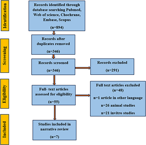
A total of seven publications including 88 intervention sites and 112 patients were included. By assessing evaluations from the baseline and follow-up procedures following the use of eggshell sourced hydroxyapatite as bone graft material, the assessment of the primary outcome aimed to determine whether there were any significant improvements in alveolar bone dimensions, density, and healing time.
Discussion
Recycling chicken eggshells benefits the environment by reducing waste and providing raw materials for nanomaterials development [8]. The majority (94%) of eggshells is made of calcium carbonate, with remainder consisting of organic matter (including insoluble proteins), magnesium carbonate, and calcium phosphate (4%, 1%, and 1%, respectively) [9, 10]. Other natural sources of hydroxyapatite, such as corals, cuttlefish, and oyster shells, as well as fish and cattle bones, have also been utilised to create biomaterials. But unlike eggshells, these sources are susceptible to depletion if they are continuously harvested, especially for slow-growing corals [8, 11].
While comparing the hydroxyapatite (HAp) made from eggshell to HAp made from other sources, the former has shown better outcomes in terms of density and hardness. The biological suitability of eggshell based HAp has been confirmed by a cytotoxicity test conducted utilising osteoblast cell culture [12]. It demonstrates that eggshell based HAp is noncytotoxic and promotes osteoblast cell attachment [12, 13]. The calcination temperature for eggshell-based HAp ranges up to 900°C. At this temperature, all disease-transmission concerns are eliminated by eliminating bacteria that can affect patients [12, 14].
In both human and animal models, eggshells have demonstrated potential as a bone regenerative material due to their high calcium content. EHA and EnHA have been shown to be very successful in promoting bone repair, providing a viable substitute for tissue regeneration. The results indicate eggshell-based materials, particularly in the nano-form, enhanced the Guided Bone Regeneration (GBR) process [15–18].
Socket preservation
In a prior study, bone width and radiographic density were measured at 1, 12, and 24 weeks after 23 sockets and 11 patients had been grafted with EnHA and coated with a platelet rich fibrin (PRF) membrane. Over the course of 24 weeks, the results revealed a substantial increase in bone density (p < 0.05), with 26.09% of sockets having a ground-glass look and 73.91% having a trabecular pattern. Micro-CT verified the creation of new trabecular bone, whereas histomorphometric analysis showed Grade-3 bone in 56.52% sockets, Grade-2 in 13.04% sockets, and Grade-1 in 8.70 sockets%. However, the absence of a control group was a major limitation [19].
In a split mouth randomized control trial (RCT),12 patients (24 extraction sites) were grafted with EnHA (12 sites) and the remaining sites with no graft after surgical removal of bilateral mandibular molars. Radiographic bone density was assessed at baseline, first, third, and sixth month; the analysis showed a significant increase in bone density in the graft group when compared to control group (p < 0.05), indicating complete bone healing in the graft group at the 3-month and 6-month recalls [20].
In a split mouth study, 40 extraction sites were equally divided and grafted with EnHA+PRF and the remaining sites with Demineralized Freeze-Dried Bone Allograft (DFDBA)+PRF. The dissimilarity in mean bone density involving the EnHA group and the DFDBA group was not statistically significant immediately post-grafting (p > 0.05), after the first month (p > 0.05), as well as after fourth months (p > 0.05). A significant difference was observed within the group at different intervals, showing an improvement in bone density [2].
Because of its high water permeability, low toxicity, stability, affordability, and ease of application, eggshell membrane has emerged as a potential biomaterial in recent years. In comparison to only a PRF membrane, eggshell membrane dramatically increases bone density and socket volume over a 6-month period while decreasing bone resorption [21]. Similarly, several authors determined that it contains excellent regenerative and osteogenic potential [22–27].
Intrabony defects
A study found that, at 6 months, the pedicle membrane with EnHA group and the EnHA alone group had significantly greater bone density and bone fill than the open flap debridement group in intrabony defects [28]. Opris et al. reported eggshell to be an attainable filler material for bone regeneration treatments, which may be utilised effectively either alone or in conjunction with PRF for alveolar preservation procedures [4]. Similarly, Wardana et al. found that, particularly after 6 months, eggshell produced full bone regeneration or improved bone density in proportion to the bovine group in the transplanted tooth region [29].
Maxillary cystic defects
After apicoectomy and/or cystic enucleation, eggshell was employed as a bone graft material in eight maxillary bone defects. By the eighth week, there was a noticeable increase in bone regeneration, with just a small amount of bone regeneration occurring in non-grafted locations [30].
Apicectomy
EHA dramatically increased bone regeneration between weeks 4 and 8, resulting in smooth wound healing and trabecular bone apparent by the 12th week [31]. In contrast, when comparing EHA with synthetic hydroxyapatite (SHA) at 1 and 6-month intervals, there was no bone regrowth. This might be because graft particles resorb either early or late [7].
However, the synthesis route and processing parameters of EHA play a critical role in determining its physicochemical and biological properties. Common synthesis techniques include wet chemical precipitation, hydrothermal processing, and calcination-based methods. Calcination temperatures ranging from 800°C to 900°C have been reported to influence crystallinity, phase purity, and particle size while ensuring sterilisation and elimination of organic contaminants. Higher calcination temperatures generally increase crystallinity but may reduce resorption rates, whereas lower temperatures yield poorly crystalline hydroxyapatite with enhanced biodegradability. Nano sized particles produced via hydrothermal or precipitation methods demonstrate superior bioactivity, improved osteoblast response, and faster bone regeneration. However, the absence of standardised synthesis protocols across studies limits direct comparison of functional outcomes. Establishing optimal synthesis parameters is essential for achieving predictable clinical performance [32].
Conclusion
EHA and its nano version are economical, environmentally benign, and biocompatible substitutes. In a variety of clinical applications, they successfully improve bone density and healing, and their results are on par with those of traditional grafts such as SHA and DFDBA. The comparable efficacy of EHA and EnHA to that of established graft materials, coupled with their non-cytotoxic nature and ability to support osteoblast activity, make them suitable for a wide range of applications including socket preservation, intrabony defect repair, and cystic defect management. Further, their environmentally friendly production from waste resources adds even more value.
Limitations
Only seven human clinical studies were eligible for inclusion, and several originated from the same research groups, which may introduce publication or investigator bias. The limited number of RCTs and the lack of long-term follow-up restrict the generalisability of the findings. In addition, heterogeneity in study design, defect types, follow-up periods, and outcome assessment methods prevented direct quantitative comparison. The absence of standardised preparation protocols for EHA further complicates interpretation of outcomes.
Future prospects
To fully realise the promise of eggshell-derived biomaterials in standard clinical practice, future research should concentrate on refining processing methods, identifying optimal therapeutic indications, and proving comparative efficacy with bigger, multi-centre trials.
Author contributions
Conceptualisation: KSVR, MNRG; Data Curation: KSVR, MNRG, NVSGS; Formal Analysis: KSVR, MNRG; Investigation: KSVR, MNRG, NVSGS; Methodology: KSVR, PSG; Software: MNRG, MVL; Validation: KSVR, PSG, NVSGS, PMK; Writing – Original Draft: KSVR, MNRG, NVSGS, PMK, KAK ; Writing – Review & Editing: KSVR, MNRG, MVL
Data availability statement
All the data related to the current review are available in the manuscript. No additional information is required to be available in other sources.
Ethics approval statement
Not applicable.
Patient consent statement
Not applicable.
References
[1] Dimitriou R, Jones E, McGonagle D, Giannoudis PV. Bone regeneration: current concepts and future directions. BMC Med. 2011;9:66. https://doi.org/10.1186/1741-7015-9-66
[2] Mkhize SC, Onwubu SC, Mokhothu TH, Mdluli PS, Mishra AK. Comparative assessment of the remineralization characteristics of nano-hydroxyapatite extracted from fish scales and eggshells. J Appl Biomater Funct Mater. 2023;21:22808000231180390. https://doi.org/10.1177/22808000231180390
[3] Nainoor N, Shobha ES, Prashanth NT, Rangan V, Malick R, Shaju N. Comparison of clinical and radiographic assessment of efficacy of eggshell-derived nano-hydroxyapatite (EnHA) and demineralized freeze-dried bone allograft (DFDBA), both covered with platelet-rich fibrin (PRF) as a barrier membrane in socket preservation: a randomized controlled clinical trial. J Maxillofac Oral Surg. 2026;25:225–37. https://doi.org/10.1007/s12663-024-02187-4
[4] Opris H, Bran S, Dinu C, Baciut M, Prodan DA, Mester A, et al. Clinical applications of avian eggshell-derived hydroxyapatite. Bosn J Basic Med Sci. 2020;20:430. https://doi.org/10.17305/bjbms.2020.4888
[5] Lee SW, Balázsi C, Balázsi K, Seo DH, Kim HS, Kim CH, et al. Comparative Study of hydroxyapatite prepared from seashells and eggshells as a bone graft material. Tissue Eng Regen Med. 2014;11:113–20. https://doi.org/10.1007/s13770-014-0056-1
[6] Lee SW, Kim SG, Balázsi C, Chae WS, Lee HO. Comparative study of hydroxyapatite from eggshells and synthetic hydroxyapatite for bone regeneration. Oral Surg Oral Med Oral Pathol Oral Radiol. 2012;113:348–55. https://doi.org/10.1016/j.tripleo.2011.03.033
[7] Kattimani V, Lingamaneni KP, Chakravarthi PS, Kumar TS, Siddharthan A. Eggshell-derived hydroxyapatite: a new era in bone regeneration. J Craniofac Surg.2016;27:112–7. https://doi.org/10.1097/SCS.0000000000002288
[8] Wu SC, Tsou HK, Hsu HC, Hsu SK, Liou SP, Ho WF. A hydrothermal synthesis of eggshell and fruit waste extract to produce nanosized hydroxyapatite. Ceram Int. 2013;39:8183–8. https://doi.org/10.1016/j.ceramint.2013.03.094
[9] Li-Chan EC, Kim HO. Structure and chemical composition of eggs. Egg bioscience and biotechnology. 2008;10:9780470181249.
[10] Rivera EM, Araiza M, Brostow W, Castano VM, Dıaz-Estrada JR, Hernández R, et al. Synthesis of hydroxyapatite from eggshells. Mater Lett. 1999;41:128–34. https://doi.org/10.1016/S0167-577X(99)00118-4
[11] Sanosh KP, Chu MC, Balakrishnan A, Kim TN, Cho SJ. Utilization of biowaste eggshells to synthesize nanocrystalline hydroxyapatite powders. Mater Lett. 2009;63:2100–2. https://doi.org/10.1016/j.matlet.2009.06.062
[12] Abdulrahman I, Tijani HI, Mohammed BA, Saidu H, Yusuf H, Ndejiko Jibrin M, et al. From garbage to biomaterials: an overview on egg shell based hydroxyapatite. J Mater. 2014;2014:802467. https://doi.org/10.1155/2014/802467
[13] Siva Rama Krishna D, Siddharthan A, Seshadri SK, Sampath Kumar TS. A novel route for synthesis of nanocrystalline hydroxyapatite from eggshell waste. J Mater Sci. 2007;18:1735–43. https://doi.org/10.1007/s10856-007-3069-7
[14] Han Y, Xu K, Montay G, Fu T, Lu J. Evaluation of nanostructured carbonated hydroxyapatite coatings formed by a hybrid process of plasma spraying and hydrothermal synthesis. J Biomed Mater Res. 2002;60:511–6. https://doi.org/10.1002/jbm.10097
[15] Brostow W, Lobland HE. Materials: introduction and applications. John Wiley & Sons; 2016.
[16] Wood RA, Mealey BL. Histologic comparison of healing after tooth extraction with ridge preservation using mineralized versus demineralized freeze-dried bone allograft. J Periodontol. 2012;83:329–36. https://doi.org/10.1902/jop.2011.110270
[17] Ho-Shui-Ling A, Bolander J, Rustom LE, Johnson AW, Luyten FP, Picart C. Bone regeneration strategies: engineered scaffolds, bioactive molecules and stem cells current stage and future perspectives. Biomaterials. 2018;180:143–62. https://doi.org/10.1016/j.biomaterials.2018.07.017
[18] Grover V, Malhotra R, Kapoor A, Sachdeva S. Bone allografts: a review of safety and efficacy. Indian J Dent Res. 2011;22:496. https://doi.org/10.4103/0970-9290.87084
[19] Kattimani VS, Lingamaneni KP, Kreedapathi GE, Kattappagari KK. Socket preservation using eggshell-derived nano-hydroxyapatite with platelet-rich fibrin as a barrier membrane: a new technique. J Korean Assoc Oral Maxillofac Surg. 2019;45:332–42. https://doi.org/10.5125/jkaoms.2019.45.6.332
[20] Kattimani V, Lingamaneni KP, Yalamanchili S, Mupparapu M. Use of eggshell-derived nano-hydroxyapatite as novel bone graft substitute – a randomized controlled clinical study. J Biomater Appl. 2019;34:597–614. https://doi.org/10.1177/0885328219863311
[21] Rachna M, Nandita S, Rashmi KS, Vidya GB, Srikant N, Ananya J, et al. Eggshell membrane as a regenerative material in alveolar bone grafting in combination with advanced platelet rich fibrin. Clin Ter. 2024;175:219–25.
[22] Chen X, Chen Y, Fu B, Li K, Huang D, Zheng C, et al. Eggshell membrane-mimicking multifunctional nanofiber for in-situ skin wound healing. Int J Biol Macromol. 2022;210:139–51. https://doi.org/10.1016/j.ijbiomac.2022.04.212
[23] Sah MK, Banerjee I, Pramanik K. Eggshell membrane protein modified silk fibroin-poly vinyl alcohol scaffold for bone tissue engineering: in vitro and in vivo study. JBBBE.2017;32:69–81. https://doi.org/10.4028/www.scientific.net/JBBBE.32.69
[24] Ruff KJ, Morrison D, Duncan SA, Back M, Aydogan C, Theodosakis J. Beneficial effects of natural eggshell membrane versus placebo in exercise-induced joint pain, stiffness, and cartilage turnover in healthy, postmenopausal women. Clin Interv Aging. 2018;13:285–95. https://doi.org/10.2147/CIA.S153782
[25] Kiers JL, Bult JH. Mildly processed natural eggshell membrane alleviates joint pain associated with osteoarthritis of the knee: a randomized double-blind placebo-controlled study. J Med Food. 2021;24:292–8. https://doi.org/10.1089/jmf.2020.0034
[26] Mensah RA, Jo SB, Kim H, Park SM, Patel KD, Cho KJ, et al. The eggshell membrane: a potential biomaterial for corneal wound healing. J Biomater Appl. 2021;36:912–29. https://doi.org/10.1177/08853282211024040
[27] Gokul MNR, Ramesh KS, Gautami PS, Sruthima NVS, Kumar PM, Anilkumar K, et al. A comparative evaluation of eggshell derived hydroxyapatite and demineralized freeze-dried bone allograft in alveolar ridge preservation – a randomized clinical trial. Biomater Investig Dent. 2025;12:44941. https://doi.org/10.2340/biid.v12.44941
[28] Vani TMS, Paramashivaiah R, Prabhuji MLV, Peeran SW etal. Regeneration of intrabony defects with nano-hydroxyapatite graft, derived from eggshell along with periosteum as barrier membrane under magnification – an interventional study. Appl Sci. 2023;13:1693. https://doi.org/10.3390/app13031693
[29] Wardana HA, Idulhaq M, Saputra RD, Ermawan R, Roshada MF. Potential use of eggshell as bone graft compared with bovine for bone defect: a systematic review study. Open-Access Maced J Med Sci. 2021;9:470–3. https://doi.org/10.3889/oamjms.2021.7103
[30] Kattimani VS, Chakravarthi PS, Kanumuru NR, Subbarao VV, Sidharthan A, Kumar TS, et al. Eggshell derived hydroxyapatite as bone graft substitute in the healing of maxillary cystic bone defects: a preliminary report. J Int Oral Health. 2014;6:15–9.
[31] Kattimani VS, Lingamaneni KP. Natural bioceramics: our experience with changing perspectives in the reconstruction of maxillofacial skeleton. J Korean Assoc Oral Maxillofac Surg. 2019;45:34–42. https://doi.org/10.5125/jkaoms.2019.45.1.34
[32] Muñoz-Sanchez ER, Arrieta-Gonzalez CD, Quinto-Hernandez A, Garcia-Hernandez E, Porcayo-Calderon J. Synthesis of hydroxyapatite from eggshell and its electrochemical characterization as a coating on titanium. Int J Electrochem Sci. 2023;18:100204. https://doi.org/10.1016/j.ijoes.2023.100204