CASE REPORTS
DEFINING PATIENT PROFILES AFTER THE 2023 KAHRAMANMARAŞ, TURKEY EARTHQUAKE
Tuba TÜLAY KOCA, MD *
*
From the Department of Physical Medicine and Rehabilitation, Sütçü İmam University, Faculty of Medicine, Kahramanmaraş, Turkey
Objective: On 6 February 2023, 2 earthquakes with magnitudes of 7.7 and 7.6 mW occurred in Pazarcık and Elbistan districts of Kahramanmaraş province (Turkey) and affected 11 provinces in total, especially Hatay, Malatya, and Adıyaman. Here, we report 3 earthquake victims in order to define patients profiles.
Case reports: Three patients who were taken to the orthopedic and neurological rehabilitation program in our inpatient Physical Medicine and Rehabilitation unit after the earthquake are presented. Our first case, a 46-year-old man, remain-ed in the debris for 8.5 h. His left leg was left in the wreckage, and he later developed a drop foot on the left due to compartment syndrome, which developed with pain and swelling in the left calf. There is no fasciotomy procedure. Venous Doppler was reported to be within normal limits. Our second case is a 52-year-old man who was in the debris for 36 h and has a left braxial plexus injury and a left drop foot. He has Buerger’s disease in his medical history. The third case is an 8-year-old girl trapped in rubble during the earthquake with a right orbital fracture and a left foot transmetarsal amputation. She has no neurological signs.
Discussion: The patients that apply to our Physical Medicine and Rehabilitation clinic are especially amputees, patients with traumatic brain injury, spinal cord injury, peripheral nerve damage, plexus damage, multiple fractures, joint limitations and soft tissue loss in the musculoskeletal system. After the earthquake, we encountered patients from a wide variety of spectrums in our clinic. In addition to primary musculoskeletal injuries, they also experience systemic problems affecting the musculoskeletal system. Additionally, thrombosis, infection, renal failure, and multiple organ failures may also occur.
Conclusion: Countries should develop guidelines for disaster preparedness and establish coordination units that can take quick action, make decisions, and communicate in times of disaster. A good definition of patient clinical profiles after the earthquake will ensure early intervention and prevent permanent disability and functional losses.
LAY ABSTRACT
There are devastating earthquake disasters that have massive effects on the world. On 6 February 2023, 2 earthquakes with magnitudes of 7.7 and 7.6 mW occurred in Turkey. According to the latest official data, the incident resulted in at least 45,968 deaths and 108,368 injuries, with 241,577 damaged buildings. Here, we described the patient profiles of our Physical Medicine and Rehabilitation Clinic after the earthquake and shared our experiences. A good definition of earthquake experiences will guide preparation for subsequent earthquakes.
Key words: earthquake; rehabilitation; nerve injuries; plexus injuries.
Citation: JRM-CC 2024; 7: jrmcc40039. DOI: https://doi.org/10.2340/jrmcc.v7.40039
Copyright: © Published by Medical Journals Sweden, on behalf of the Foundation for Rehabilitation Information. This is an Open Access article distributed under the terms of the Creative Commons Attribution 4.0 International License (https://creativecommons.org/licenses/by/4.0/)
Submitted: Feb 2, 2024; Accepted: Feb 22, 2024; Published: Apr 2, 2024
Correspondence address: Tuba Tülay Koca, Department of Physical Medicine and Rehabilitation, Sütçü İmam University, Faculty of Medicine, Kahramanmaraş, Turkey. E-mail: tuba_baglan@yahoo.com
Competing interests and funding: The author has no conflicts of interest to declare.
On 6 February 2023, 2 devastating earthquakes with magnitudes of 7.7 and 7.6 mW occurred, shaking Kahramanmaraş and 10 provinces around it. Approximately 2 million people were affected by these earthquakes (1). In 2023, when disasters hit, countries were caught unprepared for disaster coordination. Apart from earthquakes, we also encountered other disasters such as floods, volcanic eruptions, and forest fires in 2023.
Musculoskeletal problems such as bone fractures, soft tissue damage, peripheral artery and nerve injuries, plexus injuries, compartment syndrome, and amputation are observed in patients after the earthquake. Musculoskeletal complaints such as neck and low back pain, muscular strain/sprain related to housing problems after the earthquake are also seen. Functional disability is frequently encountered after natural disasters. Complaints in the musculoskeletal system are frequently encountered with or without musculoskeletal injury (2).
A good definition of earthquake experiences will provide guidance for preparation for subsequent earthquakes. Health needs and patient profiles in acute, subacute, and chronic periods vary. Here, we aimed to contribute to the definition of the patient profile that we may encounter after the earthquake by presenting 2 of our earthquake victim.
CASE REPORTS
Three patients who were taken to the orthopedic and neurological rehabilitation program in our inpatient Physical Medicine and Rehabilitation unit after the earthquake are presented. Clinical examinations of the patients were performed by the same experienced physiatrist. Electroneurophysiological reports were accessed from archive records. The patients were included in the physiotherapy program in the inpatient service. Informed consent forms were obtained from both patients.
Our first case, a 46-year-old man, remained in the debris for 8.5 h. He lives in a country house with 2 stories in the village. His left leg was crushed in the debris. He complained of pain, numbness, and numbness in his leg. Venous Doppler was found to be within normal limits. The pulses are taken. He has no associated injury. He has uncontrolled diabetes mellitus in his medical history. No fasciotomy procedure was performed.
The patient describes loss of strength in the left ankle after 3–4 months. The electroneuromyography report was consistent with left severe partial sciatic neuropathy (tibial and peroneal).
On physical examination, left foot dorsiflexion was 2/5 (Fig. 1) according to manuel muscle test, left L5–S1 dermatome was dysesthesic. There was no difference in diameter between the extremities below or above the knee.
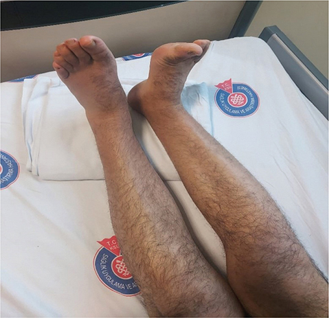
Fig. 1. He has drop foot on the left.
The patient was included in a neurological rehabilitation program including electrostimulation, transcutaneous electrical nerve stimulation (TENS) and exercises, and oral vitamin B supplementation was started for neuropathic pain. A reflex ankle foot orthosis (AFO) was prescribed for his left lower extremity. At the end of 5-week rehabilitation, his functional ambulation scale (FAS) with AFO was 5. Left foot dorsiflexor muscle strength was 3+/5 according to the manual muscle test.
Neuronal ischemic damage due to compartment syndrome was considered in the patient. Compartment syndrome is characterized by damage to the vessels and nerves surrounded by muscle and fascia in the extremity due to increased intra-tissue pressure as a result of crush injury. We may encounter symptoms such as severe pain, pallor, paleness in the extremities, coldness, immobility, absent pulse, and loss of sensation in the nerves (3, 4).
Early diagnosis and timing of fasciotomy are critical to reduce intracompartmental pressure. Rapid rescue of earthquake victims from the debris, transfer with appropriate equipment, and intervention by experienced surgeons play a role in preventing future physical disabilities, survival, and mortality (5, 6). An extremity-sparing surgical procedure in which the fascia is cut to reduce compartment pressure and thus relieve circulation. Skin graft. Recovery may take several months.
The specialist’s experience, fasciotomy decision and time are effective in saving the extremity. In order to save the extremity, the risk of late fasciotomy and crush syndrome development should be carefully evaluated. Fasciotomy risks are neurovascular damage, soft tissue infection, sepsis, surgical scar, and limitation of joint movement due to scar tissue. Abrasions and necrotic wounds due to crushing are common. Antiseptics, debridement, and negative pressure wound therapy are the most commonly applied wound care methods (7, 8).
Our second case is a 52-year-old man who was stuck in the debris for 36 h and has loss of strength in his left arm and left foot dorsiflexors (Fig. 2). He has Buerger disease in his medical history. In the physical examination of the patient, atrophy was observed in the volar side muscle groups of the left forearm. Left forearm and wrist flexor muscle strength were 3/5 in manuel muscle test. Finger adductor and flexor muscle strength were 2+/5. In the electrophysiological evaluation (21 February 2023), there were neuropathies in ulnar, radial, musculocutaneous and axillary nerves, mostly in the left median nerve; axonal loss in all cords, especially in the lateral cord, in the infraclavicular distal part of the left brachial plexus. We included our patient in a 6-week neurological rehabilitation program and organized a program that included electrostimulation and strengthening exercises for the left upper extremity. Suprascapular nerve block, 1% lidocaine, was applied for pain. According to the visual analog scale, his pain decreased from 8 cm to 5 cm. Control left upper extremity arterial Doppler was normal.
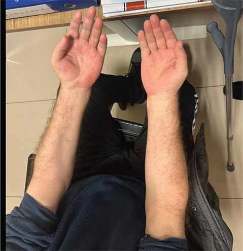
Fig. 2. He has left brachial plexus palsy.
Brachial/lumbosacral plexus or peripheral nerve lesions are common in earthquake victims. These lesions may cause temporary or lifelong disability. These lesions can occur not only due to being trapped under debris (compression), but also due to disruptions during rescue or transportation. Compartment or crush syndrome may often develop in these patients. It is known that nerve degeneration is more common in people who stay under debris for a long time. It has been reported in the literature that neuropraxia and axonomethesis are more common in the electrophysiological findings of these patients, and that signs of regeneration are observed during follow-ups with good prognosis (9–11).
Our third case is an 8-year-old girl who was trapped in the debris during the earthquake. She has a right orbital fracture and a proximal transmetarsal amputation of her left foot. There is a joint range of motion of 20 degrees. There is no accompanying neurological finding. Cranial Magnetic Resonance Imaging (MRI) is normal. On physical examination, there is a shortness in the Achilles and the joint does not come to neutral (Fig. 3). A permanent prosthesis was planned. Orthopedics was consulted for Achilles release. Pre-prosthetic training and skin care were provided. It was applied after a 3-week amputee rehabilitation program. Prosthesis use and stump care training were given.
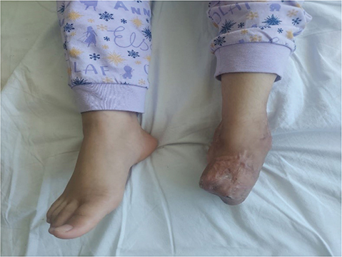
Fig. 3. She has a proximal transmetarsal amputation of her left foot.
DISCUSSION
The patients that apply to our Physical Medicine and Rehabilitation clinic are especially amputees, patients with traumatic brain injury, spinal cord injury, peripheral nerve damage, plexus damage, multiple fractures, joint limitations, and soft tissue loss in the musculoskeletal system. Our main goal in rehabilitation is to evaluate patients with their accompanying problems and include them in a comprehensive rehabilitation program. When necessary, opinions from different disciplines should be sought. In the early period, patient profiles should be well defined, and rehabilitation needs should be determined. Countries should develop guidelines for disaster preparedness and establish coordination units that can take quick action, make decisions and communicate in times of disaster. The health department of these units should include the chief coordinator, physicians, nurses, secretaries, technicians, and data processors, and a good recording system should be kept. Determining the needs well and taking coordinated action will reduce both morbidity and mortality. For this reason, good definition of patient clinical profiles after the earthquake will ensure early intervention and prevent permanent disability and functional losses.
REFERENCES
- Uluöz M, Gökmen MY. The 2023 Turkey earthquake: management of 627 pediatric musculoskeletal injuries in the first month. Children (Basel) 2023; 10: 1733. https://doi.org/10.3390/children10111733
- Yabe Y, Hagiwara Y, Sekiguchi T, Sugawara Y, Tsuchiya M, Itaya N, et al. Musculoskeletal pain and new-onset poor physical function in elderly survivors of a natural disaster: a longitudinal study after the great East Japan earthquake. BMC Geriatr 2019; 19: 274. https://doi.org/10.1186/s12877-019-1283-z
- Long B, Liang SY, Gottlieb M. Crush injury and syndrome: a review for emergency clinicians. Am J Emerg Med 2023; 69: 180–187. https://doi.org/10.1016/j.ajem.2023.04.029
- Genthon A, Wilcox SR. Crush syndrome: a case report and review of the literature. J Emerg Med 2014; 46: 313–319. https://doi.org/10.1016/j.jemermed.2013.08.052
- Görmeli G, Görmeli CA, Güner S, Ceylan MF, Dursun R. [A clinical analysis of patients undergoing fasciotomy who experienced the 2011 Van earthquake]. Eklem Hastalik Cerrahisi 2012; 23: 156–160 (In Turkish).
- Lin YM, Lee TS. Fasciotomy in crush syndrome patients: debates continue. Emerg Med J 2005; 22: 78.
- Kundakci B, Mirioglu A, Tekin M, Bagir M, Bicer OS, Arslan YK, et al. 6 February 2023, orthopedic experience in Kahramanmaraş earthquake and surgical decision in patients with crush syndrome. J Orthop Surg Res 2023; 18: 537. https://doi.org/10.1186/s13018-023-04001-2
- Ulusoy S, Kılınç İ, Oruç M, Özdemir B, Ergani HM, Keskin ÖH, et al. Analysis of wound types and wound care methods after the 2023 Kahramanmaras earthquake. Jt Dis Relat Surg 2023; 34: 488–496. https://doi.org/10.52312/jdrs.2023.1128
- Ahrari MN, Zangiabadi N, Asadi A, Sarafi Nejad A. Prevalence and distribution of peripheral nerve injuries in victims of Bam earthquake. Electromyogr Clin Neurophysiol. 2006; 46: 59–62
- Uzun N, Kiziltan ME, Savrun FK. 1999 Marmara earthquake; peripheral nerve injuries – electrophysiologic evaluation. Electromyogr Clin Neurophysiol 2005; 45: 3–13.
- Yoshida T, Tada K, Uemura K, Yonenobu K. Peripheral nerve palsies in victims of the Hanshin-Awaji earthquake. Clin Orthop Relat Res 1999; 362: 208-217. https://doi.org/10.1097/00003086-199905000-00030.