ORIGINAL REPORT
EFFECTS OF LOCAL VIBRATION COMBINED WITH EXTRACORPOREAL SHOCK WAVE THERAPY IN PLANTAR FASCIITIS: A RANDOMIZED CONTROLLED TRIAL
HyoJeong ON, MSc and JongEun YIM, DSc
From the Department of Physical Therapy, The Graduate School of Sahmyook University, Seoul, Republic of Korea.
Objective: To compare the effects of local vibration combined with extracorporeal shock wave therapy and extracorporeal shock wave therapy alone for plantar fasciitis.
Methods: A randomized controlled trial including 34 participants with a mean age of 37.5 years. Participants were randomly allocated to a group treated with extracorporeal shock wave therapy combined with local vibration (ESWT-LV group) or a group treated with extracorporeal shock wave therapy alone (ESWT-alone group). All participants in each group underwent 2 treatment sessions weekly for 5 weeks. Thickness of the plantar fascia, plantar pain, and foot function were measured using ultrasonography, numerical rating scale for pain, and Foot Function Index, respectively, at baseline and at the end of the 5-week intervention.
Results: Significant improvements were measured in both groups in the thickness of the plantar fascia, numerical rating scale, and Foot Function Index values (p < 0.05). In addition, the thickness of the plantar fascia and pain was significantly more decreased in the ESWT-LV group than in the ESWT-alone group (p < 0.05). However, the differences between groups in Foot Function Index values were not significant (p > 0.05).
Conclusion: Local vibration combined with extracorporeal shock wave therapy is an effective treatment for chronic plantar fasciitis.
LAY ABSTRACT
The aim of this study was to determine the efficacy of local vibration combined with extracorporeal shock wave therapy compared with extracorporeal shock wave therapy alone for treatment of plantar fasciitis. The study included 34 participants with a mean age of 37.5 years. The thickness of the plantar fascia, plantar pain, and foot function were measured using ultrasonography, a numerical rating scale, and Foot Function Index respectively, at the beginning and end of a 5-week intervention. The participants were randomly divided into 2 groups: 1 received extracorporeal shock wave therapy and local vibration treatment (ESWT-LV group), while the other received extracorporeal shock wave therapy alone (ESWT-alone group). Both groups underwent 2 treatment sessions weekly for 5 weeks. The results showed significant improvements in the thickness of the plantar fascia, pain level, and foot function in both groups. However, the thickness of the plantar fascia and pain were significantly more decreased in the ESWT-LV group compared with ESWT alone, while the improvement in Foot Function Index values did not differ significantly between groups. In conclusion, local vibration combined with extracorporeal shock wave therapy is an effective treatment for chronic plantar fasciitis.
Key words: plantar fasciitis; local vibration; extracorporeal shock wave therapy; ultrasonography.
Citation: J Rehabil Med 2023; 55: jrm12405. DOI: https://doi.org/10.2340/jrm.v55.12405
Copyright: © Published by Medical Journals Sweden, on behalf of the Foundation for Rehabilitation Information. This is an Open Access article distributed under the terms of the Creative Commons Attribution-NonCommercial 4.0 International License (https://creativecommons.org/licenses/by-nc/4.0/)
Accepted: Sept 26, 2023; Published: Oct 23, 2023
Correspondence address: JongEun Yim, Department of Physical Therapy, The Graduate School of Sahmyook University, 815 Hwarang-ro, Nowon-gu, Seoul 01795, Republic of Korea. E-mail: jeyim@syu.ac.kr
Competing interests and funding: The authors have no conflicts of interest to declare.
Plantar fasciitis is the commonest heel pain disorder, affecting > 2 million Americans annually (1). Clinical symptoms of plantar fasciitis include foot pain, specifically in the heel. Pain is usually worse when taking the first step in the morning, than in the afternoon, and worsens with inactivity, prolonged standing, and weight-bearing activities (2).
Some matched case-control studies have reported that a decrease in the range of ankle dorsiflexion, prolonged weight bearing, and a body mass index (BMI) > 30 kg/m2 are risk factors for plantar fasciitis (2). Plantar fasciitis is mostly treated conservatively because people want to return to work and daily activities as soon as possible (3). Treatment includes the administration of non-steroidal anti-inflammatory drugs, Achilles tendon stretching, night splint therapy, steroid injections, and extracorporeal shock wave therapy (ESWT) (4).
ESWT is widely used among these conservative treatment options in clinical settings, especially for patients with chronic plantar fasciitis. A 2017 meta-analysis of 9 randomized controlled trials with placebo treatment as the comparator concluded that focused application of ESWT should be recommended after the failure of conventional conservative therapy (5). According to Weil et al., the mechanism of pain relief is attributed to the release of enzymes, specifically targeting the nociceptors (6). Some researchers have likened an analogy of creating microtrauma to the tissues with an effect similar to tenderizing meat (7). Furthermore, during ESWT, the external stimulation increases the angiogenic factors, endothelial nitric oxide synthase, and the vessel endothelial growth factor, which increases the blood supply and induces neovascularization and tissue proliferation to hasten healing of the degenerative tissues in plantar fasciitis (8).
Similarly, vibration is known to increase blood flow (9). Some studies have reported that vibration treatment has acute effects that promote oxygen consumption and blood flow in the skin (10). According to some studies, vibration treatment is also an effective way to alleviate pain (11, 12). Patients and athletes commonly use local vibration (LV) for recovery because it reduces pain, delayed onset of muscular soreness, and fatigue. (13).
Vibration treatment can be considered an adjunct to ESWT, since the vibration may augment ESWT due to its potential in decreasing pain and increasing blood flow, which can be further amplified in combination with ESWT. To the best of our knowledge, there have been only 3 studies on plantar fasciitis treatment using a LV device (14, 15). However, 2 of these were sponsored by a company and focused on their own wearable vibration devices. An earlier study examined the use of LV for plantar fascia as a specialized treatment used as an adjunct for stretching of the triceps surae muscles.
The aim of the current study was to investigate the effects of LV combined with ESWT for treatment of plantar fasciitis, compared with ESWT alone.
METHODS
Subjects
The sample size was estimated using version 3.1.9.2 of the G*Power program (Heinrich-Heine-Universität Düsseldorf, Düsseldorf, Germany). According to a previous study on the efficacy of ESWT for plantar fasciitis (16), the effect size of plantar thickness was calculated to be 0.88. Therefore, the effect size of this study was also set at 0.88. The alpha level was set at 0.05, and the statistical power at 0.80. Thus, it was determined that at least 17 patients were required in each group. The physical therapist recruited patients with plantar fasciitis for the study. The inclusion criteria were: (i) patients aged between 20 and 60 years, (ii) had symptoms for at least 16 weeks before being diagnosed with plantar fasciitis by a medical doctor at a pain clinic (Seoul, South Korea), (iii) remained symptomatic after at least 3 months of conservative treatment, and (iv) had adequate educational background to understand the assessments and interventions. A total of 34 participants (17 men, 17 women), between the ages of 20 and 60 years volunteered for the study at the pain clinic. Prior to initiation of the study, each patient was randomly assigned using computer software (Excel 2010; Microsoft, Redmond, USA) to either the ESWT-LV or ESWT alone group. The exclusion criteria were: (i) refusal of ESWT and routine physical therapy, (ii) history of plantar fascia surgery, (iii) use of anti-inflammatory analgesic during the study, and (iv) had received manual or LV therapy on the foot and calf.
The study was approved by Sahmyook University Institutional Review Board (2-1040781-A-N-012020120HR). The aims and study procedures were explained to the participants before the commencement of the study. All participants provided written consent, in line with the principles of the Declaration of Helsinki. This study was registered at the Clinical Research Information Service (CRIS) (KCT0008634).
Experimental procedure
The thickness of the affected side of the plantar fascia, pain, and foot function were determined. Plantar fascia thickness was assessed using ultrasonography, while the pain was assessed using a numerical rating scale (NRS). Foot function was determined using the Foot Function Index (FFI). All assessments were conducted blind by the same researcher at baseline (0 weeks) and 1 week after (6 weeks) completing the 5-week interventions.
After the pre-experimental data were collected, the ESWT-LV and ESWT-alone groups received ESWT twice weekly for 5 weeks. At each session, while in the recommended prone position, 2,000 pulses with an air pressure of 3 bar were applied for 4 min (equal to a positive energy flux density of 0.12 mJ/mm2) on the point of maximal tenderness in the region of the median calcaneal tubercle. For the ESWT-LV group participants, after they had received ESWT, they additionally underwent LV therapy. At each session, while in the prone position, 30 Hz of LV was applied to the same area for 3 min (Fig. 1).
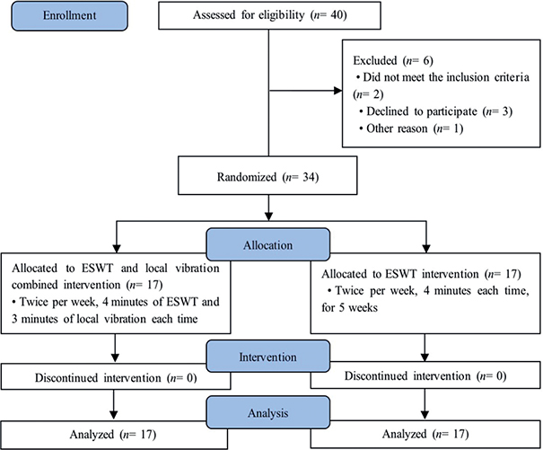
Fig. 1. Study flow diagram. ESWT: extracorporeal shockwave therapy.
Intervention
Extracorporeal shock wave therapy (ESWT). After applying the specific ultrasound coupling gel, a radial ESWT gun (OPTIMUS-Pro, REMED Co., Seongnam, Korea) was positioned on the point of maximal tenderness in the region of the medial calcaneal tubercle, as determined by the participants. During ESWT, the participants were positioned comfortably on a bed in the prone position with their feet hanging freely over the edge. Based on a previous study by Rompe et al. (17), at each session, 2,000 pulses with an air pressure of 3 bar were applied (equivalent to a positive energy flux density of 0.12 mJ/mm2). Therefore, the total positive energy flux density per treatment was 320 mJ/mm2. The treatment frequency was 9 pulses per second, and no local anaesthesia was administered.
Local vibration (LV). As for the LV treatment, a vibratory device (Massage gun, Liwell, Korea) was positioned on the point of maximal tenderness in the median calcaneal tubercle region. Participants were positioned comfortably on a bed in the prone position with their feet hanging freely over the edge. Based on a previous study (10, 18), a systematic review of 22 studies by Mahbub et al. (19), and the manufacturer’s instructions, the speed 3 mode (frequencies ≤ 30 Hz, 1,100 rpm, and 12-mm amplitude) was activated for 3 min, which created a pulsing effect, and no local anaesthesia was administered.
Outcome measures
Plantar fascia thickness. To measure plantar fascia thickness, all participants underwent B-mode ultrasound imaging evaluation using a high-resolution 10-MHz linear probe (SONON, Healcerion, Seongnam, South Korea 2019), according to the guidelines issued by the European Society of Musculoskeletal Radiology (20). Prior to the assessment, participants’ heel pads were gently wiped with alcohol swabs. Subsequently, while the patients were in the prone position, the plantar fascia was evaluated at full knee extension and at 90° dorsiflexion of the ankle. For increased accuracy, the median calcaneal tubercle was highlighted with a marker that is harmless to humans. In addition, a sufficient amount of gel was applied to minimize the probe’s friction on the skin. According to a 2005 study by Ozdemir et al. (21) the thickness was measured at 10 mm distal to the calcaneal insertion of the plantar aponeurosis from the medial aspect on the sagittal plane. To ensure the accuracy of the measurements, all ultrasound examinations were performed by the same researcher. In addition, a beam, located perpendicularly to the plantar fascia, was maintained at all times to avoid artefactually reduced echogenicity due to incident beam obliquity (22). Measurements were repeated twice for each foot. The sonography of the plantar fascia was saved directly into the application (SONON, Healcerion) of the tablet PC and measured with the measure function in the application. The mean value was then recorded (Fig. 2).
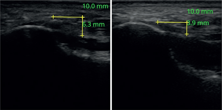
Fig. 2. Ultrasonographic longitudinal appearance (linear scanner) of the plantar fascia thickness. Symptomatic side (left) and asymptomatic side (right). With the yellow horizontal line as the baseline, the vertical line represents the thickness of the plantar fascia.
Pain. Numerical rating scales (NRS) is the simplest and most commonly used pain measurement scale. NRS is an 11-point scale with integers ranging from 0 to 10 (0 representing “No pain” and 10 representing “The worst imaginable pain”). The patient would then select a single number that best characterized their pain intensity (23).
Foot function. Functional assessment was performed using the Foot Function Index (FFI). This questionnaire measures the impact of foot pathology on function in terms of 3 categories: pain, disability, and activity restriction. The pain category, consisting of 9 items, was recorded using the VAS. The disability category, which described the difficulty experienced by the patients, also consisted of 9 items. The activity restriction category consisted of 5 items that focused on activity restrictions experienced by patients with foot pain. For the total score, the minimum score is 0% (no pain or difficulty), and the maximum score is 100% (worst pain and extreme difficulty requiring assistance). The total FFI equals the mean score of the 3 subscale scores. The subscale scores for pain, disability, and activity restriction range from 0 to 100.
Statistical analysis
All statistical analyses were performed using the Statistical Package for the Social Sciences version 22.0 for Windows (version 22, IBM, New York, USA). The normal distribution of samples was tested using the Shapiro–Wilk test. Furthermore, the pre-and post-plantar fascia thicknesses were calculated and assessed using the paired t-test, and the numerical rating scale and FFI values were calculated and assessed using the Wilcoxon signed-rank test to determine whether there was a significant difference between the variable after each intervention. In addition, the independent t-test and Mann–Whitney U test were used to determine if there was a significant difference between the ESWT-LV and ESWT alone groups for the variables listed above. The significance level was set at a p-value < 0.05.
RESULTS
Comparison of thickness of plantar fascia
The ESWT-LV group decreased significantly from 6.13 ± 1.82 mm to 5.39 ± 1.64 mm (p < 0.05). The ESWT-alone group showed a statistical significant decrease from 5.44 ± 1.54 mm to 4.97 ± 1.43 mm (p < 0.05). In the intergroup comparison, the differences in the values of plantar fascia thickness were significantly greater in the ESWT-LV group than in the ESWT-alone group (p < 0.05) (Table I, Fig. 3).
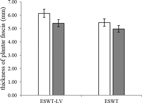
Fig. 3. Comparison of plantar fascia thickness between pre- and post-test. White bars: pre-test; grey bars: post-test. Error bars represent percentage. ESWT-LV: extracorporeal shockwave therapy and local vibration combined group; ESWT-alone: extracorporeal shockwave therapy alone group.
Comparison of pain
The NRS results showed a statistically significant decrease from 7.05 ± 1.08 to 2.41 ± 0.87 points in the ESWT-LV group and from 6.94 ± 1.43 to 3.05 ± 0.96 points in the ESWT-alone group (p < 0.05). In the intergroup comparison, the differences in the NRS were significantly greater in the ESWT-LV group than in the ESWT-alone group (p < 0.05) (Table II, Fig. 4).
| ESWT-LV | ESWT-alone | Za (p) | |
| Pre-test, mean ± SD | 7.06 ± 1.09 | 6.94 ± 1.44 | –0.270 (0.787) |
| Post-test, mean ± SD | 2.41 ± 0.87 | 3.06 ± 0.97 | |
| Post–Pre, mean ± SD | –4.65 ± 0.99 | –3.88 ± 0.99 | –1.988 (0.047) |
| Zb (p) | –3.655 (0.000) | –3.691 (0.000) | |
| aMann–Whitney U test. bWilcoxon signed-rank test. SD: standard deviation; ESWT-LV: extracorporeal shockwave therapy and local vibration combined group; ESWT-alone: extracorporeal shockwave therapy alone group. |
|||
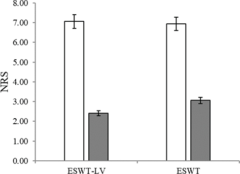
Fig. 4. Comparison of numerical rating scale (NRS) between pre- and post-test. White bars: pre-test; grey bars: post-test. Error bars represent percentage. ESWT-LV: extracorporeal shockwave therapy and local vibration combined group; ESWT-alone: extracorporeal shockwave therapy alone group.
Comparison of foot function
The FFI results decreased significantly from 61.70 ± 12.33 to 51.82 ± 12.86 points in the ESWT-LV group and from 57.29 ± 13.13 to 47.00 ± 11.82 points in the ESWT-alone group (p < 0.05). In the intergroup comparison, however, there was no significant difference between the groups (p = 0.73) (Table III, Fig. 5).
| ESWT-LV | ESWT-alone | Za (p) | |
| Pre-test, mean ± SD | 61.70 ± 12.33 | 57.29 ± 13.13 | –0.862 (0.389) |
| Post-test, mean ± SD | 51.82 ± 12.86 | 47.00 ± 11.82 | |
| Post–Pre, mean ± SD | –9.88 ± 3.03 | –10.29 ± 3.82 | –0.607 (0.544) |
| Zb (p) | –3.633 (0.000) | –3.629 (0.000) | |
| aMann–Whitney U test. bWilcoxon signed-rank test. SD: standard deviation; ESWT-LV: extracorporeal shockwave therapy and local vibration combined group; ESWT-alone: extracorporeal shockwave therapy alone group. |
|||
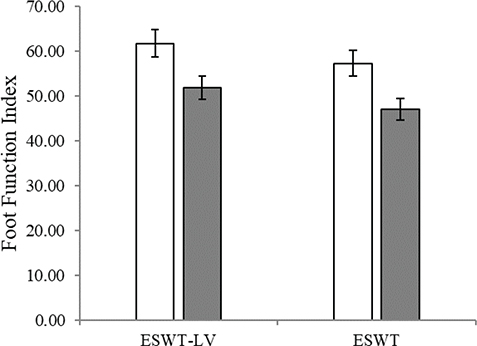
Fig. 5. Comparison of Foot Function Index between pre- and post-test. White bars: pre-test; grey bars: post-test. Error bars represent percentage. ESWT-LV: extracorporeal shockwave therapy and local vibration combined group; ESWT-alone: extracorporeal shockwave therapy alone group.
DISCUSSION
The aim of this study was to investigate the effects of LV combined with ESWT on plantar fascia thickness, pain, and foot function in patients with plantar fasciitis. ESWT results in neovascularization and is associated with increased tissue growth factors within locally injured structures (24). However, it is more painful and relatively expensive than other conservative treatments. Treatments may result in local swelling, ecchymosis, and numbness with dysesthesia (25). Meanwhile, LV treatment is relatively more affordable and less painful. Several previous studies have shown that vibration treatment alleviates pain and enhances peripheral circulation (11, 26). This study examined the use of LV combined with ESWT compared with ESWT alone to ease the burden of ESWT and to suggest a more efficient way to treat plantar fasciitis.
Both groups showed improvements after 5 weeks of intervention, with all patients exhibiting decreased plantar fascia thickness and improved NRS values and foot function. In addition, LV combined with ESWT was significantly more efficacious than ESWT alone for plantar fasciitis, according to the plantar fascia thickness and NRS values but not foot function. Such results can be attributed to various factors.
First, the results of the decreased thickness of the plantar fascia in both groups were consistent with those of several previous studies (27, 28). Through a histopathological study of the tendons of ponies, Bosch et al. (29) determined the cause of the morphological changes associated with plantar fasciitis. They stated that the plantar fascia is a fibrous tissue composed primarily of type I collagen, similar to tendons. After 3 h of ESWT, the number of irregularly distributed tenocytes, loss of the regular collagen wave pattern, loss of the organization of fibrils, and the percentage of degraded collagen were significantly increased (29). In addition, Dizon et al. (30) reported that ESWT creates a controlled local tissue injury that leads to neovascularization and is associated with increased tissue growth factors within the locally injured structures (30, 31). Therefore, it is hypothesized that ESWT resumes the healing process (30).
Furthermore, the intergroup differences can be explained by a previous study by Kerschan-Schindl et al. (9), which showed that a 26-Hz vibration had positive effects on peripheral blood flow. It seems that enhanced peripheral circulation, through vibration treatment, created a synergistic effect with the ESWT. Thus, the combined treatment resulted in a greater change than the ESWT alone. In addition, increased peripheral blood flow may deliver more nutrients, such as oxygen and minerals, to the treatment area, facilitating physiological changes.
Secondly, regarding pain improvement, the results of this study showed that ESWT and vibration combined treatment was more efficacious than ESWT alone. Previous studies have demonstrated that ESWT alone significantly improved pain management for plantar fasciitis. The study by Dizon et al. suggested that the chemical alteration of pain receptor neurotransmission prevents pain perception and hyper-stimulates activation of the gate control mechanism, which then also leads to analgesia (30).
In addition, vibration treatment is known to have an analgesic mechanism. In a study by Broadbent et al., whole-body vibration significantly improved pain and inflammatory marker values, such as interleukin-6, histamine, neutrophils, and lymphocytes. They also suggested that vibration treatment may reduce inflammation, accounting for a decreased percentage of activated macrophages and reduced interleukin-6 release. It may also activate satellite cells through compression and increased blood flow (12). In addition, a study by Lobre et al. (32) reported that increased blood flow in response to vibratory stimulation at regular intervals may effectively reduce pain perception.
Thirdly, consistent with the results of the previous studies, the FFI values improved in both groups (33, 34). However, no significant difference between the groups was noted. A period of 5 weeks possibly improved foot function, but was not enough to translate significant differences in morphological changes into functionality changes between the 2 groups. Nevertheless, due to the significant morphological differences in the thickness of the plantar fascia between the 2 groups, we could expect a significant difference in functionality from a long-term perspective.
To the best of our knowledge, this was the first study to show a significantly different result using LV combined with ESWT for plantar fasciitis.
This study had some limitations. First, there was no placebo group. Including other modalities can evoke a placebo effect in ESWT-LV group patients, even though participants were not given any information about the algorithm used in this study. Secondly, the study did not directly assess blood circulation, even though it emphasized the importance of blood supply to improvement of the illness. The study indirectly assumed that blood supply was enhanced by observing the changes in the dependent variables, based on previous studies. Lastly, the assessments and interventions were all performed by a physical therapist, so the effect of bias could not be completely dismissed entirely. Hence, further studies investigating the effectiveness of LV are required.
CONCLUSION
This is the first clinical study of the effects of LV combined with ESWT compared with ESWT alone for treatment of plantar fasciitis. LV devices are portable, relatively easy to access, and widely used in general populations. However, while ESWT is undoubtedly beneficial, it is more expensive and is associated with some side-effects, which may be intolerable for patients with chronic plantar fasciitis who receive several sessions weekly. Treatment with ESWT and LV combined showed a significant difference between the 2 groups in the thickness of the plantar fascia and the NRS (p < 0.05). Based on these results, LV can be considered an effective adjunct to ESWT for plantar fasciitis.
Further studies are necessary to determine the best parameters for LV combined with ESWT treatment in muscular skeletal disorders, in order to establish a treatment protocol.
ACKNOWLEDGEMENTS
This paper was supported by the Academic Research Fund of Dr Myung Ki(MIKE) Hong in 2022.
REFERENCES
- Doty JF, Coughlin MJ. Hallux valgus and hypermobility of the first ray: facts and fiction. Int Orthop 2013; 37: 1655–1660. DOI: 10.1007/s00264-013-1977-3
- Riddle DL, Pulisic M, Pidcoe P, Johnson RE. Risk factors for plantar fasciitis: a matched case-control study. J Bone Joint Surg Am 2003; 85: 872–877. DOI: 10.2106/00004623-200305000-00015
- Scher CDL, Belmont Jr LCPJ, Bear MR, Mountcastle SB, Orr JD, Owens MBD. The incidence of plantar fasciitis in the United States military. J Bone Joint Surg Am 2009; 91: 2867–2872. DOI: 10.2106/jbjs.i.00257
- Wang C-J, Wang F-S, Yang KD, Weng L-H, Ko J-Y. Long-term results of extracorporeal shockwave treatment for plantar fasciitis. Am J Sports Med 2006; 34: 592–596. DOI: 10.1177/0363546505281811
- Lou J, Wang S, Liu S, Xing G. Effectiveness of extracorporeal shock wave therapy without local anesthesia in patients with recalcitrant plantar fasciitis: a meta-analysis of randomized controlled trials. Am J Phys Med Rehabil 2017; 96: 529–534. DOI: 10.1097/phm.0000000000000666
- Weil Jr LS, Roukis TS, Weil Sr LS, Borrelli AH. Extracorporeal shock wave therapy for the treatment of chronic plantar fasciitis: indications, protocol, intermediate results, and a comparison of results to fasciotomy. J Foot Ankle Surg 2002; 41: 166–172. DOI: 10.1016/s1067-2516(02)80066-7
- Steinbach P, Hofstädter F, Nicolai H, Rössler W, Wieland W. In vitro investigations on cellular damage induced by high energy shock waves. Ultrasound Med Biol 1992; 18: 691–699. DOI: 10.1016/0301-5629(92)90120-y
- Kerschan-Schindl K, Grampp S, Henk C, Resch H, Preisinger E, Fialka-Moser V, et al. Whole-body vibration exercise leads to alterations in muscle blood volume. Clin Physiol 2001; 21: 377–382. DOI: 10.1046/j.1365-2281.2001.00335.x
- Cochrane DJ, Stannard SR, Sargeant AJ, Rittweger J. The rate of muscle temperature increase during acute whole-body vibration exercise. Eur J Appl Physiol 2008; 103: 441–448. DOI: 10.1007/s00421-008-0736-4
- Kakigi R, Shibasaki H. Mechanisms of pain relief by vibration and movement. J Neurol Neurosurg Psychiatry 1992; 55: 282–286. DOI: 10.1136/jnnp.55.4.282
- Broadbent S, Rousseau JJ, Thorp RM, Choate SL, Jackson FS, Rowlands DS. Vibration therapy reduces plasma IL6 and muscle soreness after downhill running. Br J Sports Med 2010; 44: 888–894. DOI: 10.1136/bjsm.2008.052100
- García-Gutiérrez MT, Guillén-Rogel P, Cochrane DJ, Marín PJ. Cross transfer acute effects of foam rolling with vibration on ankle dorsiflexion range of motion. J Musculoskelet Neuronal Interact 2018; 18: 262.
- Sarmast F. Design of a wearable vibration therapy device for relieving plantar fasciitis pain, based on the input of clinicians. Auckland: Auckland University of Technology; 2019.
- Vidyadhar RP, Kiran PG, Pranavi E, Ashwitha J, Ravindra J. Noninvasive design of low-cost foot therapy BOT for plantar fasciitis. Int J 2020; 9: 4153–4157. DOI: 10.30534/ijatcse/2020/59932020
- Vahdatpour B, Sajadieh S, Bateni V, Karami M, Sajjadieh H. Extracorporeal shock wave therapy in patients with plantar fasciitis. A randomized, placebo-controlled trial with ultrasonographic and subjective outcome assessments. J Res Med Sci 2012; 17: 834–838.
- Rompe JD, Furia J, Cacchio A, Schmitz C, Maffulli N. Radial shock wave treatment alone is less efficient than radial shock wave treatment combined with tissue-specific plantar fascia-stretching in patients with chronic plantar heel pain. Int J Surg. 2015; 24: 135–142. DOI: 10.1016/j.ijsu.2015.04.082.
- Souron R, Zambelli A, Espeit L, Besson T, Cochrane DJ, Lapole T. Active versus local vibration warm-up effects on knee extensors stiffness and neuromuscular performance of healthy young males. J Sci Med Sport 2019; 22: 206–211. DOI: 10.1016/j.jsams.2018.07.003
- Mahbub M, Hiroshige K, Yamaguchi N, Hase R, Harada N, Tanabe T. A systematic review of studies investigating the effects of controlled whole-body vibration intervention on peripheral circulation. Clin Physiol Funct Imaging 2019; 39: 363–377. DOI: 10.1111/cpf.12589
- Martinoli C. Musculoskeletal ultrasound: technical guidelines. Insights Imaging 2010; 1: 99–141. DOI: 10.1007/s13244-010-0032-9
- Ozdemir H, Yilmaz E, Murat A, Karakurt L, Poyraz AK, Ogur E. Sonographic evaluation of plantar fasciitis and relation to body mass index. Eur J Radiol 2005; 54: 443–447. DOI: 10.1016/j.ejrad.2004.09.004
- Abul K, Ozer D, Sakizlioglu SS, Buyuk AF, Kaygusuz MA. Detection of normal plantar fascia thickness in adults via the ultrasonographic method. J Am Podiatr Med Assoc 2015; 105: 8–13. DOI: 10.7547/8750-7315-105.1.8
- Trevethan R. Evaluation of two self-referent foot health instruments. Foot 2010; 20: 101–108. DOI: 10.1016/j.foot.2010.07.001
- Gerdesmeyer L, Frey C, Vester J, Maier M, Lowell Jr W, Weil Sr L, et al. Radial extracorporeal shock wave therapy is safe and effective in the treatment of chronic recalcitrant plantar fasciitis: results of a confirmatory randomized placebo-controlled multicenter study. Am J Sports Med 2008; 36: 2100–2109. DOI: 10.1177/0363546508324176
- Roerdink R, Dietvorst M, van der Zwaard B, Van der Worp H, Zwerver J. Complications of extracorporeal shockwave therapy in plantar fasciitis: systematic review. Int J Surg 2017; 46: 133–145. DOI: 10.1016/j.ijsu.2017.08.587
- Ren W, Pu F, Luan H, Duan Y, Su H, Fan Y, et al. Effects of local vibration with different intermittent durations on skin blood flow responses in diabetic people. Front Bioeng Biotechnol 2019; 7: 1–8. DOI: 10.3389/fbioe.2019.00310
- Cardinal E, Chhem RK, Beauregard CG, Aubin B, Pelletier M. Plantar fasciitis: sonographic evaluation. Radiology 1996; 201: 257–259. DOI: 10.3389/fbioe.2019.00310
- Chew KTL, Leong D, Lin CY, Lim KK, Tan B. Comparison of autologous conditioned plasma injection, extracorporeal shockwave therapy, and conventional treatment for plantar fasciitis: a randomized trial. PM R 2013; 5: 1035–1043. DOI: 10.1016/j.pmrj.2013.08.590
- Bosch G, de Mos M, Van Binsbergen R, Van Schie H, Van De Lest C, van Weeren PR. The effect of focused extracorporeal shock wave therapy on collagen matrix and gene expression in normal tendons and ligaments. Equine Vet J 2009; 41: 335–341. DOI: 10.2746/042516409x370766
- Dizon JNC, Gonzalez-Suarez C, Zamora MTG, Gambito ED. Effectiveness of extracorporeal shock wave therapy in chronic plantar fasciitis: a meta-analysis. Am J Phys Med Rehabil 2013; 92: 606–620. DOI: 10.1097/phm.0b013e31828cd42b
- Malay DS, Pressman MM, Assili A, Kline JT, York S, Buren B, et al. Extracorporeal shockwave therapy versus placebo for the treatment of chronic proximal plantar fasciitis: results of a randomized, placebo-controlled, double-blinded, multicenter intervention trial. J Foot Ankle Surg 2006; 45: 196–210. DOI: 10.1053/j.jfas.2006.04.007
- Lobre WD, Callegari BJ, Gardner G, Marsh CM, Bush AC, Dunn WJ. Pain control in orthodontics using a micropulse vibration device: a randomized clinical trial. Angle Orthod 2016; 86: 625–630. DOI: 10.2319/072115-492.1
- Kumnerddee W, Pattapong N. Efficacy of electro-acupuncture in chronic plantar fasciitis: a randomized controlled trial. Am J Chin Med 2012; 40: 1167–1176. DOI: 10.1142/s0192415x12500863
- Rathleff M, Molgaard C, Fredberg U, Kaalund S, Andersen K, Jensen T, et al. High-load strength training improves outcome in patients with plantar fasciitis: a randomized controlled trial with 12-month follow-up. Scand J Med Sci Sports 2014; 20: 284–285. DOI: 10.1111/sms.12313