REVIEW ARTICLE
SHOULDER ULTRASOUND IMAGING IN THE POST-STROKE POPULATION: A SYSTEMATIC REVIEW AND META-ANALYSIS
Ting-Yu LIN, MD1, Peng-Chieh SHEN, MD1, Ke-Vin CHANG, MD, PhD2–4, Wei-Ting WU, MD2,3 and Levent ÖZÇAKAR, MD5
From the 1Department of Physical Medicine and Rehabilitation, Lo-Hsu Medical Foundation, Inc., Lotung Poh-Ai Hospital, Yilan, 2Department of Physical Medicine and Rehabilitation, National Taiwan University Hospital, College of Medicine, National Taiwan University, 3Department of Physical Medicine and Rehabilitation, National Taiwan University Hospital, Bei-Hu Branch, 4Center for Regional Anesthesia and Pain Medicine, Wang-Fang Hospital, Taipei Medical University, Taipei, Taiwan and 5Department of Physical and Rehabilitation Medicine, Hacettepe University Medical School, Ankara, Turkey
Objective: Post-stroke shoulder pain is a serious challenge for stroke survivors. The aim of this meta-analysis was to review the literature to confirm information on structural changes in post-stroke shoulders detected by ultrasound examination.
Methods: PubMed, Embase, Web of Science and ClinicalTrials.gov were searched until 7 December 2022, for studies describing shoulder sonographic findings in stroke patients. Two independent authors selected the studies, extracted the data, and performed the critical appraisal.
Results: A total of 23 clinical studies were included. The most prevalent pathologies in hemiplegic shoulders pertained to the biceps long head tendon (41.4%), followed by the supraspinatus tendon (33.2%), subdeltoid bursa (29.3%), acromioclavicular joint (15.0%), and subscapularis tendon (9.2%). The common pathological findings encompassed bicipital peritendinous effusion (39.2%), biceps tendinopathy (35.5%), subdeltoid bursitis (29.3%) and supraspinatus tendinopathy (24.6%). Biceps long head tendon and supraspinatus tendon abnormalities were observed significantly more in the hemiplegic (vs contralateral) shoulders, with odds ratios of 3.814 (95% confidence interval 2.044–7.117) and 2.101 (95% confidence interval 1.257–3.512), respectively. No correlation was observed between motor function and shoulder pathology.
Conclusion: Ultrasonography enabled the identification of common shoulder pathologies after stroke. Further research is needed to establish the association between these changes and the clinical course of stroke patients.
LAY ABSTRACT
Stroke is a debilitating disease affecting thousands of people worldwide. Post-stroke shoulder pain is a prevailing complication among stroke survivors that negatively impacts their rehabilitative endeavours and quality of life. Musculoskeletal ultrasound is a practical tool for investigating soft-tissue pathologies. Through comprehensive literature review and analysis, this study found that abnormalities in the biceps long head tendon, supraspinatus tendon, subdeltoid bursa, acromioclavicular joint, and subscapularis tendon were common. Specifically, the most frequent pathologies were bicipital peritendinous effusion, biceps tendinopathy, subdeltoid bursitis, supraspinatus tendinopathy, and supraspinatus partial thickness tear. Ultrasound is useful in uncovering these lesions and guiding relevant interventions in case of painful shoulder after stroke.
Key words: hemiplegia; rotator cuff; pain; ultrasonography; rehabilitation.
Citation: J Rehabil Med 2023; 55: jrm13432. DOI: https://doi.org/10.2340/jrm.v55.13432.
Copyright: © Published by Medical Journals Sweden, on behalf of the Foundation for Rehabilitation Information. This is an Open Access article distributed under the terms of the Creative Commons Attribution-NonCommercial 4.0 International License (https://creativecommons.org/licenses/by-nc/4.0/)
Accepted: Jul 12, 2023; Published: Aug 21, 2023
Correspondence address: Ke-Vin Chang, Department of Physical Medicine and Rehabilitation, National Taiwan University Hospital, Bei Hu Branch and National Taiwan University College of Medicine, Taipei, Taiwan. E-mail: kvchang011@gmail.com
Competing interests and funding: The authors have no conflicts of interest to declare.
Stroke is a major cause of mortality and morbidity, with over 100 million survivors worldwide (1). The musculoskeletal system is affected at multiple levels following a cerebrovascular event, such as weakness, impaired sensation, abnormal muscle tone, autonomic dysfunction, and pain. The incidence of post-stroke hemiplegic shoulder pain is particularly high, ranging from 34% to 84% (2). Tenderness at the bicipital groove and supraspinatus muscle appears to be the most common complaint (3).
The everyday functioning of the shoulder joint depends upon an intrinsic framework of articulations, muscles, and connective tissues. However, the anatomical structure of the shoulder, which allows freedom of movement of the shoulder, makes it vulnerable to injuries (4). Likewise, numerous pathologies have been described in hemiplegic shoulder pain, including rotator cuff disorders, adhesive capsulitis, shoulder subluxation, spasticity, shoulder-hand syndrome, and post-stroke central pain (5). These problems can simultaneously occur in stroke patients, undermining their rehabilitation process and quality of life (6). Data regarding the aforementioned shoulder pathologies after stroke are insufficient (3, 7–9).
Ultrasonography has emerged as an accessible tool to diagnose diverse soft-tissue problems owing to its portability, non-invasiveness, economic efficiency, and zero radiation (10). The imaging technique also allows for real-time guided interventions, which is an advantage over magnetic resonance imaging (MRI). Furthermore, comparable diagnostic accuracy of ultrasonography and MRI in detecting shoulder pathologies has been demonstrated (11, 12). Although several studies have reported ultrasonographic findings of hemiplegic shoulders, their sample sizes were relatively small (13–15). To the best of our knowledge, no systematic review has investigated this subject. Therefore, the aim of this meta-analysis is to provide insights into the pathological changes in post-stroke hemiplegic shoulders using ultrasonography. Such data could be used to determine the relevant treatment strategies for hemiplegic shoulder pain.
METHODS
Protocol registration
This meta-analysis was performed in accordance with the PRISMA 2020 guidelines (Table SI) (16). The study protocol was registered on inplasy.com (registration number: INPLASY2022120075).
Database search
To identify potentially eligible studies, 3 electronic databases (PubMed, Embase, and Web of Science) were systematically searched from inception to 7 December 2022, without any language restrictions. The following key words were used: (“ultrasound” OR “sonography” OR “ultrasonography”) AND (“stroke” OR “post-stroke” OR “hemiplegic”) AND (“shoulder” OR “upper limb” OR “arm”). The reference lists of the retrieved articles were manually checked for additional studies. ClinicalTrials.gov was searched for unpublished data from the ongoing trials. The search strategy is described in the Table SII.
Inclusion and exclusion criteria
The PICO (population, intervention, comparison, and outcome) question was formulated as follows: What were the ultrasound findings of hemiplegic shoulders (outcome) in post-stroke patients (population), and how did those structural abnormalities differ between hemiplegic and non-hemiplegic shoulders (comparison) ? Clinical studies that examined hemiplegic shoulders with ultrasound and reported at least 1 pathological finding were included. Articles were excluded if ultrasound was employed as a treatment modality rather than as a diagnostic tool or if only shoulder subluxation was described. Case reports, letters, editorials, commentaries, posters, and unpublished articles were also excluded. Two independent authors (TY-L. and PC-S.) evaluated the titles and abstracts of the papers. Eligible articles were read in the full text to ensure that all inclusion/exclusion criteria were met. Any disagreement during the entire procedure was resolved through discussion with the corresponding author.
Methodology quality appraisal
The methodological quality of the included studies was graded using the Newcastle-Ottawa scale (17). The checklist, designed for assessing the quality of non-randomized studies, contained 7 items in 3 domains: selection, comparability, and outcome. A maximum of 5 stars could be awarded to the selection domain, 2 to the comparability domain, and 3 to the outcome domain. Study quality was judged as very high with a score of ≥ 9, high with a score of 7 or 8, satisfactory with a score of 5 or 6, and unsatisfactory with a score of ≤ 4.
Data extraction
Two authors (TY-L. and PC-S.) separately collected the pathological shoulder findings from each included study and sorted them into an Excel spreadsheet (Microsoft Corp., Redmond, WA, USA). All differences were inspected and adjudicated by the corresponding author. Other information extracted included the first author, year of publication, study design, number of participants, age and hemiplegic side of the participants, and mean time elapsed since stroke onset.
Statistical analysis
The primary outcome was the prevalence of pathological structures and findings on ultrasound imaging of the hemiplegic shoulders (in comparison with the non-hemiplegic side). The effect sizes were the percentage for reporting the prevalence and odds ratios for quantifying the association of categorical variables. Subgroup analysis was conducted to determine differences in motor function of the affected upper extremities. Data were pooled using the random effects model, considering significant variations in ultrasound scanning protocols and different stages of stroke (18). Between-study heterogeneity was assessed using I2 and Cochran’s Q statistics. An I2 value > 50% was considered as significant heterogeneit (19). Publication bias was evaluated using Egger’s test and visual inspection of the funnel plot in the pooled analysis, with more than 10 studies included (20). The analysis was conducted using Comprehensive Meta-Analysis software (version 3; Biostat, Englewood, NJ, USA), with a 2-tailed p-value < 0.05 deemed statistically significant.
RESULTS
Literature search
The search identified 970 publications. After removing duplicates and title/abstract readings, 928 articles were discarded. The full texts of the remaining 42 studies were then assessed carefully. A further 19 articles were excluded: 13 did not report specific shoulder pathological findings, 4 did not evaluate shoulder structures, and 2 included only patients with low motor function (Table SIII). A total of 23 studies (13–15, 21–40) were included in the final quantitative analysis (Fig. 1). Details of data extraction from the included trials are listed in Table SIV.
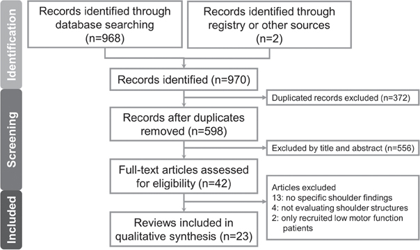
Fig. 1. Flow diagram for the literature search.
Study characteristics
The 23 included studies included 1,509 participants, age range 20–96 years. Among them, 14 studies (13–15, 21–31) evaluated only the hemiplegic shoulders, whereas 9 (32–40) reported comparative/bilateral ultrasound examinations. The majority of the studies were published in Asia, 8 in Taiwan (13, 14, 23, 26–28, 32, 36), 7 in South Korea (15, 21, 24, 33–35, 38), 2 in Turkey (25, 29) and 1 in India (30). The mean duration since stroke onset ranged widely between 17 days (13) and 26 months (30). More than half of the studies analysed more than 50 participants (14, 15, 22–25, 29, 31, 32, 35, 37–39). The characteristics of the studies are shown in Table I.
| Reference | Country | Study type | Duration since stroke onset, days(mean) | Participants n | Age, years Mean (range) | Hemiplegic side (right/left) | Ultrasound machine/transducer | |
| Studies evaluating only the hemiplegic shoulder | ||||||||
| Pong et al., 2009 (13) | Taiwan | Prospective, case-control | 17.3 | 34 | 66.6 (42–85) | 15/19 | Sequoia 512 ultrasound/(Siemens, Germany) 5–10 MHz linear array transducer | |
| Huang et al., 2010 (14) | Taiwan | Crosssectional | 19.7 | 57 | 59.9 (47–74) | 20/37 | Terason t3000 (Terason, USA) /5–12 MHz linear array transducer | |
| Kim et al., 2011 (15) | South Korea | Retrospective | 43.8 | 82 | 63.5 (41–85) | 33/49 | EnVisor HD (Philips, Netherlands)/7–15 MHz high resolution linear probe | |
| Pompa et al, 2011 (21) | South Korea | Retrospective | 35.4 | 41 | 72.3 (41–85) | 24/17 | NA/5–12 MHz linear transducer | |
| Zaiton et al, 2011 (22) | Egypt | Prospective, case-control | 68.2 | 106 | 57.0 (40–78) | 39/67 | Nemio XG (Toshiba, Japan)/6–12 MHz linear phased array transducer | |
| Pong et al., 2012 (23) | Taiwan | Prospective, longitudinal | 46.1 | 76 | 59.7 (30–87) | NA | Terason t3000 (Terason, USA)/5–12 MHz linear array transducer | |
| Rah et al., 2012 (24) | South Korea | Multicentre, randomized, triple-blind, placebo-controlled | 23.6 | 58 | 56.6 (20–70) | Dominant: 33 Non-dominant: 25 |
Logiq P6 (GE Healthcare, US), Accuvix XQ (Samsung Medison, Korea), SonoAce X8 (Samsung Medison, Korea), HD 11XE (Philips, Netherlands)/5–13 MHz linear array transducers | |
| Doğun et al., 2014 (25) | Turkey | Retrospective | 49.0 | 68 | 63.7 (43–82) | 36/32 | ATL HDI 1500 (Philips, Netherlands)/superficial probe | |
| Huang et al., 2017 (26) | Taiwan | Double-blind, placebo-controlled | 71.1 | 21 | 57.4 (43–72) | 9/12 | Aplio 300 TUS-A300 (Toshiba, Japan)/5–12 MHz high-resolution linear scanner | |
| Huang et al., 2018 (27) | Taiwan | Randomized, double-blinded controlled | 89.0 | 27 | 50.5 (49– 72) | 11/16 | ACUSON S2000 (Siemens, Germany)/9–14 MHz linear array transducer | |
| Lin et al., 2018 (28) | Taiwan | Retrospective | 78.2 | 26 | 66.5 (38–91) | 10/16 | Acuson X300 (Siemens, Germany)/4.4–13.0 MHz linear transducer | |
| Korkmaz et al. 2020 (29) | Turkey | Cross-sectional | 390.0 | 64 | 63.1 (18–75) | 31/33 | Logic e portable (GE HealthCare, US)/5–12 MHz linear array transducer | |
| Arya et al., 2021 (30) | India | Prospective, case-series | 792.0 | 8 | 53.3 (35–76) | 4/4 | LOGIQ S8 (GE HealthCare, USA)/4–15 MHz linear-array probe | |
| El-Sonbaty et al., 2022 (31) | Egypt | Cross-sectional | 254.3 | 210 | 61.7 (59–67) | 97/113 | Xario 200 (Toshiba, Japan)/7–14 MHz. linear phased array transducer | |
| Studies comparing bilateral shoulders | ||||||||
| Lee et al., 2002 (32) | Taiwan | Prospective, case-control | 308.7 | 82 | 61.0 (20–87) | 37/45 | ATL HDI 1500 (Philips, Netherlands)/5–12 MHz high-resolution linear scanner | |
| Park et al, 2007 (33) | South Korea | Prospective, single blind | 30.0 | 41 | 56.0 (34–78) | 15/26 | Sonoace 9900 (Samsung Medison, Korea)/5–12 MHz linear array transducer | |
| Baek et al., 2009 (34) | South Korea | Prospective, case-control | 43.0 | 36 | 62.9 (49–77) | 9/27 | SonoAce 8800 (Samsung Medison, Korea)/5–9 MHz linear array transducer | |
| Lee et al., 2009 (35) | South Korea | Prospective, case-control | 91.0 | 71 | 60.0 (23–78 ) | 34/38 | iU22 (Philips, Netherlands)/1217 MHz high-resolution electronic linear array transducer | |
| Huang et al., 2012 (36) | Taiwan | Cross-sectional | 22.0 | 39 | 65.0 (54–76 ) | NA | HD 11XE (Philips, Netherlands)/5–12 MHz high-resolution linear scanner | |
| Pop et al., 2013 (37) | Poland | Retrospective | 243.8 | 182 | 63.0 (21–96) | 90/92 | EUB-565 (Hitachi, Japan)/7.5 MHz high-resolution linear scanner | |
| Yi et al., 2013 (38) | South Korea | Cross-sectional | 54.0 | 55 | 63.5 (45–80) | 16/39 | Accuvix V20 (Samsung Medison, Korea)/NA | |
| Mohamed et al., 2014 (39) | Egypt | Prospective, case-control | 61.9 | 80 | 62.3 (35–75) | 63/17 | Model unspecified (Biomedical, Denmark)/12 MHz linear array transducer | |
| Idowu et al., 2017 (40) | Nigeria | Prospective, case-control | NA | 45 | 62.0 (50–77) | 26/19 | Mindray DC-7 (Mindray, China)/7–12 MHz linear transducer | |
| NA: not available | ||||||||
Quality assessment
Only 3 studies (22, 24, 25) performed power calculations and therefore fulfilled Item 3 (justified and satisfactory sample size). One point was deducted from Item 6 (validated method) in 8 articles (21, 22, 25, 27, 28, 30, 37, 38) due to the lack of a detailed description of the ultrasound findings. Lee et al. (32) did not elaborate on the stroke type of the participants and lost 1 point in Item 4 (ascertainment of diagnosis). Full marks were scored for all other items for all recruited papers. According to the Newcastle-Ottawa scale, 16 studies (13–15, 22–26, 29, 31, 33–36, 39, 40) were of very high quality and 7 (21, 27, 28, 30, 32, 37, 38) of high quality (Table II).
| Author, year | Selection | Comparability | Outcome | Total score | ||||
| Representativeness | Selection | Sample size | Diagnosis | Cofounders | Validated method | Statistical test | ||
| Studies evaluating only the hemiplegic shoulder | ||||||||
| Pong et al., 2009 (13) | * | * | ** | ** | ** | * | 9 | |
| Huang et al., 2010 (14) | * | * | ** | ** | ** | * | 9 | |
| Kim et al., 2011 (15) | * | * | ** | ** | ** | * | 9 | |
| Pompa et al, 2011 (21) | * | * | ** | ** | * | * | 8 | |
| Zaiton et al, 2011 (22) | * | * | * | ** | ** | * | * | 9 |
| Pong et al., 2012 (23) | * | * | ** | ** | ** | * | 9 | |
| Rah et al., 2012 (24) | * | * | * | ** | ** | ** | * | 10 |
| Doğun et al., 2014 (25) | * | * | * | ** | ** | * | * | 9 |
| Huang et al., 2017 (26) | * | * | ** | ** | ** | * | 9 | |
| Huang et al., 2018 (27) | * | * | ** | ** | * | * | 8 | |
| Lin et al., 2018 (28) | * | * | ** | ** | * | * | 8 | |
| Korkmaz et al., 2020 (29) | * | * | ** | ** | ** | * | 9 | |
| Arya et al., 2021 (30) | * | * | ** | ** | * | * | 8 | |
| El-Sonbaty et al., 2022 (31) | * | * | ** | ** | ** | * | 9 | |
| Studies comparing bilateral shoulders | ||||||||
| Lee et al., 2002 (32) | * | * | * | ** | ** | * | 8 | |
| Park et al, 2007 (33) | * | * | ** | ** | ** | * | 9 | |
| Baek et al., 2009 (34) | * | * | ** | ** | ** | * | 9 | |
| Lee et al, 2009 (35) | * | * | ** | ** | ** | * | 9 | |
| Huang et al., 2012 (36) | * | * | ** | ** | ** | * | 9 | |
| Pop et al., 2013 (37) | * | * | ** | ** | * | * | 8 | |
| Yi et al., 2013 (38) | * | * | ** | ** | * | * | 8 | |
| Mohamed et al, 2014 (39) | * | * | ** | ** | ** | * | 9 | |
| Idowu et al, 2017 (40) | * | * | ** | ** | ** | * | 9 | |
| *Number of points earned in each item. | ||||||||
Ultrasound findings (general)
Hemiplegic shoulder pathologies pertained to various structures, most commonly being the biceps long head tendon (41.4%; 95% confidence interval (95% CI) 33.3–50.1%; I2 86.2%; Fig. S1), followed by the supraspinatus tendon (33.2%; 95% CI 24.13–43.6%; I2 86.1%; Fig. S2), subdeltoid bursa (29.3%; 95% CI 21.9–38.0%; I2 87.4%; Fig. S3), acromioclavicular joint (15.0%; 95% CI, 3.5–46.0%; I2 93.4%; Fig. S4), and subscapularis tendon (9.2%; 95% CI 3.4–22.4%; I2 87.6%; Fig. S5). Regarding the prevalence of specific pathological findings, bicipital peritendinous effusion ranked first (39.2%; 95% CI, 35.1–43.5%; I2 87.3%; Fig. S6), followed by biceps tendinopathy (35.5%; 95% CI 18.0–57.9%; I2 92.6%; Fig. S7), subdeltoid bursitis (29.3%; 95% CI, 21.9–38.0%; I2 87.4%; Fig. S3), supraspinatus tendinopathy (24.6%; 95% CI 13.8–39.9%; I2 88.5%; Fig. S8), supraspinatus partial-thickness tear (14.5%, 95% CI, 6.8–28.2%; I2 89.0%; Fig. S9), infraspinatus tendinopathy (14.1%; 95% CI 7.4–25.3%; I2 79.4%; Fig. S10), biceps tendon tear (8.1%; 95% CI 2.7–22.1%; I2 68.3%; Fig. S11), supraspinatus full-thickness tear (3.5%; 95% CI 1.9–6.5%; I2 < 0.01%; Fig. S12), and infraspinatus tendon tear (2.0%; 95% CI 0.6–6.6%; I2 < 0.01%; Fig. S13).
Ultrasound findings (hemiplegic vs non-hemiplegic shoulders)
Compared with the non-hemiplegic side, hemiplegic shoulders had an increased risk of developing pathologies in the biceps long head tendon (odds ratio (OR) 3.814; 95% CI 2.044–7.117; I2 70.1%; Fig. 2A) and supraspinatus tendon (OR 2.101; 95% CI 1.257–3.512; I2 27.0%; Fig. 2B). A similar increased risk was not observed for the subscapularis tendon (OR 1.808; 95% CI 0.285–11.479; I2 35.0%; Fig. S14), acromioclavicular joint (OR 1.556; 95% CI 0.530–4.570; I2 < 0.01%; Fig. S15), and infraspinatus tendon (OR 1.764; 95% CI 0.294–10.591; I2 35.0%; Fig. S16).
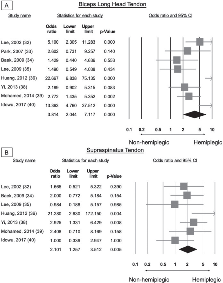
Fig. 2. Comparison of the prevalence of pathologies regarding (A) the biceps long head and (B) supraspinatus tendons (hemiplegic vs non-hemiplegic sides).
Regarding specific pathologies, hemiplegic shoulders had an increased risk of developing biceps peritendinous effusion (OR 3.081; 95% CI 1.801–5.272; I2 56.2%; Fig. 3A), biceps tendinopathy (OR 12.894; 95% CI 1.718–96.798; I2 76.9%; Fig. 3B), subdeltoid bursitis (OR 3.747; 95% CI 2.242–6.264; I2 26.6%; Fig. 4A), supraspinatus tendinopathy (OR 4.722; 95% CI 1.482–15.046; I2 47.4%; Fig. 4B), and supraspinatus partial-thickness tear (OR 2.302; 95% CI 1.467–3.612; I2 < 001%; Fig. 4C). No increased risk was observed for supraspinatus full-thickness tear (OR 0.695; 95% CI 0.184–2.621; I2 < 001%; Fig. S17).
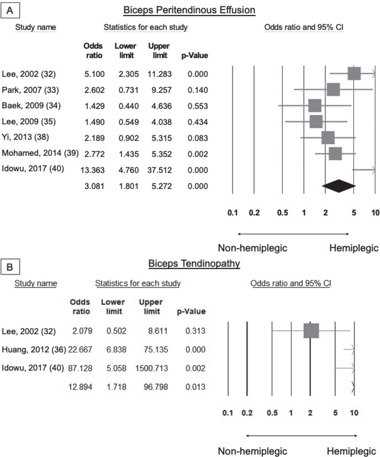
Fig. 3. Comparison of the prevalence regarding biceps (A) peritendinous effusion and (B) tendinopathy (hemiplegic vs non-hemiplegic sides).
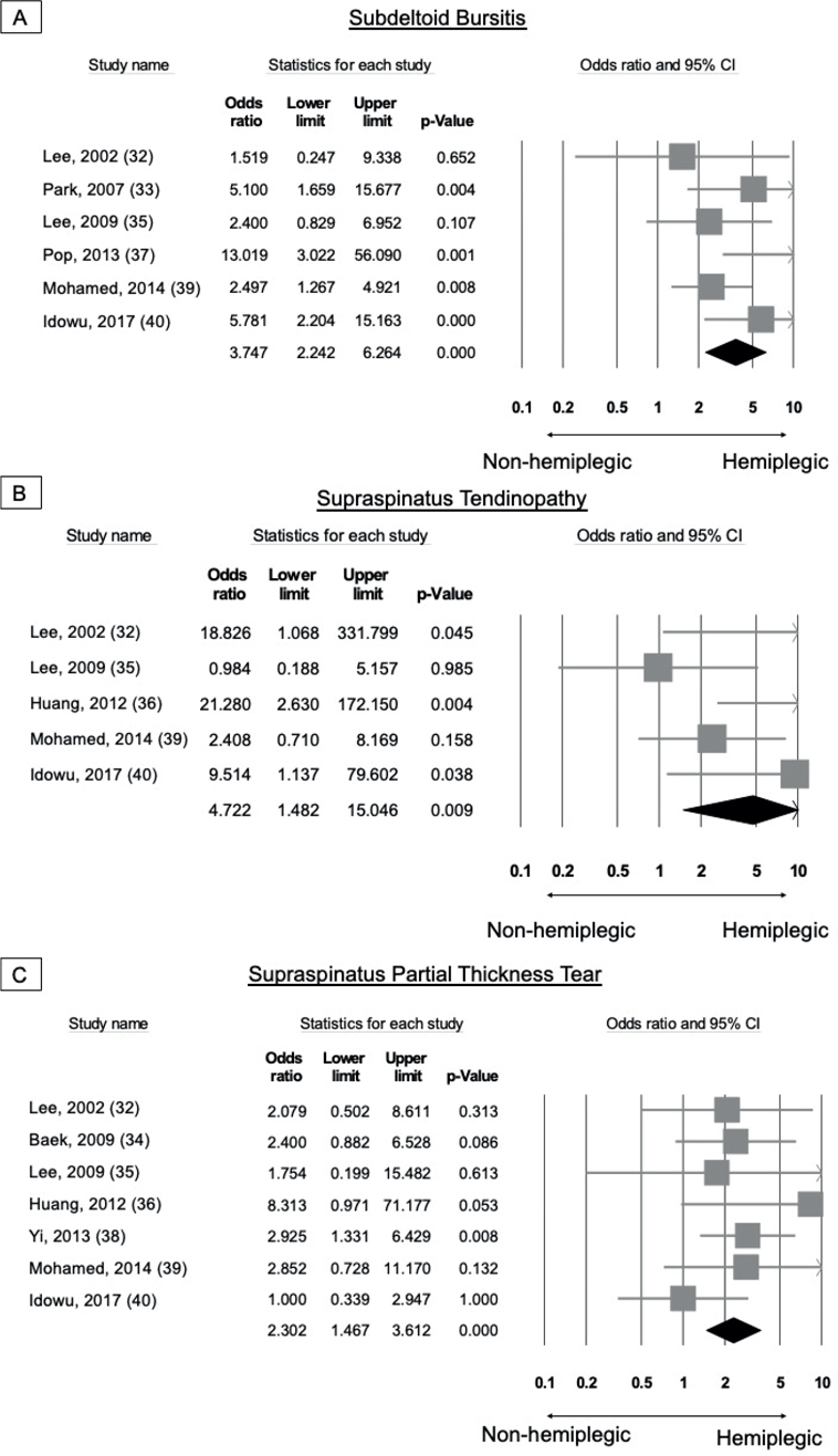
Fig. 4. Comparison of the prevalence regarding (A) subdeltoid bursitis, (B) supraspinatus tendinopathy and (C) partial thickness tear (hemiplegic vs non-hemiplegic sides).
Ultrasound findings (shoulders with high vs low motor function)
Based on the Brunnstrom stage, the categorization of hemiplegic shoulders into high and low motor functions was available in 3 studies. Compared with shoulders with high motor function, those with low motor function did not have an increased risk of developing pathologies in the biceps tendon (OR 0.769; 95% CI 0.196–3.020; I2 < 001; Fig. S18), subscapularis tendon (OR 0.784; 95% CI 0.155–3.961; I2 < 001; Fig. S19), subdeltoid bursa (OR 0.627; 95% CI 0.070–5.573; I2 75.2%; Fig. S20) and supraspinatus tendon (OR, 2.206; 95% CI 0.951–5.116; I2 < 001; Fig. S21).
Publication bias
Publication bias was analysed in the pooled analysis for the prevalence of pathologies of the biceps long head tendon, subdeltoid bursa, and supraspinatus tendon, and the existence of biceps peritendinous effusion. None of the p-values examined by Egger’s test were less than 0.05 (Figs S22–S25).
DISCUSSION
In this meta-analysis, several ultrasound findings were common in post-stroke hemiplegic shoulders. The top-ranking structure with pathologies was the biceps long head tendon, followed by the supraspinatus tendon, subdeltoid bursa, acromioclavicular joint, and subscapularis tendon. The most frequent hemiplegic shoulder pathologies were bicipital peritendinous effusion, biceps tendinopathy, subdeltoid bursitis, supraspinatus tendinopathy, and supraspinatus partial-thickness tear. The prevalence of pathologies was significantly higher in the hemiplegic (vs non-hemiplegic) shoulders, but only for the biceps long head and supraspinatus tendons. In addition, impaired upper-limb motor function was not associated with an increased incidence of shoulder pathologies.
The biceps long head tendon was the primary lesion site in hemiplegic shoulders, with a pooled prevalence of 41.4% according to the current meta-analysis. Peritendinous effusion and tendinopathy were the first and second most commonly reported biceps pathologies, respectively. This tendon originates intra-articularly from the supraglenoid tubercle of the scapula, exits the joint, and enters the rotator cuff interval (41). Proximal to the intertubercular groove of the humeral head, it is enveloped by the coracohumeral and superior glenohumeral ligaments as well as fibres from the supraspinatus and subscapularis tendons (42, 43). These soft-tissue restraints maintain it in place (43). Hence, it is not surprising that injuries in the surrounding structures can cause secondary irritation and disrupt its anatomical integrity. Studies reported associations between peritendinous effusion and supraspinatus/subscapularis tears, subscapularis tendinopathy, subdeltoid bursitis, and dynamic subacromial impingement (44, 45). In addition, as the tendon sheath extends directly from the glenohumeral cavity, bicipital peritendinous effusion may reflect intra-articular abnormalities. Similarly, biceps tendinopathy rarely occurs alone, and is often accompanied by other shoulder pathologies (43). Mechanical stress from the neighbouring structures or the coracoacromial arch can result in tendon inflammation, degeneration, fraying, tearing, and ultimately, rupture. Since the biceps long head tendon helps anchor the humeral head in the glenoid fossa, its derangement can lead to further shoulder instability and tissue damage, exacerbating dysfunctional biomechanics (41).
Pathologies of the supraspinatus tendon was present in one-third (33.2%) of the hemiplegic shoulders according to the current meta-analysis. The supraspinatus muscle has 2 main tasks: to perform arm abduction along with the deltoid, and to maintain the stability of the glenohumeral joint (46). Several factors can make it susceptible to injury in stroke. First, shoulder kinematics are altered by weakness, spasticity, contracture, and proprioception deficiency. The supraspinatus would have to compensate for the weakened deltoid during shoulder movements. Failure of scapular stabilizers (e.g. trapezius, rhomboid, and serratus anterior muscles) generates shearing forces on the rotator cuff muscles (23, 47). Likewise, the dominant flexor tone of the upper extremity results in depression of the scapula and exposes the supraspinatus to tension. Secondly, suboptimal positioning and traction forces from gravity, transfer, or inappropriate exercises also contribute to potential trauma. In particular, shoulder subluxation quickly appears in a large proportion of patients with stroke (48). Third, a cadaveric study demonstrated short muscle fibres and long resting sarcomeres in the supraspinatus, which was ideal for maintaining the humeral head in the shallow glenoid due to large passive tension (49). However, this exquisite architectural arrangement also implies that minor stretches would exert great restoring forces. Multifactorial tensile stress, coupled with the sensitive nature of the muscle, can lead to fatigue, stiffening, and tears after stroke.
The third most widely seen pathological site in hemiplegic shoulders was the subdeltoid bursa, with a prevalence of 29.3% among the enrolled studies. The extra-articular space is situated atop the rotator cuff tendon and beneath the deltoid muscle, acromioclavicular joint, and acromion (50). The physiological fluid within thin, slippery synovial membranes helps reduce friction between adjacent tissues with normal thicknesses within 2 mm (51). Thickening of the subdeltoid bursa could be a consequence of chronic impingement by the proliferation of protective bursal fluid, as well as cellular infiltration and collagen deposition in its wall (52, 53). Another origin of subdeltoid effusion is injury to the compromised rotator cuff muscles after stroke. Under normal circumstances, the bursa is separated from the glenohumeral joint; nonetheless, a full-thickness rotator cuff tear could create a communication between them (54). In fact, concurrent joint effusion is a highly specific sign of rotator cuff tears (sensitivity 22%; specificity 99%; positive predictive value 95%) (55).
Of the 9 studies that listed bilateral ultrasonographic findings, there was a significantly higher incidence of biceps long head tendon and supraspinatus tendon lesions in the hemiplegic shoulders. Biceps peritendinous effusion, biceps tendinopathy, subdeltoid bursitis, supraspinatus tendinopathy, and supraspinatus partial-thickness (but not full-thickness) tears were more common in hemiplegic shoulders. Full-thickness supraspinatus tears were observed in only 3.5% of post-stroke shoulders. This might indicate opportunities for successful non-operative treatment of hemiplegic shoulder pain due to commonly encountered rotator cuff tendon disorders.
Only 3 studies divided the patients according to their Brunnstrom stage. There was no statistical difference in the incidence of pathological shoulder structures between the high and low motor function groups. This could possibly be attributed to the small sample sizes. In addition, ultrasound examinations in the current meta-analysis were performed in the relatively early stages of post-stroke (range of mean duration since stroke 17–44 days). Therefore, discrepancies between the hemiplegic shoulders with high vs low motor function might not have been apparent. Moreover, although shoulders with worse function experienced greater musculoskeletal imbalances, those with better recovery might have been more actively used, potentially making them prone to overuse injuries.
Some limitations should be considered when interpreting the results of this meta-analysis. First, the scanning protocols and reporting formats varied among studies. For example, not all studies differentiated between partial- and full-thickness tears; therefore, their respective prevalence could not always be promptly extracted. Secondly, the timing of the ultrasound examinations was inconsistent; mostly being in the subacute phase. Due to the vastly different time-points of evaluation, both within each study and between the included papers, it was not possible to perform in-depth analysis of the influence of stroke chronicity on lesion types. The development of pertinent pathologies and their relationship with hemiplegic shoulder pain should be explored in future studies. Finally, few studies have categorized hemiplegic shoulders by motor function or by the presence/severity of pain. More research on such issues is needed to improve patient care and facilitate the use of ultrasound in stroke.
In conclusion, abnormalities of the biceps long head tendon, supraspinatus tendon, subdeltoid bursa, acromioclavicular joint, and subscapularis tendon are commonly observed in post-stroke hemiplegic shoulders. Likewise, bicipital peritendinous effusion, biceps tendinopathy, subdeltoid bursitis, supraspinatus tendinopathy, and supraspinatus partial-thickness tears were the most frequent pathological findings. Of note, ultrasound is useful in uncovering these lesions and guiding relevant interventions in case of painful shoulders. Further research is needed to decipher the clinical course of structural changes in the post-stroke shoulder and their correlation with pain and motor recovery.
ACKNOWLEDGEMENTS
This work was funded by National Taiwan University Hospital, Bei-Hu Branch; Ministry of Science and Technology (MOST 106-2314-B-002-180-MY3 and 109-2314-B-002-114-MY3); and the Taiwan Society of Ultrasound in Medicine.
REFERENCES
- Feigin VL, Stark BA, Johnson CO, Roth GA, Bisignano C, Abady GG, et al. Global, regional, and national burden of stroke and its risk factors, 1990–2019: a systematic analysis for the Global Burden of Disease Study 2019. Lancet Neurol 2021; 20: 795–820.
- Murie-Fernández M, Iragui MC, Gnanakumar V, Meyer M, Foley N, Teasell R. Painful hemiplegic shoulder in stroke patients: causes and management. Neurologia 2012; 27: 234–244.
- Dromerick AW, Edwards DF, Kumar A. Hemiplegic shoulder pain syndrome: frequency and characteristics during inpatient stroke rehabilitation. Arch Phys Med Rehabil 2008; 89: 1589–1593.
- Prescher A. Anatomical basics, variations, and degenerative changes of the shoulder joint and shoulder girdle. Eur J Radiol 2000; 35: 88–102.
- Kalichman L, Ratmansky M. Underlying pathology and associated factors of hemiplegic shoulder pain. Am J Phys Med Rehabil 2011; 90.
- Adey-Wakeling Z, Liu E, Crotty M, Leyden J, Kleinig T, Anderson CS, et al. Hemiplegic shoulder pain reduces quality of life after acute stroke: a prospective population-based study. Am J Phys Med Rehabil 2016; 95: 758–763.
- Turner-Stokes L, Jackson D. Shoulder pain after stroke: a review of the evidence base to inform the development of an integrated care pathway. Clin Rehabil 2002; 16: 276–298.
- Lo S-F, Chen S-Y, Lin H-C, Jim Y-F, Meng N-H, Kao M-J. Arthrographic and clinical findings in patients with hemiplegic shoulder pain. Arch Phys Med Rehabil 2003; 84: 1786–1791.
- Kim YH, Jung SJ, Yang EJ, Paik NJ. Clinical and sonographic risk factors for hemiplegic shoulder pain: a longitudinal observational study. J Rehabil Med 2014; 46: 81–87.
- Özçakar L, Kara M, Chang K-V, Çarl AB, Akkaya N, Tok F, et al. Nineteen reasons why physiatrists should do musculoskeletal ultrasound: EURO-MUSCULUS/USPRM recommendations. Am J Phys Med Rehabil 2015; 94: e45–e49.
- De Jesus JO, Parker L, Frangos AJ, Nazarian LN. Accuracy of MRI, MR arthrography, and ultrasound in the diagnosis of rotator cuff tears: a meta-analysis. AJR Am J Roentgenol 2009; 192: 1701–1707.
- Zappia M, Di Pietto F, Aliprandi A, Pozza S, De Petro P, Muda A, et al. Multi-modal imaging of adhesive capsulitis of the shoulder. Insights Imaging 2016; 7: 365–371.
- Pong YP, Wang LY, Wang L, Leong CP, Huang YC, Chen YK. Sonography of the shoulder in hemiplegic patients undergoing rehabilitation after a recent stroke. J Clin Ultrasound 2009; 37: 199–205.
- Huang YC, Liang PJ, Pong YP, Leong CP, Tseng CH. Physical findings and sonography of hemiplegic shoulder in patients after acute stroke during rehabilitation. J Rehabil Med 2010; 42: 21–26.
- Kim N-H, Pyun S-B, Kwon H-K. Ultrasonographic finding of painful hemiplegic shoulder associated with motor recovery after stroke. Brain Neurorehabil 2011; 4: 44–49.
- Page MJ, McKenzie JE, Bossuyt PM, Boutron I, Hoffmann TC, Mulrow CD, et al. The PRISMA 2020 statement: an updated guideline for reporting systematic reviews. Int J Surg 2021; 88: 105906.
- Stang A. Critical evaluation of the Newcastle-Ottawa scale for the assessment of the quality of nonrandomized studies in meta-analyses. Eur J Epidemiol 2010; 25: 603–605.
- Borenstein M, Hedges LV, Higgins JP, Rothstein HR. Fixed-effect versus random-effects models. Introduction to Meta-analysis 2009; 77: 85.
- Higgins JP, Li T, Deeks JJ. Choosing effect measures and computing estimates of effect. Cochrane Handbook for Systematic Reviews of Interventions. Chichester (UK): John Wiley & Sons, 2019: 143–176.
- Higgins JP, Thomas J, Chandler J, Cumpston M, Li T, Page MJ, et al. Assessing risk of bias due to missing results in a synthesis. Cochrane Handbook for Systematic Reviews of Interventions: John Wiley & Sons; 2019.
- Pompa A, Clemenzi A, Troisi E, Di Mario M, Tonini A, Pace L, et al. Enhanced-MRI and ultrasound evaluation of painful shoulder in patients after stroke: a pilot study. Eur Neurol 2011; 66: 175–181.
- Zaiton F, Tantawy HI, Elsharkawy KAM, Sanad G. Painful poststroke shoulder: Comparison of magnetic resonance imaging and high frequency ultrasonography. Egypt J Radiol Nucl Med 2011; 42: 47–55.
- Pong YP, Wang LY, Huang YC, Leong CP, Liaw MY, Chen HY. Sonography and physical findings in stroke patients with hemiplegic shoulders: a longitudinal study. J Rehabil Med 2012; 44: 553–557.
- Rah UW, Yoon SH, Moon DJ, Kwack KS, Hong JY, Lim YC, et al. Subacromial corticosteroid injection on poststroke hemiplegic shoulder pain: a randomized, triple-blind, placebo-controlled trial. Arch Phys Med Rehabil 2012; 93: 949–956.
- Doğun A, Karabay İ, Hatipoğlu C, Özgirgin N. Ultrasound and magnetic resonance findings and correlation in hemiplegic patients with shoulder pain. Top Stroke Rehabil 2014; Suppl 1: S1–7.
- Huang YC, Chang KH, Liou TH, Cheng CW, Lin LF, Huang SW. Effects of Kinesio taping for stroke patients with hemiplegic shoulder pain: a double-blind, randomized, placebo-controlled study. J Rehabil Med 2017; 49: 208–215.
- Huang YC, Leong CP, Tso HH, Chen MJ, Liaw MY, Hsieh HC, et al. The long-term effects of hyaluronic acid on hemiplegic shoulder pain and injury in stroke patients A randomized controlled study. Medicine (Baltimore) 2018; 97: 7.
- Lin PH. Sonographic findings of painful hemiplegic shoulder after stroke. J Chin Med Assoc 2018; 81: 657–661.
- Korkmaz N, Yaşar E, Demir Y, Tezen Ö, Gurcay E. Sonographic predictors in patients with hemiplegic shoulder pain: a cross-sectional study. J Stroke Cerebrovasc Dis 2020; 29: 105170.
- Arya KN, Pandian S, Bhatnagar N, Sharma A. Rehabilitation of the shoulder subluxation based on ultrasonographic findings among post stroke subjects: a case series. Neurol India 2021; 69: 1309–1317.
- El-Sonbaty HAE, Abou Elmaaty AA, Zarad CA, El-Bahnasawy AS. Clinical and radiological assessment of hemiplegic shoulder pain in stroke patients. Egypt J Neurol Psychiatr Neurosurg 2022; 58: 15.
- Lee CL, Chen TW, Weng MC, Wang YL, Cheng HS, Huang MH. Ultrasonographic findings in hemiplegic shoulders of stroke patients. Kaohsiung J Med Sci 2002; 18: 70–76.
- Park GY, Kim JM, Sohn SI, Shin IH, Lee MY. Ultrasonographic measurement of shoulder subluxation in patients with post-stroke hemiplegia. J Rehabil Med 2007; 39: 526–530.
- Baek S-R, Jung SH, Oh B-M, Chung SG, Sim YJ, Han TR. Ultrasonographic and physical examination to investigate the cause of painful hemiplegic shoulder. Brain Neurorehabil 2009; 2: 140–145.
- Lee IS, Shin YB, Moon TY, Jeong YJ, Song JW, Kim DH. Sonography of patients with hemiplegic shoulder pain after stroke: correlation with motor recovery stage. Am J Roentgenol 2009; 192: W40–44.
- Huang SW, Liu SY, Tang HW, Wei TS, Wang WT, Yang CP. Relationship between severity of shoulder subluxation and soft-tissue injury in hemiplegic stroke patients. J Rehabil Med 2012; 44: 733–739.
- Pop T. Subluxation of the shoulder joint in stroke patients and the influence of selected factors on the incidence of instability. Ortop Traumatol Rehabil 2013; 15: 259–267.
- Yi Y, Shim JS, Kim K, Baek SR, Jung SH, Kim W, et al. Prevalence of the rotator cuff tear increases with weakness in hemiplegic shoulder. Ann Rehabil Med 2013; 37: 471–478.
- Mohamed RE, Amin MA, Aboelsafa AA. Ultrasonographic and clinical study of post-stroke painful hemiplegic shoulder. Egypt J Radiol Nucl Med 2014; 45: 1163–1170.
- Idowu BM, Ayoola OO, Adetiloye VA, Komolafe MA. Sonographic evaluation of structural changes in post-stroke hemiplegic shoulders. Pol J Radiol 2017; 82: 141–148.
- Elser F, Braun S, Dewing CB, Giphart JE, Millett PJ. Anatomy, function, injuries, and treatment of the long head of the biceps brachii tendon. Arthroscopy 2011; 27: 581–592.
- Favorito PJ, Harding WG, 3rd, Heidt RS, Jr. Complete arthroscopic examination of the long head of the biceps tendon. Arthroscopy 2001; 17: 430–432.
- Friedman DJ, Dunn JC, Higgins LD, Warner JJP. Proximal biceps tendon: injuries and management. Sports Med Arthrosc Rev 2008; 16.
- Chang K-V, Wu W-T, Özçakar L. Association of bicipital peritendinous effusion with subacromial impingement: a dynamic ultrasonographic study of 337 shoulders. Sci Rep 2016; 6: 38943.
- Chang K-V, Chen W-S, Wang T-G, Hung C-Y, Chien K-L. Associations of sonographic abnormalities of the shoulder with various grades of biceps peritendinous effusion (BPE). Ultrasound Med Biol 2014; 40: 313–321.
- de Witte PB, Werner S, ter Braak LM, Veeger HEJ, Nelissen RGHH, de Groot JH. The supraspinatus and the deltoid – not just two arm elevators. Hum Mov Sci 2014; 33: 273–283.
- Kibler BW, McMullen J. Scapular dyskinesis and its relation to shoulder pain. J Am Acad Orthop Surg 2003; 11: 142–151.
- Paci M, Nannetti L, Taiti P, Baccini M, Pasquini J, Rinaldi L. Shoulder subluxation after stroke: relationships with pain and motor recovery. Physiother Res Int 2007; 12: 95–104.
- Ward SR, Hentzen ER, Smallwood LH, Eastlack RK, Burns KA, Fithian DC, et al. Rotator cuff muscle architecture: implications for glenohumeral stability. Clin Orthop Relat Res 2006; 448: 157–163.
- Draghi F, Scudeller L, Draghi AG, Bortolotto C. Prevalence of subacromial-subdeltoid bursitis in shoulder pain: an ultrasonographic study. J Ultrasound 2015; 18: 151–158.
- van Holsbeeck M, Strouse PJ. Sonography of the shoulder: evaluation of the subacromial-subdeltoid bursa. Am J Roentgenol 1993; 160: 561–564.
- Awerbuch MS. The clinical utility of ultrasonography for rotator cuff disease, shoulder impingement syndrome and subacromial bursitis. Med J Aust 2008; 188: 50–53.
- Daghir AA, Sookur PA, Shah S, Watson M. Dynamic ultrasound of the subacromial–subdeltoid bursa in patients with shoulder impingement: a comparison with normal volunteers. Skeletal Radiol 2012; 41: 1047–1053.
- Precerutti M, Garioni E, Madonia L, Draghi F. US anatomy of the shoulder: pictorial essay. J Ultrasound 2010; 13: 179–187.
- Hollister MS, Mack LA, Patten RM, Winter TC, 3rd, Matsen FA, 3rd, Veith RR. Association of sonographically detected subacromial/subdeltoid bursal effusion and intraarticular fluid with rotator cuff tear. Am J Roentgenol 1995; 165: 605–608.