ORIGINAL REPORT
EFFECTIVENESS OF MOTOR IMAGERY COMBINED WITH STRUCTURED PROGRESSIVE CIRCUIT CLASS TRAINING ON FUNCTIONAL MOBILITY IN POST-STROKE INDIVIDUALS: A RANDOMIZED CONTROLLED TRIAL
Nilar AUNG, PhD  1,2, Vimonwan HIENGKAEW, PhD 1, Jarugool TRETRILUXANA, PhD 1, Mon S. BRYANT, PhD 3, and Sunee BOVONSUNTHONCHAI, PhD 1
From the 1Faculty of Physical Therapy, Mahidol University, Nakhon Pathom, Thailand, 2Department of Physiotherapy, University of Medical Technology, Mandalay, Myanmar and 3Department of Physical Medicine and Rehabilitation, Baylor College of Medicine, Houston, TX, USA
Objective: To compare the effect of motor imagery combined with structured progressive circuit class therapy vs health education combined with structured progressive circuit class therapy on dynamic balance, endurance, and functional mobility in post-stroke individuals.
Design: Randomized controlled trial.
Methods: A total of 40 post-stroke individuals were randomly assigned to experimental and control groups. The experimental group was trained using motor imagery combined with structured progressive circuit class therapy, while the control group received health education combined with structured progressive circuit class therapy, 3 times a week for 4 weeks with an overall of 12 sessions. Outcomes included the step test for affected and unaffected limbs, the 6-Minute Walk Test, and the Timed Up and Go test. Assessments were performed at baseline, 2 weeks, and 4 weeks after the intervention
Results: There were significant effects (p < 0.05) of: group on the step test for unaffected limb; of time on all outcomes; and of their interaction effect on the step test for affected limb, 6-Minute Walk Test, and Timed Up and Go test. Inter-group comparison showed significant differences (p < 0.05) in the step test for unaffected limb at 2 weeks after the intervention. At 4 weeks after the intervention, significant differences (p < 0.05) were found in the step test for affected and unaffected limbs and in the Timed Up and Go test.
Conclusion: Motor imagery combined with structured progressive circuit class therapy was more effective on the step test, 6-Minute Walk Test, and Timed Up and Go test than training with structured progressive circuit class therapy alone. This suggest that that motor imagery should be incorporated into training programmes for restoring dynamic balance, endurance, and functional mobility in post-stroke individuals.
LAY ABSTRACT
Survival rates after stroke have increased, with the frequency of mobility impairments remaining high. Therefore, a low-cost and effective stroke rehabilitation technique must be developed and its efficacy proved. This study compared the effect of motor imagery combined with structured progressive circuit class therapy vs health education combined with structured progressive circuit class therapy on dynamic balance, endurance, and functional mobility in post-stroke individuals. Outcomes included the step test, 6-Minute Walk Test, and Timed Up and Go test. Significant improvements occurred in all outcomes when training with motor imagery combined with structured progressive circuit class therapy compared with structured progressive circuit class therapy alone. In conclusion, these results suggest that incorporating motor imagery into the post-stroke training programme may result in greater improvement.
Key words: exercise; stroke; motor imagery; mobility limitation.
Citation: J Rehabil Med 2022; 54: jrm00297. DOI: https://dx.doi.org/10.2340/jrm.v54.1390
Copyright: © Published by Medical Journals Sweden, on behalf of the Foundation for Rehabilitation Information. This is an Open Access article distributed under the terms of the Creative Commons Attribution-NonCommercial 4.0 International License (https://creativecommons.org/licenses/by-nc/4.0/)
Accepted: Jun 6, 2022; Epub ahead of print: Jul 7, 2022; Published: Jul 27, 2022
Correspondence address: Sunee Bovonsunthonchai, Faculty of Physical Therapy, Mahidol University, 999 Phuttamonthon 4 Road, Salaya, Phuttamonthon, Nakhon Pathom, 73170, Thailand. E-mail: sunee.bov@mahidol.edu
Competing interests and funding: The authors have no conflicts of interest to declare.
This research was funded by grants from Norway Scholarship (Mahidol-Norway Capacity Building Initiative for ASEAN) and Mahidol University.
Stroke is one of the major medical conditions and a leading cause of disability and mortality (1). There has been an 11.3% reduction in the age-standardized stroke incidence rate in countries worldwide from 1990 to 2017. Furthermore, there has been a 33.4% decrease in the age-standardized stroke mortality rate and a 34% decrease in the stroke death rate, especially in developed countries (1). In contrast, the annual stroke incidence, mortality rate, and disability-adjusted life-years lost have increased in low- and middle-income countries (2).
Impaired functional mobility is one of the earliest and most common characteristics observed in many post-stroke individuals. The ability to get out in the community was considered essential by 74.6% of patients (3). A longitudinal cohort study showed that individuals with mild stroke encountered difficulty in terms of community mobility (30%) and indoor activities (10%) 1 year after stroke (4). Mobility difficulty was increased in individuals with a moderate severity of stroke; 75% encountered community mobility difficulties, and 35% encountered difficulty in indoor activities. It was also found that physical performance, assessed by considering lower limb strength, and functional mobility at discharge, could predict health-related quality of life (HRQoL) and reintegration into the community 6 months later (5).
Dynamic balance is one of the crucial factors for mobility, and is a significant problem in post-stroke individuals, who often have decreased equilibrium, exaggerated postural sway, and altered weight distribution patterns. Maintaining balance while standing and ambulating is necessary to successfully perform various daily activities, and impaired balance is related to fall risk in stroke patients (6, 7). Another factor positively influencing mobility is walking endurance ability at the submaximal level, which is usually assessed using the submaximal treadmill walking test, cycle ergometer, and the 6-Minute Walk Test (6MWT) (8–11). Post-stroke individuals can regain their ability to walk, but their walking endurance is often limited, even in individuals with high functional ability (11). Dynamic balance and walking endurance are associated with HRQoL and participation in post-stroke individuals (9, 10).
To restore functional mobility appropriately, a task-oriented training regimen including augmented feedback tuning can be created for post-stroke individuals for enhancing limited functional mobility (12). With the benefits of circuit class training in the improvement of cardiovascular fitness, muscle strength, and endurance, post-stroke individuals can improve physical performance by incorporating specific exercises into the circuit class training (13–17). In addition, the advantages of circuit class training are related to increased social interaction during workouts and exercise adherence.
Various training strategies can be used to improve patients’ motor control and learning process. Motor imagery (MI) method is one of the training strategies that has gained in interest, facilitating the recovery of neurological deficits of upper and lower limb functions for stroke patients in the past decade (18–20). The operational definition of MI is as follows: “a dynamic mental state during which the representation of a given motor act or movement is rehearsed in working memory without any overt motor output” (21). The motor simulation theory introduced by Jeannerod in 1994 (22) is the most common reference for explaining the efficacy of MI training. This theory attempted to explain the relationship between distinct action-related cognitive states and actual motor execution. Three notions of the theory are summarized as follows: firstly, actions are postulated to include a covert stage in which they are mentally simulated. The goal and consequence of the action are included in this covert stage. As a result, it consists of a representation of the future, which includes the action’s objective, the means to achieve it, and its effect on the environment. Secondly, the theory claims that both imagined and actual actions share a motor representation of a desire to act. In the event of overt actions, this desire is translated into an actual movement; nevertheless, it is not carried out in the case of imagined actions. Finally, the theory proposes that the simulation and execution of actions are functionally equivalent (22, 23). Previous studies (13, 16, 24–27) have reported a greater improvement in gait, balance, and functional mobility from the combined effect of MI with the exercise programme in post-stroke individuals. A finding showing the efficacy of a combined MI with circuit class training was reported in a previous study (16). With an overall 40 min of training, 7 days a week for 2 weeks, several outcomes, such as the Functional Ambulatory Classification (FAC), Rivermead Visual Gait Assessment (RVGA), walking speed, and 6MWT, were improved significantly. The benefits of this training on reported variables were found to persist at a 6-month follow-up. However, the variety of training protocols both in MI and exercise parts among studies causes controversial results. Systematic reviews (18, 20, 28) reported high heterogeneity protocols that led to the different findings between studies, as well as the need to explore the ideal treatment protocol. Sub-group analysis by Guerra et al. (18) showed that MI was associated with balance gain, based on the functional reach test, and improved performance, based on the Timed Up and Go (TUG) test and gait speed. In particular, very low certainty evidence was reported on endurance and functional mobility (20). Due to the subjective nature of MI, the facilitation action and engagement monitoring method should be carried out.This can be done by counting the number of movement actions during imagery, enquiring about MI movement at the end of the session, and movement command during imagery periodically (29, 30). For an objective measurement of engagement, the investigation of autonomic changes was recommended in the clinic. The study of autonomic response to MI reported a decrease in skin resistance and an increase in heart rate and respiration (31). However, it was reported that most studies did not perform these methods in a protocol (18) and took only verbal agreement for training engagement (7, 32).
Given the above, the aim of this study was to examine the effect of MI combined with structured progressive circuit class therapy (SPCCT) on functional mobility in post-stroke individuals. It was hypothesized that there would be a greater improvement in functional mobility in the group that received MI combined with SPCCT compared with training with SPCCT alone.
METHODS
Sample size calculation
The sample size was estimated using the mean and standard deviation of 6MWT from the previous study (44), which investigated the effect of task-oriented training in improving mobility in post-stroke individuals. By using the G*Power program version 3.1.9.2 with an α value of 0.05 and a β value of 0.02, the total sample size was estimated at 30. Therefore, a sample of 40 would provide sufficient statistical power for this study.
Participants and design
The study used a randomized double-blind balanced parallel-group (1:1) design. The recruitment of the subjects took place from January 2018 to May 2018. The trial was registered at clinicaltrials.gov (NCT03436810). Participants were recruited from the National Rehabilitation, North Okkalapa General, and East Yangon General Hospitals, Yangon, Myanmar. Candidates were screened by physiotherapists according to the selection criteria. Informed consent was obtained from participants who met the selection criteria. The study targeted patients with mild and moderate motor severity who were able to perform the study intervention.
Inclusion criteria were: (i) first stroke with unilateral hemiparesis; (ii) age between 18 and 75 years; (iii) male and female; (iv) stroke onset of 3 months to 1 year; (v) ability to walk at least 10 m with or without using gait aids; (vi) Functional Ambulation Category (FAC) score ≥ 3 (33); (vii) Mini-Mental State Examination (MMSE) score ≥ 24 (34); (viii) National Institutes of Health Stroke Scale (NIHSS) score < 14 (35); and (ix) Kinesthetic and Visual Imagery Questionnaire (KVIQ-10) score ≥ 3 (36).
Exclusion criteria were: (i) communication deficits and unable to follow instructions; (ii) a history of serious or unstable cardiac condition; (iii) severe musculoskeletal problems and unable to stand and walk; and (iv) a history of other neurological diseases or unilateral neglect. Eligible participants who agreed to take part in the study signed the informed consent approved by the institutional ethics committee (Certificate of Approval: Mahidol University Central Institutional Review Board 2017/178.1010).
Randomization and blinding
Four permuted blocks of 10 participants were created using a computer-generated program and allocated with the concealment method in the envelopes. The outcome measure assessments were performed by a blinded assessor. All participants were blinded regarding the group they were assigned for training.
Intervention
Both the experimental and control groups underwent a 90-min training session, 3 sessions a week continuously for 4 weeks. The experimental group received the MI protocol for 25 min, while the control group received HE for 25 min. Then, both groups underwent the same SPCCT for 65 min. The SPCCT involved 7 functional mobility tasks including: (i) forward and backward step up; (ii) lateral step up; (iii) heel raise and lower; (iv) reaching in standing; (v) transition from sit to stand, walk, and then back to sit; (vi) walking with even steps; and (vii) quick walk. SPCCT tasks 1, 5, 6, and 7 were the functional mobility aspects used in the MI training method.
To identify the most effective training, MI was provided at a quiet place in the training centre. MI involved familiarization with the task for 2 days in the first week and practice from the third day of the first week to the fourth week of the training programme. It was performed in 3 stages: relaxation (3 min), MI practice involving both kinesthetic imagery (KI) and visual imagery (VI) (8.5 min in each imagery), and refocusing (3 min). Between the 2 imagery practices, a rest period (2 min) was allowed to avoid mental fatigue. Both KI and VI were provided from the first-person perspective; the participants were instructed to visualize the movements they were performing from inside their body, i.e. via their own eyes for VI and to feel the sensation of movements they were performing, i.e. to perceive muscle tension, and stretch for KI. In the stepping task, a metronome was used to adjust the step rhythm in the progression phase of the training. Participants were instructed to bear weight on the affected limb in the walking tasks. Furthermore, they were asked: (i) to avoid movement and remain in a relaxed position; (ii) close their eyes throughout the practice; (iii) count the repetition of the practice with fingers; and (iv) open their eyes if their concentration was reduced. The engagement was checked by observing the finger movement while counting the practice repetition and by monitoring the pulse rate using the oximeter attached to the unaffected finger. To ensure that the training was standardized and consistent in every session, the participants had to practice MI according to a script written by a trained researcher.
The protocol of the SPCCT included warm-up exercises 3 min prior to training the 7 functional mobility tasks (62 min). The SPCCT was provided as group therapy with the distributed pattern. Participants were trained in pairs; while 1 participant performed the tasks (4 min), the other observed and encouraged the partner’s performance (4 min). There was 1 min to transfer to the next task. The training progression for SPCCT was performed by increasing the number of repetitions, weight, task complexity, and metronome speed in tasks 1, 2, 6 and 7. Progression was adjusted based on the participant’s performance assessed by the physiotherapist, and the self-efficacy score given by the participant themselves.
HE comprised the topics related to the necessary information for self-care after stroke, as well as preventive details of recurrent stroke. One HE session consisted of an explanation of the topics concerned and a discussion with post-stroke individuals. Details of the treatments were as in our previous work (13). MI and HE were provided by trained research staff, and the SPCCT was provided by a physiotherapist specialized in neurological rehabilitation.
Outcome measures
The step test (n) for the affected and the unaffected limb, the TUG (s), and the 6MWT (m) were assessed at baseline (T0), 2 weeks (T1) and 4 weeks (T2) after the intervention.
Step test. The step test is a reliable and valid measure for dynamic balance standing balance in post-stroke individuals (37, 38). Dynamic balance is positively associated with physical function in the first month post-stroke (6). A cut-off score of 13 on the step test for the affected limb was reported as a value used to distinguish post-stroke individuals from healthy individuals over 50 years with a sensitivity of 87% and specificity of 87%. For the unaffected limb, it was found that a cut-off score of 11 could distinguish, with a sensitivity of 100% and specificity of 67% (38). The participants were asked to step onto a 7.5 cm stool with the affected or unaffected limb repeatedly as fast as possible while the other limb stance. The step test for the affected and the unaffected limbs was recorded separately for 15 s.
6-Minute Walk Test. The 6MWT is a test of walking endurance. A previous study reported the excellent intra-tester reliability of this test in post-stroke individuals (39). Participants were asked to walk along a 20-m walkway as rapidly as possible. Walking devices were allowed during the test, and the same type of walking device was used at all assessment time-points. Participants were allowed to rest if needed. There were 2 trials of the test with a 30-min rest period between trials. The mean value of the 2 trials was used in data analysis.
Timed Up and Go test. The TUG is used to measure mobility and as a predictor of long-term ability level and HRQoL in post-stroke individuals (40–42). It is a reliable and valid tool to measure mobility in post-stroke individuals (43). The time taken to stand up from a chair, walk 3 m at a comfortable and safe pace, turn around, walk back, and sit back on the chair was recorded. One practice trial was performed to familiarize the participants with the test. During testing, walking aids were allowed, depending on the mobility of each patient.
Statistical analysis
Data analysis was performed using the SPSS program version 20.0 (IBM Corp., Armonk, NY, USA), and the significant p-value was set at < 0.05 for all comparisons. Data distribution was checked with the Kolmogorov–Smirnov Goodness-of-Fit test, and it showed normal distribution. The baseline characteristics were analysed using descriptive statistics according to the type of scales. A 2-way mixed analysis of variance (ANOVA) was used to analyse the effect of group and time and interaction effect on the outcome measures. Further sub-analyses using the independent sample t-test and repeated measure ANOVA were performed for inter- and intra-group comparisons. A Bonferroni-corrected post-hoc test was used for pairwise comparison.
RESULTS
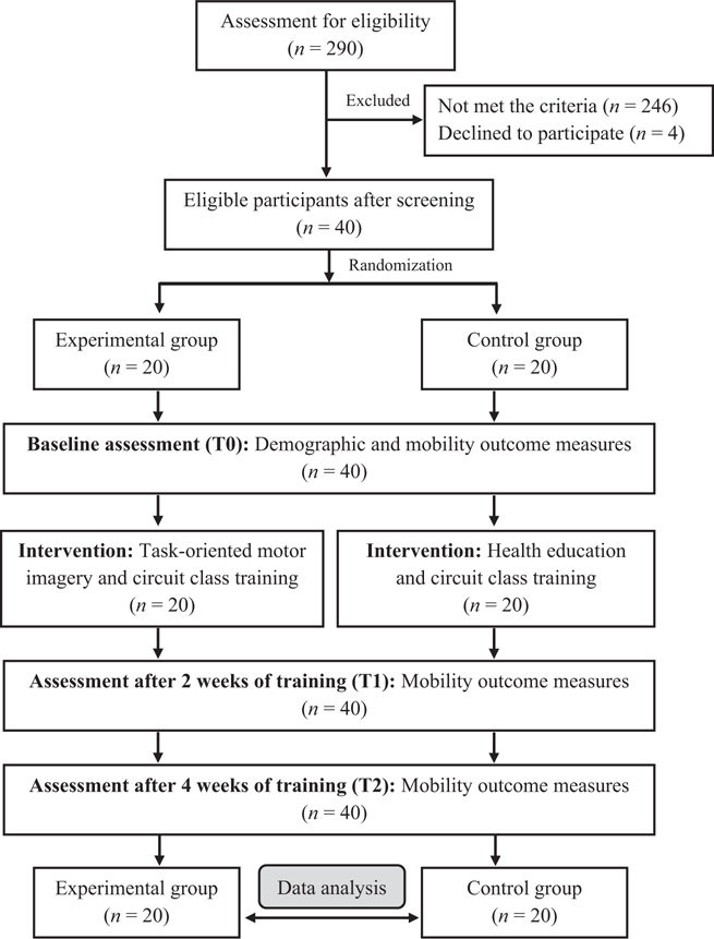
A total of 290 post-stroke individuals were screened, based on the criteria set by a physiotherapist; 246 did not meet the inclusion criteria, and 4 declined to participate in the study (Fig. 1). Thus, 40 post-stroke individuals participated in the study, which was conducted over 4 weeks. There were no dropouts and no reports of any hazardous situations for the participants.
The demographics of all participants are shown in Table I. There were no significant differences (p > 0.05) in the baseline demographics between the groups. The mean age of the experimental and control groups were 49.40 and 55.55 years, respectively. There were 15 males and 5 females in the experimental group and 11 males and 9 females in the control group. Both groups had a high walking capacity, monitored by FAC scores of 3 and 4, and low severity, indicated by the NIHSS score of 3. Furthermore, no problems of imagination ability were reported in either group.
| Clinical Characteristics | Total participants (n = 40) | Experimental group (n = 20) | Control group (n = 20) | p-value |
| Age, mean (SEM) (years) | 52.48 (1.81) | 49.40 (2.59) | 55.55 (2.40) | 0.090* |
| Weight, mean (SEM) (kg) | 63.49 (1.61) | 63.64 (1.48) | 63.34 (2.92) | 0.927* |
| Height, mean (SEM) (cm) | 162.98 (1.11) | 163.89 (1.46) | 162.06 (1.67) | 0.416* |
| Timed since stroke, mean (SEM) (weeks) | 26.93 (2.25) | 25.00 (3.46) | 28.85 (2.89) | 0.398* |
| Sex, n (%) | ||||
| Male | 26 (65) | 15 (75) | 11 (55) | 0.185† |
| Female | 14 (35) | 5 (25) | 9 (45) | |
| Type of stroke, n (%) | ||||
| Ischaemic | 18 (45) | 8 (40) | 10 (30) | 0.525† |
| Haemorrhagic | 22 (55) | 12 (60) | 10 (70) | |
| Side of involvement, n (%) | ||||
| Left | 22 (55) | 10 (50) | 12 (60) | 0.525† |
| Right | 18 (45) | 10 (50) | 8 (40) | |
| FAC, n (%) | ||||
| 3: Dependent for supervision | 18 (45) | 10 (50) | 8 (40) | 0.525† |
| 4: Independent level surfaces only | 22 (55) | 10 (50) | 12 (60) | |
| 5: Independent | 0 (0) | 0 (0) | 0 (0) | |
| NIHSS, mean (SEM) (scores) | 3.30 (0.21) | 3.45 (0.31) | 3.15 (0.29) | 0.487* |
| MMSE, mean (SEM) (scores) | 26.83 (0.16) | 26.80 (0.25) | 26.85 (0.21) | 0.878* |
| KVIQ-10, mean (SEM) (scores) | 37.75 (0.84) | 38.50 (1.03) | 37.00 (1.33) | 0.378* |
| p-values between the experimental and control groups tested by †χ2 test and *independent samples t-test. NIHSS: National Institutes of Health Stroke Scale; MMSE: Mini-Mental State Examination; FAC: Functional Ambulation Category; KVIQ-10: Kinesthetic and Visual Imagery Questionnaire; SEM: standard error of the mean. |
||||
Comparisons of functional mobility outcomes
Influence of the group on functional mobility outcomes. There was a significant main effect of group on the step test for the unaffected limb [F (1, 38) = 4.127, p = 0.049]. Meanwhile, no effects of group were found for the step test for the affected limb [F (1, 38) = 1.691, p = 0.201], 6MWT [F (1, 38) = 1.000, p = 0. 324], and TUG [F (1, 38) = 4.041, p = 0.052]. These findings indicated that only the step test for the unaffected limb was different between the groups throughout the testing periods.
Influence of time on functional mobility outcomes. There were significant main effects of time on all functional mobility outcomes, which included the step test for the affected limb [F (1.243, 47.241) = 141.481, p < 0.001], the step test for the unaffected limb [F (1.103, 41.929) = 139.211, p < 0.001], 6MWT [F (2, 76) = 145.095, p < 0.001], and TUG [F (1.044, 39.691) = 95.524, p < 0.001]. Over the time-points of assessment, significant changes were found in all functional mobility outcomes for both the groups that either received MI with SPCCT or education with SPCCT.
Interaction effect of the group by time on functional mobility outcomes. There were significant interaction effects of group and time in the step test for the affected limb [F (1.243, 47.241) = 16.988, p < 0.001], 6MWT [F (2, 76) = 26.887, p < 0.001], and TUG [F (1.044, 39.691) = 7.284, p = 0. 009], while no interaction effect was found in the step test for the unaffected limb [F (1.103, 41.929) = 1.762, p = 0.192]. The type of the training significantly influenced all functional mobility outcomes, except for the step test for the unaffected limb over the time-points of assessment (T0, T1 and T2).
Between-group and within-group sub-comparisons
Between-group and within-group comparisons in terms of functional mobility are shown in Table II. For the between-group comparisons, no difference was found at T0, but significant differences were found for the step test for the unaffected limb at T1 (p = 0.039), step test for the affected limb (p = 0.017), step test for the unaffected limb (p = 0.026), and TUG (p = 0.012) at T2.
| Outcome measures | Groups | Before (T0) | After 2 weeks (T1) | After 4 weeks (T2) | Difference between T0 and T1 | Difference between T0 and T2 | p-value† | p-valuea | p-valueb | p-valuec |
| Step test: affected limb (n) | Exp | 6.79 (0.56) | 8.90 (0.60) | 11.35 (0.64) | 2.20 (0.30) | 4.65 (0.45) | <0.001 | <0.001 | <0.001 | <0.001 |
| Con | 6.94 (0.61) | 7.74 (0.61) | 9.15 (0.61) | 0.80 (0.14) | 2.30 (0.30) | <0.001 | <0.001 | <0.001 | <0.001 | |
| p-value* | 0.858 | 0.183 | 0.017 | <0.001 | <0.001 | |||||
| Step test: unaffected limb (n) | Exp | 9.25 (0.67) | 11.05 (0.58) | 12.60 (0.57 | 1.80 (0.19) | 3.35 (0.37) | < 0.001 | <0.001 | <0.001 | <0.001 |
| Con | 7.95 (0.57) | 9.25 (0.61) | 10.65 (0.62) | 1.30 (0.16) | 2.70 (0.33) | <0.001 | <0.001 | <0.001 | <0.001 | |
| p-value* | 0.148 | 0.039 | 0.026 | 0.051 | 0.201 | |||||
| 6MWT (m) | Exp | 187.49 (16.97) | 217.18 (18.17) | 256.75 (19.49) | 29.69 (5.19) | 69.26 (5.36) | <0.001 | <0.001 | <0.001 | <0.001 |
| Con | 176.01 (24.90) | 188.00 (25.64) | 203.60 (26.28) | 11.99 (1.75) | 27.59 (3.01) | <0.001 | <0.001 | <0.001 | <0.001 | |
| p-value* | 0.706 | 0.360 | 0.113 | 0.004 | <0.001 | |||||
| TUG (s) | Exp | 22.02 (1.76) | 16.82 (1.35) | 11.64 (1.12) | 5.20 (0.62) | 10.38 (1.16) | <0.001 | <0.001 | <0.001 | <0.001 |
| Con | 29.24 (4.96) | 26.26 (4.48) | 23.35 (4.13) | 2.98 (0.62) | 5.89 (1.16) | <0.001 | <0.001 | <0.001 | <0.001 | |
| p-value* | 0.182 | 0.056 | 0.012 | 0.015 | 0.010 | |||||
| 6MWT: 6-Minute Walk Test, TUG: Timed Up and Go test, Exp: Experimental group, Con: Control group, Data reported in mean (standard error of mean; SEM), significance tested at p<0.05. p-value*independent sample t-test. p-value †repeated measure analysis of variance (ANOVA). p-value aBonferroni-corrected post hoc test between T0 and T1. p-value bBonferroni-corrected post hoc test between T0 and T2. p-value cBonferroni-corrected post hoc test between T1 and T2. |
||||||||||
For within-group comparisons, significant improvements were found for both the experimental and control groups over the testing times in all functional mobility outcomes, which included the step test for the affected limb (p < 0.001), step test for the unaffected limb (p < 0.001), 6MWT (p < 0.001), and TUG (p < 0.001). Moreover, the Bonferroni-corrected post hoc test showed significant differences for both groups in all pairwise comparisons (p < 0.001).
DISCUSSION
Previous review studies have reported that more intensive exercise therapy with repetitive task training can improve activities of daily living (ADL) and lower limb functions for post-stroke individuals (45, 46). The aim of this study was to examine the effects of MI combined with SPCCT on the dynamic balance, endurance, and functional mobility in post-stroke individuals. The tasks being trained in this study focused on increasing dynamic balance and functional mobility to increase an individual’s confidence using a progressive training method. No hazardous effects were reported. The large improvement in all outcomes in the study participants could be due to the tasks used in the MI strategy, specifically the tasks training in the SPCCT protocol. Four tasks in MI were identical to the 7 tasks used in the SPCCT. According to motor simulation theory (22), this priming effect of the MI method may reinforce practice capability in the SPCCT. In addition, the training enhances motor learning and prompts neuroplasticity. This reforms the brain’s physical structure through repeated training experience (47).
The step test was used to measure dynamic balance. The ability to step for the affected or unaffected limb indicates improvement of dynamic postural balance and lower limb strength when performing either the supportive or movable functions. When comparing the data between the 2 groups, the step test for the unaffected limb was improved at T1, while the improvement in the affected limb was found at T2. This improvement could be from carrying out the task being trained in MI involving a similar stepping motion. The key point that should be noted was that the instruction used in both MI and SPCCT was emphasized with prolonged weight-bearing on the affected limb. Improvement in step performance probably relates to the step forwards and backwards onto a stool, which was used in the MI training. The consistency of training and measurement may be a factor that greatly improved patients’ performance. The study by Vats (48) reported the benefits of task-specific step-up exercise in improving the spatiotemporal gait parameters of chronic stroke individuals. Likewise, the current study provided step tasks for the affected and unaffected sides, as well as alternately between both limbs. However, training progression was carried out by increasing the distance between the feet and stool in the previous study (48), while the current study progressed the step rhythm using a metronome. Stepping imagery was investigated in a previous study that found activation in the brain (49). This reinforces the usefulness of practicing different forms of MI on motor functioning. Furthermore, the benefits of reciprocal stepping and aerobic overground training have been reported to improve gait speed and endurance after training for 40 sessions in post-stroke individuals (50). Multidirectional step up-down practicing was not only useful in improving balance and gait performances, but also in improving falls efficacy (51). To step up on a stool successfully, post-stroke individuals had to lift the movable limb while alternately shifting weight onto the supporting limb (52). In the current study, the experimental group had an improvement of 4.7 steps for the affected limb and 3.4 steps for the unaffected limb at T2 compared with T0. Meanwhile, the controls had an improvement of only 2.3 and 2.7 steps for the affected and unaffected limbs, respectively.
For the 6MWT, slight, but not significant, improvement was found at T1 after training in both groups. However, a significant improvement was identified by a mean difference of 69.3 m in the experimental group with the ability to walk up to 256.8 m after the training at T2. The previous study on MI combined with the task-oriented circuit class training by Verma et al. (16) found an improvement of 6MWT for 84 m after training for 60 min, 7 days a week for only 2 weeks. This difference found may be due to the difference in the tasks and training protocols being used. Previous studies (14, 53) reported that massed practice pattern may yield a better effect on walking endurance, although our study used the distributed pattern. When considering the MI perspective, the current study provided a first-person perspective, while the previous study (16) provided an external perspective. Another point of consideration may be related to the stroke onset. The current study included patients with onset ranging between 12 and 52 weeks (mean onset 25 and 29 weeks for the experimental and control groups, respectively), while individuals with an onset of 4–12 weeks (mean onset 6.3 weeks) were included in the previous study (16). Regarding the type of stroke between studies, the current study has 55% haemorrhagic strokes, while the previous study (16) reported inclusion of only 20% haemorrhagic strokes. However, the minimal detectable change (MDC) for 6MWT has been estimated at 61 m for early stroke (54) and 34.4 m for late stroke (55). Furthermore, the minimally clinically important difference (MCID) of 6MWT was reported by the correlational study of 6MWT with the perceived change score after 3 months of the training in stroke individuals and reported a determining threshold of 34.4 m (56). Therefore, the experimental group could indicate a clinically change in 6MWT after 4 weeks of training.
Comparing the TUG between the groups, the experimental group showed a significant improvement by 0.47% at T2 compared with T0, while the control group showed an improvement of only 0.20%. A greater time reduction in performing TUG may be due to the same TUG task being used in the MI process and the SPCCT programme. Compared with the previous studies (7, 26, 32, 57), a 6.7 s improvement in TUG was found in post-stroke individuals after 5 weeks training of MI with conventional therapy, including muscle stretching, positioning, facilitating normal patterns of movement, facilitatory and inhibitory techniques, reflex inhibitory patterns, facilitating higher-level reflexes, and muscle-tone normalization (7). Another study (32) reported TUG improvement of 7.7 s after undergoing sit-to-stand MI with task-specific training for 6 weeks. The study by Cho et al. (26) found TUG improvement of 8.3 s after 6 weeks of MI training combined with treadmill training in chronic stroke patients. In addition, Kim et al. (57) identified significant effects of KI and VI training on TUG using a metronome, after 1 treatment session of 15 min. However, kinesthetic imagery training showed a better effect than VI training, with mean differences of 6.9 vs 6.8 s. It can be seen that the differences in outcomes between studies were due to training particulars and duration. The minimal detectable change (MDC) for the TUG recommended by the previous review study (15) was at least 8 s. Therefore, it is reasonable to infer that there was a clinically significant improvement of TUG in post-stroke individuals after training with the current study programme.
As the protocol details used in MI play an important role in training success, the first-person perspective has usually been used and reported, in order to facilitate greater cognitive function stimulation (58, 59) and autonomic response (60). Relationships between MI, autonomic nervous system, and movement observation have been reported through sensorimotor coupling, mediated by memory systems (61). Autonomic responses during MI training showed a change in heart rate, respiratory rate, skin resistance, and skin blood flow (31, 60). The sentiment regarding the use of heart rate for imaginary monitoring was presented in previous studies (31, 62), finding that heart rate was highly responsive to MI training. In addition, an in-depth study of the altered heart rate during MI training alongside the electroencephalogram (EEG) measurement was performed by Pfurtscheller et al. (63). The findings showed that both with and without MI conditions caused a rapid centrally localized beta event-related desynchronization (ERD) shortly with a maximum of 400 ms and a concurrent heart rate deceleration. Subsequently, the response pattern was changed to substantially increase ERD and heart rate acceleration. Nonetheless, many alternative methods have been proposed for tracking training engagement and reducing the concealed nature of the MI, such as counting the number of repetitions, verbal commanding to move a certain part of the body, monitoring an autonomic response, recalling the training details, and using VI (29–31). The current study used heart rate monitoring and periodic action commands to engage post-stroke individuals during the MI protocol, whereas some previous studies used explanation and verbal agreement between the trainer and trainee (7, 32). The current study MI programme shows evidence of more improvement because it is based on specificity, initiating from thinking (imaginary) to the exercise processes. Appropriate clinical training should not be too difficult or too easy to challenge functional ability in post-stroke individuals. The structured progressive training protocol was used to stepwise the exercise based on individuals’ performance. Thus, the protocol is feasible and provides a promising outcome in terms of several aspects of functional mobility.
Study limitations
This study has several limitations. First, the selection criteria were strict about including only the participants who were expected to be suitable for the proposed training programme and were able to walk a certain distance, leading to a large number of exclusions. Secondly, 55% of participants enrolled in the study had haemorrhagic stroke. The distribution of the type of stroke in this study differed from previous studies which found that the majority of participants were ischemic stroke. Both of these limitations may affect the generalizability of the results. Thirdly, the similarity of training task and measurement may favour the task specificity to the outcome. Limitations in terms of the learning challenge and transfer effect were also not assessed. Fourthly, a long-term effect of the intervention was not evaluated in this study. Further studies should investigate the long-term effect as well as other variables, such as quality of life and psychological factors.
CONCLUSION
This study validates the effects of MI with SPCCT in enhancing functional mobility in post-stroke individuals. The training is simple, low-cost, and appropriate for post-stroke individuals with mild to moderate levels of severity without problems of unilateral neglect. After mastering the training method, post-stroke individuals can practice it on their own. The additional effects of MI could facilitate better mobility function than did the education combined with SPCCT.
ACKNOWLEDGEMENTS
Authors’ contributions
Nilar Aung contributed to data curation, investigation, and writing the original draft of this paper, Vimonwan Hiengkaew and Jarugool Tretriluxana contributed to conceptualization, Mon S. Bryant contributed to writing review and editing, and Sunee Bovonsunthonchai contributed to conceptualization, writing the original draft, review and editing, and correspondence. All authors read and approved the final version of the manuscript.
REFERENCES
- Avan A, Digaleh H, Di Napoli M, Stranges S, Behrouz R, Shojaeianbabaei G, et al. Socioeconomic status and stroke incidence, prevalence, mortality, and worldwide burden: an ecological analysis from the Global Burden of Disease Study 2017. BMC Med 2019; 17: 1–30.
- Benjamin EJ, Muntner P, Alonso A, Bittencourt MS, Callaway CW, Carson AP, et al. Heart disease and stroke statistics-2019 update: a report from the American Heart Association. Circulation 2019; 139: e56–e528.
- Lord SE, McPherson K, McNaughton HK, Rochester L, Weatherall M. Community ambulation after stroke: how important and obtainable is it and what measures appear predictive? Arch Phys Med Rehabil 2004; 85: 234–239.
- DePaul VG, Moreland JD, deHueck AL. Physiotherapy needs assessment of people with stroke following discharge from hospital, stratified by acute functional independence measure score. Physiother Can 2013; 65: 204–214.
- Cohen JW, Ivanova TD, Brouwer B, Miller KJ, Bryant D, Garland SJ. Do performance measures of strength, balance, and mobility predict quality of life and community reintegration after stroke? Arch Phys Med Rehabil 2018; 99: 713–719.
- Mercer VS, Freburger JK, Chang S-H, Purser JL. Step test scores are related to measures of activity and participation in the first 6 months after stroke. Phys Ther 2009; 89: 1061–1071.
- Hosseini SA, Fallahpour M, Sayadi M, Gharib M, Haghgoo H. The impact of mental practice on stroke patients’ postural balance. J Neurol Sci 2012; 322: 263–267.
- Yoon HM, Han EY, Joo SJ. Significance of cycle ergometer as a measure of peak aerobic capacity in the disabled. J Stroke Cerebrovasc Dis 2021; 30: 105477.
- de Rooij IJM, Riemens MMR, Punt M, Meijer JG, Visser-Meily JMA, van de Port IGL. To what extent is walking ability associated with participation in people after stroke? J Stroke Cerebrovasc Dis 2021; 30: 106081.
- Danielsson A, Willen C, Sunnerhagen KS. Is walking endurance associated with activity and participation late after stroke? Disabil Rehabil 2011; 33: 2053–2057.
- Kelly JO, Kilbreath SL, Davis GM, Zeman B, Raymond J. Cardiorespiratory fitness and walking ability in subacute stroke patients. Arch Phys Med Rehabil 2003; 84: 1780–1785.
- Jonsdottir J, Cattaneo D, Recalcati M, Regola A, Rabuffetti M, Ferrarin M, et al. Task-oriented biofeedback to improve gait in individuals with chronic stroke: motor learning approach. Neurorehabil Neural Repair 2010; 24: 478–485.
- Bovonsunthonchai S, Aung N, Hiengkaew V, Tretriluxana J. A randomized controlled trial of motor imagery combined with structured progressive circuit class therapy on gait in stroke survivors. Sci Rep 2020; 10: 1–11.
- Dean CM, Richards CL, Malouin F. Task-related circuit training improves performance of locomotor tasks in chronic stroke: a randomized, controlled pilot trial. Arch Phys Med Rehabil 2000; 81: 409–417.
- English C, Hillier SL, Lynch EA. Circuit class therapy for improving mobility after stroke. Cochrane Database Syst Rev 2017.
- Verma R, Narayan Arya K, Garg R, Singh T. Task-oriented circuit class training program with motor imagery for gait rehabilitation in poststroke patients: a randomized controlled trial. Top Stroke Rehabil 2011; 18: 620–632.
- Wevers L, Van De Port I, Vermue M, Mead G, Kwakkel G. Effects of task-oriented circuit class training on walking competency after stroke: a systematic review. Stroke 2009; 40: 2450–2459.
- Guerra ZF, Lucchetti AL, Lucchetti G. Motor imagery training after stroke: a systematic review and meta-analysis of randomized controlled trials. J Neurol Phys Ther 2017; 41: 205–214.
- Polli A, Moseley GL, Gioia E, Beames T, Baba A, Agostini M, et al. Graded motor imagery for patients with stroke: a non-randomized controlled trial of a new approach. Eur J Phys Rehabil Med 2016; 53: 14–23.
- Silva S, Borges LR, Santiago L, Lucena L, Lindquist AR, Ribeiro T. Motor imagery for gait rehabilitation after stroke. Cochrane Database Syst Rev 2020; 9: CD013019.
- MacIntyre TE, Madan CR, Moran AP, Collet C, Guillot A. Chapter 9 - Motor imagery, performance and motor rehabilitation. In: Marcora S, Sarkar M, editors. Progress in brain research. Cambridge: Elsevier; 2018, p. 141–159.
- Jeannerod M. The representing braIn: Neural correlates of motor intention and imagery. Behav Brain Sci 1994; 17: 187–202.
- Jeannerod M. Neural simulation of action: a unifying mechanism for motor cognition. Neuroimage 2001; 14: S103–109.
- Kumar VK, Chakrapani M, Kedambadi R. Motor imagery training on muscle strength and gait performance in ambulant stroke subjects-a randomized clinical trial. J Clin Diagn Res 2016; 10: YC01.
- Bae Y-H, Ko Y, Ha H, Ahn SY, Lee W, Lee SM. An efficacy study on improving balance and gait in subacute stroke patients by balance training with additional motor imagery: a pilot study. J Phys Ther Sci 2015; 27: 3245–3248.
- Cho H-y, Kim J-s, Lee G-C. Effects of motor imagery training on balance and gait abilities in post-stroke patients: a randomized controlled trial. Clin Rehabil 2013; 27: 675–680.
- Oostra KM, Oomen A, Vanderstraeten G, Vingerhoets G. Influence of motor imagery training on gait rehabilitation in sub-acute stroke: a randomized controlled trial. J Rehabil Med 2015; 47: 204–209.
- Carrasco DG, Cantalapiedra JA. Effectiveness of motor imagery or mental practice in functional recovery after stroke: a systematic review. Neurologia 2016; 31: 43-52.
- Sharma N, Pomeroy VM, Baron JC. Motor imagery: a backdoor to the motor system after stroke? Stroke 2006; 37: 1941–1952.
- Hall C, Moore J, Annett J, Rodgers W. Recalling demonstrated and guided movements using imaginary and verbal rehearsal strategies. Res Q Exerc Sport 1997; 68: 136-144.
- Oishi K, Kasai T, Maeshima T. Autonomic response specificity during motor imagery. J Physiol Anthropol Appl Human Sci 2000; 19: 255–261.
- Helmy H, Elrewainy RM, Elbalawy Y, Sabbah A. Effect of adding motor imagery to task specific training on facilitation of sit to stand in hemiparetic patients. Arch Neurosci 2020; 7: e102053.
- Holden MK, Gill KM, Magliozzi MR, Nathan J, Piehl-Baker L. Clinical gait assessment in the neurologically impaired: reliability and meaningfulness. Phys Ther 1984; 64: 35–40.
- Folstein MF, Folstein SE, McHugh PR. “Mini-mental state”: a practical method for grading the cognitive state of patients for the clinician. J Psychiatr Res 1975; 12: 189–198.
- Dancer S, Brown AJ, Yanase LR. National Institutes of Health Stroke Scale reliable and valid in plain English. J Neurosci Nurs 2009; 41: 2–5.
- Malouin F, Richards CL, Jackson PL, Lafleur MF, Durand A, Doyon J. The Kinesthetic and Visual Imagery Questionnaire (KVIQ) for assessing motor imagery in persons with physical disabilities: a reliability and construct validity study. J Neurol Phys Ther 2007; 31: 20–29.
- Hill KD, Bernhardt J, McGann AM, Maltese D, Berkovits D. A new test of dynamic standing balance for stroke patients: reliability, validity and comparison with healthy elderly. Physiother Can 1996; 48: 257–262.
- Hong S-J, Goh EY, Chua SY, Ng SS. Reliability and validity of step test scores in subjects with chronic stroke. Arch Phys Med Rehabil 2012; 93: 1065–1071.
- Flansbjer U-B, Holmbäck AM, Downham D, Patten C, Lexell J. Reliability of gait performance tests in men and women with hemiparesis after stroke. J Rehabil Med 2005; 37: 75–82.
- Persson CU, Danielsson A, Sunnerhagen KS, Grimby-Ekman A, Hansson P-O. Timed Up & Go as a measure for longitudinal change in mobility after stroke–Postural Stroke Study in Gothenburg (POSTGOT). J Neuroeng Rehabil 2014; 11: 1–7.
- Rand D. Mobility, balance and balance confidence–correlations with daily living of individuals with and without mild proprioception deficits post-stroke. NeuroRehabilitation 2018; 43: 219–226.
- Nakao M, Izumi S, Yokoshima Y, Matsuba Y, Maeno Y. Prediction of life-space mobility in patients with stroke 2 months after discharge from rehabilitation: a retrospective cohort study. Disabil Rehabil 2020; 42: 2035–2042.
- Alghadir AH, Al-Eisa ES, Anwer S, Sarkar B. Reliability, validity, and responsiveness of three scales for measuring balance in patients with chronic stroke. BMC Neurol 2018; 18: 1–7.
- Blennerhassett J, Dite W. Additional task-related practice improves mobility and upper limb function early after stroke: a randomised controlled trial. Aust J Physiother 2004; 50: 219–224.
- Kwakkel G, van Peppen R, Wagenaar RC, Wood Dauphinee S, Richards C, Ashburn A, et al. Effects of augmented exercise therapy time after stroke: a meta-analysis. Stroke 2004; 35: 2529–2539.
- Thomas LH, French B, Coupe J, McMahon N, Connell L, Harrison J, et al. Repetitive task training for improving functional ability after stroke: a major update of a Cochrane review. Stroke 2017; 48: e102–e103.
- Moran A, O’Shea H. Motor imagery practice and cognitive processes. Front Psychol 2020; 11: 394.
- Vats M. Efficacy of task specific step-up exercises on the gait parameters of chronic hemiparetic stroke individuals. Int J Physiother Res 2013; 1: 130–137.
- Hsu W-C, Lin L-F, Chou C-W, Hsiao Y-T, Liu Y-H. EEG classification of imaginary lower limb stepping movements based on fuzzy support vector machine with kernel-induced membership function. Int J Fuzzy Syst 2017; 19: 566–579.
- Holleran CL, Straube DD, Kinnaird CR, Leddy AL, Hornby TG. Feasibility and potential efficacy of high-intensity stepping training in variable contexts in subacute and chronic stroke. Neurorehabil Neural Repair 2014; 28: 643–651.
- Park G-D, Choi J-U, Kim Y-M. The effects of multidirectional stepping training on balance, gait ability, and falls efficacy following stroke. J Phys Ther Sci 2016; 28: 82–86.
- Dean CM, Rissel C, Sherrington C, Sharkey M, Cumming RG, Lord SR, et al. Exercise to enhance mobility and prevent falls after stroke: the community stroke club randomized trial. Neurorehabil Neural Repair 2012; 26: 1046–1057.
- Salbach N, Mayo N, Wood-Dauphinee S, Hanley J, Richards C, Cote R. A task-orientated intervention enhances walking distance and speed in the first year post stroke: a randomized controlled trial. Clin Rehabil 2004; 18: 509–519.
- Perera S, Mody SH, Woodman RC, Studenski SA. Meaningful change and responsiveness in common physical performance measures in older adults. J Am Geriatr Soc 2006; 54: 743–749.
- Eng JJ, Dawson AS, Chu KS. Submaximal exercise in persons with stroke: test-retest reliability and concurrent validity with maximal oxygen consumption. Arch Phys Med Rehabil 2004; 85: 113–118.
- Tang A, Eng JJ, Rand D. Relationship between perceived and measured changes in walking after stroke. J Neurol Phys Ther 2012; 36: 115–121.
- Kim J-S, Oh D-W, Kim S-Y, Choi J-D. Visual and kinesthetic locomotor imagery training integrated with auditory step rhythm for walking performance of patients with chronic stroke. Clin Rehabil 2011; 25: 134–145.
- Berthoz A. The role of inhibition in the hierarchical gating of executed and imagined movements. Brain Res Cogn Brain Res 1996; 3: 101–113.
- Jeannerod M. Mental imagery in the motor context. Neuropsychologia 1995; 33: 1419–1432.
- Brown R, Kemp U, Macefield VG. Increases in muscle sympathetic nerve activity, heart rate, respiration, and skin blood flow during passive viewing of exercise. Front Neurosci 2013; 7: 102.
- Collet C, Di Rienzo F, El Hoyek N, Guillot A. Autonomic nervous system correlates in movement observation and motor imagery. Front Hum Neurosci 2013; 7: 415.
- Lanata A, Sebastiani L, Di Modica S, Scilingo EP, Greco A, editors. Classifying human motor imagery abilities from heart rate variability analysis: a preliminary study. 11th Conference of the European Study Group on Cardiovascular Oscillations (ESGCO); 2020, July 15-15; Pisa, Italy: IEEE; 2020.
- Pfurtscheller G, Solis-Escalante T, Barry RJ, Klobassa DS, Neuper C, Muller-Putz GR. Brisk heart rate and EEG changes during execution and withholding of cue-paced foot motor imagery. Front Hum Neurosci 2013; 7: 379.