ORIGINAL REPORT
EFFECTS OF DIFFERENT MONTAGES OF TRANSCRANIAL DIRECT CURRENT STIMULATION ON HAEMODYNAMIC RESPONSES AND MOTOR PERFORMANCE IN ACUTE STROKE: A RANDOMIZED CONTROLLED TRIAL
Wanalee KLOMJAI, PhD, PT1,2*, Benchaporn ANEKSAN, PhD, PT1,2, Songkram CHOTIK-ANUCHIT, MD3,4, Pentida JITKAEW, MSc, PT1,2, Kasina CHAICHANUDOMSUK, MSc, PT1,2, Pagamas PIRIYAPRASARTH, PhD, PT1,2, Roongtiwa VACHALATHITI, PhD, PT1, Yongchai NILANON, MD3,4 and Vimonwan HIENGKAEW, PhD, PT1
From the 1Faculty of Physical Therapy, 2Neuro Electrical Stimulation Laboratory (NeuE Lab), Faculty of Physical Therapy, Mahidol University, Salaya, Phutthamonthon, Nakhon Pathom, 3Department of Medicine and 4Siriraj Stroke Center, Faculty of Medicine Siriraj Hospital, Mahidol University, Bangkok, Thailand
Objective: Transcranial direct current stimulation (tDCS) has shown positive results in neurorehabilitation. However, there is limited evidence on its use in acute stroke, and unclear evidence regarding the best tDCS montage (anodal-, cathodal-, or dual-tDCS) for stroke recovery. This study investigated the effects of these montages combined with physical therapy on haemodynamic response and motor performance.
Methods: Eighty-two eligible acute stroke participants were allocated randomly into anodal, cathodal, dual, and sham groups. They received 5 consecutive sessions of tDCS combined with physical therapy for 5 days. Cerebral mean blood flow velocity (MFV) and motor outcomes were assessed pre- and post-intervention and at a 1-month follow-up.
Results: None of the groups showed significant changes in the MFV in the lesioned or non-lesioned hemispheres immediately post-intervention or at a 1-month follow-up. For motor performance, all outcomes improved over time for all groups; between-group comparisons showed that the dual-tDCS group had significantly greater improvement than the other groups for most of the lower-limb performance measures. All 5-day tDCS montages were safe.
Conclusion: MFV was not modulated following active or sham groups. However, dual-tDCS was more efficient in improving motor performance than other groups, especially for lower-limb performance, with after-effects lasting at least 1 month.
LAY ABSTRACT
Different methods (anodal, cathodal, dual, or sham) of transcranial direct current stimulation (tDCS) combined with physical therapy were randomly provided to patients with acute stroke for 5 consecutive daily sessions. The outcome measures were evaluated before the intervention, post-intervention, and at 1-month follow-up using clinical motor assessments and transcranial colour-coded Doppler ultrasonography to measure blood flow velocity in the middle cerebral artery. The results showed no significant changes in cerebral blood flow in the lesioned and non-lesioned brain in all groups. For motor outcomes, dual-tDCS was more efficient for improving motor performance, especially lower-limb performance, than other montages, with a positive effect lasting for at least 1 month. All tDCS montages combined with physical therapy were safe for use in patients with acute stroke.
Key words: acute stroke; ischaemic; transcranial direct current stimulation; unilateral; dual; cerebral blood flow; physical therapy.
Citation: J Rehabil Med 2022; 54: jrm00331. DOI: https://dx.doi.org/10.2340/jrm.v54.3208
Copyright: © Published by Medical Journals Sweden, on behalf of the Foundation for Rehabilitation Information. This is an Open Access article distributed under the terms of the Creative Commons Attribution-NonCommercial 4.0 International License (https://creativecommons.org/licenses/by-nc/4.0/)
Accepted: Aug 8, 2022; Epub ahead of print: Aug 29, 2022; Published: Sep 13, 2022
Correspondence address: Wanalee Klomjai, Faculty of Physical Therapy, Mahidol University, 999 Phuttamonthon 4 Road, Salaya, Nakhon Pathom 73170, Thailand. E-mail: wanalee.klo@mahidol.edu
Competing interests and funding: The authors have no conflicts of interest to declare.
After a unilateral stroke, the excitability of the affected hemisphere is decreased. This is coupled with an increase in the excitability of the unaffected hemisphere and abnormally high interhemispheric inhibition (IHI) from the intact to the lesioned hemisphere (1, 2). This reorganization of neuronal plasticity begins in the early stages after stroke (3). Transcranial direct current stimulation (tDCS) can induce changes in cortical excitability, which modulates brain plasticity in humans, and positive and safe results have been reported for tDCS in stroke neurorehabilitation (4, 5).
Unilateral and dual-tDCS are used to induce post-stroke motor recovery. Anodal-tDCS has been shown to increase cortical excitability, and cathodal-tDCS decreases cortical excitability, based on polarity-specific effects with limited doses (i.e. 0.5–1.5 mA) (8–13). Unilateral anodal-tDCS is used to restore excitability in the ipsilesional hemisphere by anodal stimulation, while cathodal-tDCS is used to decrease excitability in the contralesional hemisphere and to rebalance the IHI. Dual-tDCS can stimulate both hemispheres simultaneously. However, the evidence is unclear regarding the best tDCS montage to perform for stroke recovery, especially in the early phase (12).
One of the signs of a change in cortical activity is a subsequent variation in the haemodynamic response (13). Cerebral blood flow velocity is a haemodynamic evaluation that can be measured by transcranial Doppler ultrasonography through the major intracranial vessels and relatively thin bone windows. This method is a non-invasive, relatively inexpensive, safe, and portable bedside method that is convenient in the intensive care setting (14).
Five consecutive days of tDCS applied over the primary motor cortex (M1) area is reportedly safe in acute stroke treatment (15) and at least 5 sessions of physical therapy (PT) are required to induce clinical changes in motor performance (16). The aim of the current study was to investigate the haemodynamic and motor responses immediately following different montages of 5-session tDCS applied over the M1, and at a 1-month follow-up.
MATERIAL AND METHODS
Participants
A total of 82 eligible patients (see Table I for characteristics) were recruited by convenience sampling from the acute stroke unit of Siriraj Hospital, Bangkok, Thailand. Each patient was diagnosed with first unilateral ischaemic stroke in the anterior circulation (confirmed by magnetic resonance imaging/computed tomography (MRI/CT)), had unilateral weakness, stable vital signs, was able to follow commands, and had a modified Rankin Scale score ≤ 4. Participants were excluded if they had a National Institute of Health Stroke Scale (NIHSS) score > 20, hemineglect, presence of a contraindication to tDCS (17), or moderate-to-severe pain in any limb. Hemineglect and cognitive problems were screened by subscale of the NIHSS (inattention subscale for neglect (18), and cognitive subscale (Cog-4 : orientation, command, language and inattention) for cognitive problem (19, 20)). None of the participants had a history of mental health conditions or received psychological drugs (e.g. antidepressants) during study participation.
Experimental protocol
The study protocol is registered at ClinicalTrials.gov (NCT04051658). The participants provided written informed consent before the study commenced, and the study was approved by the ethics committee of the Faculty of Medicine at Siriraj Hospital. This study was a double-blind (participants, assessor) randomized controlled trial. Participants were allocated randomly to the anodal group (n = 21), cathodal group (n = 20), dual group (n = 20), or sham group (n = 21) by a third party. Match-paired design was used to match age (± 5 years), lesions, and motor subscale from the NIHSS (upper extremities (UE): 0–1, 2, 3–4 or lower extremities (LE): 0–1, 2). Randomization was performed using sealed envelopes marked “active,” “cathodal,” “dual,” or “sham” when it was necessary to start a new pair. The assessments were performed at the baseline (pre-), immediately after day-5 intervention (post-), and at 1-month follow-up (Fig. 1).
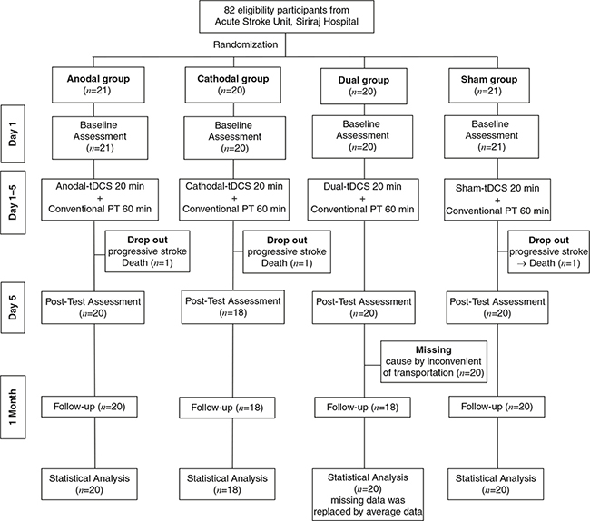
Fig. 1. Study flow chart. PT: physical therapy; tDCS: transcranial direct current stimulation.
Assessment
Haemodynamic response. Transcranial colour-coded Doppler (TCCD) ultrasonography (EPIQ 5C Ultrasound System, Philips, Bothell, Washington, USA) was used to measure the blood flow velocity in each middle cerebral artery (MCA) through the temporal bone, using a 5-1 MHz hand-held probe. The participants underwent TCCD in the supine position. The mean blood flow velocity (MFV) is a consistent measure used to document changes in cerebral blood flow; it is minimally affected by high flow velocity and low-amplitude signals from small vessels surrounding the major vessels being studied (21). The assessment was performed by the first researcher, (PJ) who was blinded to the group allocation.
Motor performance. The motor section of the Fugl–Meyer Assessment (FMA), FMA-UE and FMA-LE were used to assess motor function. Performance times on the Wolf Motor Function Test (WMFT) (lifting a can and lifting a pencil) were used to assess upper-limb function (22). The Five-Times Sit-To-Stand (FTSTS) and Timed Up and Go (TUG) tests were used to assess lower-limb function. The muscle strength of the elbow extensor, wrist extensor, hip extensor, hip flexor, knee extensor, and ankle dorsiflexor were measured using a hand-held dynamometer (Lafayette Manual Muscle Test System, model 01165; Lafayette, Indiana, USA). All assessments were performed using 1 trial, except for muscle strength, in which the better of 2 trials was used. All motor assessments were performed by the second researcher (PJ), who was blinded to the group allocation. Blood pressure, heart rate, and the adverse effects of tDCS were monitored throughout the experiment.
Intervention
tDCS. tDCS (Ybrain, MINDD STIM; Seongnam-si, Gyeonggi-do, Republic of Korea) was applied before the PT session (23). tDCS applied a direct current via 2 rectangular saline-soaked sponge-pad electrodes (35 cm2) that were attached firmly using a head cap. For the anodal group, the anodal electrode was placed at the M1 position of the ipsilesional hemisphere using the international 10–20 electrode placement system (C3 or C4), and the reference electrode was positioned over the contralateral supraorbital area (Fp1 or Fp2). For the cathodal group, the cathodal electrode was positioned over the M1 of the contralesional hemisphere, and the reference electrode was positioned over Fp1 or Fp2. For the dual group, the anodal electrode was placed over the M1 of the ipsilesional hemisphere, and the cathode was positioned over the M1 of the contralesional hemisphere (C3 or C4). The current intensity was 1.5 mA for 20 min for the active group (anodal/cathodal/dual); for the sham group 1.5 mA was applied for 30 s, and the electrodes remained in place for 20 min. The total charge density was 0.07 mAh/cm2. If electrode displacement occurred, an auto-alarm system was triggered, and the current was stopped immediately. The tDCS application was performed by the third researcher (KC), who was not involved in the outcome assessment or treatment. The participants were seated in a comfortable position during the stimulation.
PT session. A 1-h PT session was provided immediately after the tDCS by a physical therapist. The treatment programme followed the stroke rehabilitation guidelines for each individual impairment (24), with 30 min spent on each of the upper and lower limbs. The programme consisted of passive stretching, range of motion exercises, active/active-assisted/active-resisted exercises, weight-bearing on the arm, hand-function training (reaching to an object), sitting and standing balance exercises, and gait training.
Statistical analysis
Sample size was calculated based on the determination of input parameters for comparison of 4 groups (effect size f = 0.4, α error probability p = 0.05, power = 0.8), based on 1-way analysis of variance (ANOVA). The determined effect size is the intermediate effect size for statistically significant results reported for tDCS studies (25).The 0.8 power was selected as it is the minimal requirement for clinical research (26). Results showed that a sample size of 76 was adequate to attain reliable effects. Therefore, 82 participants were enrolled and, after dropout, data from 78 participants were used for statistical analysis.
Shapiro–Wilk test was performed to analyse parametric assumptions. Between-group comparisons of baseline data and demographic characteristics were performed using 1-way ANOVA and the χ2 test. All motor performance outcome data were transformed to percentage changes prior to analysis. Comparisons between groups (anodal vs cathodal vs dual vs sham) were performed using the Kruskal–Wallis test (post hoc by Dunnett’s test), and within-group comparisons (baseline (Pre) vs post-intervention (Post) vs follow-up (F/U)) were performed using the Friedman test (post hoc by Tukey’s test). Statistical significance was set at p < 0.05. All statistical analyses were performed using SPSS software. Data are reported as the mean (SD) for haemodynamic response data and the median (Q1, Q3) for motor performance data.
RESULTS
There were similarities in the characteristics and baseline data between the groups except for the number of participants who received thrombolytic therapy in each group (p = 0.011) (Table I). Minor adverse effects were reported only in the active group: tingling (50%), mild dizziness (20%), itching (10%), burning sensation (10%), and sleepiness (10%). However, the symptoms disappeared within 1 h. No significant changes in blood pressure or heart rate were observed during the experiment.
| Characteristics | Anodal | Cathodal | Dual | Sham | p-value | |||
| Age (years), mean (SD) | 59.94 (9.80) | 61.11 (9.70) | 57.20 (12.54) | 60.18 (10.20) | 0.599a | |||
| NIHSS (/42 score), mean (SD) | 3.38 (2.50) | 3.00 (2.62) | 3.12 (2.70) | 4.01 (2.28) | 0.895a | |||
| Time since stroke (days), Mean (SD) | 4.71 (1.38) | 3.56 (1.04) | 3.58 (1.42) | 3.62 (1.22) | 0.554a | |||
| BMI (kg/m2), mean (SD) | 24.12 (2.73) | 26.63 (2.98) | 24.59 (2.78) | 24,69 (2.37) | 0.054a | |||
| Sex | ||||||||
| Male | 17 | 9 | 11 | 12 | 0.120b | |||
| Female | 4 | 9 | 9 | 8 | ||||
| Artery occlusion (n) | ||||||||
| ACA | 4 | 2 | 1 | 0 | 0.086b | |||
| MCA | 17 | 16 | 19 | 20 | ||||
| Lesion (n) | ||||||||
| Subcortical | 19 | 11 | 17 | 18 | 0.088b | |||
| Cortical | 2 | 7 | 3 | 2 | ||||
| Handedness (n) | ||||||||
| Right | 20 | 17 | 20 | 20 | 0.968b | |||
| Left | 1 | 1 | 0 | 0 | ||||
| Affected limb (n) | ||||||||
| Right | 11 | 9 | 11 | 12 | 0.840b | |||
| Left | 10 | 9 | 9 | 8 | ||||
| Thrombolytic therapy (n) | ||||||||
| Received | 11 | 4 | 15 | 12 | 0.011b | |||
| Not received | 10 | 14 | 5 | 8 | ||||
| Rehabilitation post-discharge (n) | ||||||||
| Continue rehabilitation at hospital or centre | 6 | 6 | 7 | 5 | 0.906b | |||
| Continue home programme exercise by themselves | 15 | 12 | 13 | 15 | ||||
| CCB treatment during 5 days intervention (n) | ||||||||
| Received | 10 | 9 | 7 | N/A | 0.75b | |||
| Not received | 11 | 9 | 13 | N/A | ||||
| p-value from (a) one-way ANOVA and (b) x2 test. | ||||||||
| NIHSS: National Institutes of Health Stroke Scale; ACA: anterior cerebral artery; MCA: middle cerebral artery; CCB: calcium channel blocker drug; N/A: not assessed. | ||||||||
| p-value significant level at < 0.05. | ||||||||
MFV
Data were missing for 11 participants (3, 4, 2, and 2 from the anodal, cathodal, dual, and sham groups, respectively) due to the poor temporal acoustic window. Friedman’s test showed that Pre vs Post vs F/U were non-significant in anodal/cathodal/dual/sham groups. Kruskal–Wallis test showed that differences between groups (anodal vs cathodal vs dual vs sham) for both the lesioned and non-lesioned hemispheres were non-significant (Table II).
| Outcome measures (MCV) | Group | Mean (SD) (cm/s) | p-value | |||||
| Within-group comparison: Time effect | Between-group comparison: Stimulation effect | |||||||
| Pre | Post | F/U | Overall | Pre | Post | F/U | ||
| Lesion | Anodal | 74.71(35.46) | 75.28(25.45) | 72.83(22.17) | 0.664a | 0.730b | 0.825b | 0.183b |
| Cathodal | 71.71(38.44) | 76.48(44.86) | 76.97(39.97) | 0.789a | ||||
| Dual | 78.37(30.04) | 73.92(24.20) | 71.62(29.00) | 0.486a | ||||
| Sham | 67.27(26.84) | 70.31(33.64) | 59.01(20.99) | 0.179a | ||||
| Non-Lesion | Anodal | 65.00(28.93) | 83.33(39.38) | 78.26(28.68) | 0.152a | 0.574b | 0.475b | 0.447b |
| Cathodal | 79.12(28.16) | 74.76(24.46) | 77.34(28.35) | 0.396a | ||||
| Dual | 68.85(21.90) | 66.19(22.64) | 70.45(21.69) | 0.249a | ||||
| Sham | 68.31(31.76) | 67.29(26.56) | 65.72(26.95) | 0.348a | ||||
| aTesting by Friedman test; btesting by Kruskal–Wallis test. | ||||||||
| MCV: mean cerebral velocity; A: anodal; C: cathodal; Pre: baseline; Pre: pre-intervention; Post: post-intervention; F/U: follow-up. Significant level at *p < 0.005. | ||||||||
Motor outcomes (Table III)
- FMA-UE: Significant within-group differences were found in all groups (p < 0.001) with no significant differences between the groups (Fig. 2a).
- FMA-LE: Significant within-group differences were found in anodal/dual/sham (p < 0.001), and cathodal (p = 0.002) groups. Significant differences between the groups were found at F/U (H(3) = 10.95, p = 0.012). Post hoc comparisons revealed that the dual treatment induced greater improvement than the anodal and sham groups (Fig. 2b).
- WMFT-pencil: Significant within-group differences were found in anodal/cathodal/dual (p < 0.001) and sham (p = 0.001). Significant differences between groups were found at Post (H(3) = 10.95, p = 0.011) and F/U (H(3) = 11.32, p = 0.01). Post hoc comparisons revealed that the dual treatment induced greater improvement than the other treatments at Post and F/U (Fig. 2c).
- WMFT-can: Significant within-group differences were found in dual (p < 0.001), anodal/cathodal (p = 0.001), and sham (p = 0.043). Significant differences between the groups were found at Post (H(3) = 14.83, p = 0.011) and F/U (H(3) = 9.19, p = 0.014). Post hoc comparisons revealed that the dual treatment induced greater improvement than the other treatments at Post and F/U (Fig. 2d).
- FTSTS: Significant within-group differences were found in all groups (p < 0.001) with no significant differences between the groups (Fig. 2e).
- TUG: Significant within-group differences were found in anodal/cathodal/dual groups (p < 0.001). Significant differences were found at Post (H(3) = 12.04, p = 0.007) and F/U (H(3) = 18.01, p < 0.001). Post hoc comparisons revealed that the dual treatment induced greater improvement than the other treatments at Post and F/U (Fig. 2f).
Muscle strength
- Wrist extensor: Significant within-group differences were found in anodal (p < 0.001), dual (p = 0.001), and cathodal/sham (p = 0.002) groups, while the differences between groups were non-significant (Fig. 2g).
- Elbow extensor: Significant within-group differences were found in dual (p < 0.001), cathodal (p = 0.001), anodal (p = 0.005), and sham (p = 0.008) groups, while the differences between groups were non-significant (Fig. 2h).
- Hip flexor: Significant within-group differences were found in dual (p < 0.001), cathodal (p = 0.002), anodal (p = 0.005), and sham (p = 0.012) groups. Significant differences between the groups were found at Post (H(3) = 7.92, p = 0.048). Post hoc comparisons revealed that the dual treatment induced greater improvement than the other treatments (Fig. 2i).
- Hip extensor: Significant within-group differences were found in dual (p < 0.001), anodal (p = 0.001), cathodal (p = 0.005), and sham (p = 0.024). Significant differences between the groups were found at F/U (H(3) = 8.34, p = 0.039). Post hoc comparisons suggested that the dual treatment induced greater improvement than the other treatments (Fig. 2j).
- Knee extensor: Significant within-group differences were found in dual (p < 0.001), anodal (p = 0.005), and sham (p = 0.007). Significant differences between the groups were found at Post (H(3) = 12.01, p = 0.007) and F/U (H(3) = 11.10, p = 0.011). Post hoc comparisons revealed that the dual treatment induced greater improvement than the other treatments at Post and F/U (Fig. 2k).
- Ankle dorsiflexor: Significant within-group differences were found in anodal (p < 0.001), dual (p = 0.001), and cathodal (p = 0.007). Significant differences between groups were found at Post (H(3) = 9.19, p = 0.027) and F/U (H(3) = 12.08, p = 0.007). Post hoc comparisons revealed that the anodal treatment induced greater improvement than the cathodal and sham treatments at Post, and the dual treatment showed greater improvement than the sham treatment at F/U (Fig. 2l).
DISCUSSION
The main finding of this study was that there were no significant within- or between-group changes in MFV in the lesioned or non-lesioned brain for all montages (anodal, cathodal, dual, or sham) of tDCS combined with PT provided for patients with acute stroke for 5 consecutive daily sessions. For motor performance, all outcome measures improved over time (Pre vs Post vs F/U) in all groups. For between-group comparisons, the FMA-LE, WMFT, TUG, and muscle strength of the lower-limb muscles (hip, knee, and ankle) showed greater improvement after dual-tDCS than the other groups at Post and F/U; there were no significant differences between groups for FMA-UE, FTSTS, and muscle strength of the upper-limb muscles (wrist and elbow) (Table III).
| Outcome measures | Group | p-value | Post hoc | ||||||
| Within-group comparison: Time effect | Between-group comparison: Stimulation effect | ||||||||
| Overall | Post hoc | Post | F/U | ||||||
| Pre vs Post | Pre vs F/U | Overall | Post hoc | Overall | |||||
| Motor performance | FMA-UE | Anodal | < 0.001a*** | 0.001** | 0.001** | 0.228b | not-test | 0.186b | not-test |
| Cathodal | < 0.001a*** | 0.023* | 0.002** | ||||||
| Dual | < 0.001a*** | 0.005** | < 0.001** | ||||||
| Sham | < 0.001a*** | 0.001** | 0.010* | ||||||
| FMA-LE | Anodal | < 0.001a*** | 0.001** | 0.002** | 0.551b | not-test | 0.018b* | D > C (0.003**) | |
| Cathodal | 0.002a** | 0.032* | 0.002** | D > S (0.008**) | |||||
| Dual | < 0.001a*** | 0.061 | < 0.001*** | ||||||
| Sham | < 0.001a*** | 0.001** | 0.010* | ||||||
| Motor function | WMFT-pencil | Anodal | < 0.001a*** | 0.004** | 0.006** | 0.011b* | D > A (0.003**) | 0.010b* | D > A (<0.001***) |
| Cathodal | < 0.001a*** | 0.007** | 0.003** | D > S (0.024*) | D > C (0.003**) | ||||
| Dual | < 0.001a*** | 0.001** | < 0.001** | ||||||
| Sham | 0.001a** | 0.001** | 0.263 | ||||||
| WMFT-can | Anodal | 0.001a** | 0.008** | 0.008** | 0.002b* | D > A (<0.001***) | 0.011b* | D > A (0.003**) | |
| Cathodal | 0.001a** | 0.006** | 0.001** | D > C (0.001**) | D > C (0.019*) | ||||
| Dual | < 0.001a*** | < 0.001** | < 0.001** | ||||||
| Sham | 0.043a* | 0.001** | 0.478 | ||||||
| FTSTST | Anodal | < 0.001a*** | 0.003** | 0.001** | 0.774b | not-test | 0.369b | not-test | |
| Cathodal | < 0.001a*** | 0.016** | < 0.001*** | ||||||
| Dual | < 0.001a*** | 0.001** | < 0.001*** | ||||||
| Sham | < 0.001a*** | 0.001* | 0.002** | ||||||
| TUG | Anodal | < 0.001a*** | 0.017** | 0.001** | 0.007b** | D > C (0.006**) | < 0.001b** | A > S (0.001**) | |
| Cathodal | < 0.001a*** | 0.001** | < 0.001*** | D > S (<0.001***) | C > S (0.010*) | ||||
| Dual | < 0.001a*** | 0.001** | < 0.001** | D > S (<0.001***) | |||||
| Sham | 0.086 | not-test | not-test | ||||||
| Muscle strength | Wrist E | Anodal | < 0.001a*** | 0.001** | < 0.001*** | 0.201b | not-test | 0.505b | not-test |
| Cathodal | 0.002a** | 0001** | 0.001** | ||||||
| Dual | 0.001a** | 0.001** | 0.001** | ||||||
| Sham | 0.002a** | 0.030* | 0.002** | ||||||
| Elbow E | Anodal | 0.005a** | 0.022* | 0.003** | 0.852b | not-test | 0.495b | not-test | |
| Cathodal | 0.001a** | 0.022* | 0.001** | ||||||
| Dual | < 0.001a*** | 0.031* | 0.002** | ||||||
| Sham | 0.008a** | 0.005** | 0.009** | ||||||
| Hip F | Anodal | 0.005a** | 0.039* | 0.001** | 0.048b* | D > C (0.008**) | 0.055b | not-test | |
| Cathodal | 0.002a** | 0.094 | 0.001** | D > S (0.026*) | |||||
| Dual | < 0.001a*** | < 0.001*** | < 0.001*** | ||||||
| Sham | 0.012a* | 0.052 | 0.011* | ||||||
| Muscle strength | Hip E | Anodal | 0.001a** | 0.002** | 0.001** | 0.420b | not-test | 0.039b* | D > C (0.006**) |
| Cathodal | 0.005a** | 0.001** | 0.001** | D > S (0.024*) | |||||
| Dual | < 0.001a*** | 0.001** | < 0.001*** | ||||||
| Sham | 0.024a* | 0.012* | 0.002** | ||||||
| Knee E | Anodal | 0.005a** | 0.011* | 0.002** | 0.007b** | D > S (0.009**) | 0.011b* | D > A (0.007**) | |
| Cathodal | 0.059 | not test | not test | D > A (0.048*) | |||||
| Dual | < 0.001a*** | < 0.001*** | < 0.001*** | ||||||
| Sham | 0.007a** | 0.037* | 0.006** | ||||||
| Ankle DF | Anodal | < 0.001a*** | 0.001** | < 0.001 | 0.027b* | A > S (0.008**) | 0.007b** | A > C (0.001**) | |
| Cathodal | 0.007a** | 0.005** | 0.001** | A > S (0.004**) | |||||
| Dual | 0.001a** | 0.001** | 0.001** | D > S (0.024*) | |||||
| Sham | 0.076 | not test | not test | ||||||
| aTesting by Friedman test (post hoc by Tukey Test), bTesting by Kruskal-Wallis test (post hoc by Dunnett’s test). | |||||||||
| FMA-UE: Fugl-Meyer assessments upper extremity; FMA-LE: Fugl-Meyer assessments lower extremity; WMFT: Wolf Motor Function Test; TUG: Timed Up and Go Test; FTSTS: Five Times Sit to Stand Test; Muscles strength; F: flexor; E: extensor; DF: dorsiflexor; A: anodal; C: cathodal; D: dual; S: sham; Pre: pre-intervention; Post: post-intervention; F/U: follow-up. | |||||||||
| Significant level at *p < 0.05; **p < 0.01; ***p < 0.001. | |||||||||
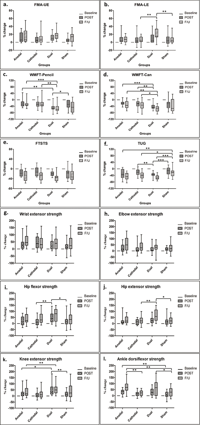
Fig. 2. Box plots represent percentage change of motor outcomes. The data are reported as median (Q1,Q3) at baseline, post-test (POST), and follow-up (F/U).
Considering participants’ characteristics, no between-group differences were observed except for the number of participants who received thrombolytic therapy that was low in the cathodal group (see Table I). However, it was shown that stroke patients who did not receive thrombolytic therapy had no significantly different function outcomes at 3 months post-stroke compared with those who received thrombolytic therapy. Age, premorbid functional status, the NIHSS score, Modified Ranking Scale score on the first day of admission at the hospital were shown to be significant predictors for the improvement of motor recovery within 3 months post-stroke (27). In the current study, age, the NIHSS score and motor performance at baseline were similar between groups.
Haemodynamic response
It has been shown that cerebral blood flow is continuously coupled to neural activity (28), and changes in blood flow velocity correlate with blood flow changes in the area supplied by the monitored arteries (29). Thus, changes in haemodynamic responses after ischaemic stroke are important pathophysiological measures. The current results showed that the MFVs of MCA in both hemispheres were significantly unchanged within 1-month post-stroke in all groups; however, we hypothesize that active tDCS might be able to induce changes in the cerebral blood flow. A longitudinal study reported that the MFV response to neural activation showed a triphasic trend from 72 h to 3 months post-stroke. The initially reduced bilateral MFV response was followed by an increase in MFV in the lesioned brain after 1 month, progressively returning to the control level by 3 months (30). A study showed that the cerebral blood flow response was poor in the lesioned brain during movement of the affected hand in the first stage of stroke (during 1-month post-stroke), progressively improving to a normal pattern at 6 months post-stroke (31). This could explain the unchanged MFVs we found, since the 1-month follow-up period had the first triphasic pattern. The MFVs of both hemispheres may continue to decrease during this phase (30); thus, it might be difficult to modulate the MFV by tDCS or training. The current study showed a slight decrease in the MFV in the lesioned brains of the sham group at 1-month post-stroke compared with that in the active-tDCS groups (Table II). The MFV correlates with oxygen delivery to the cortical tissue (32) during the first month of the recovery period and for long-term changes, including the sprouting of fibres and building new synaptic connections (33). However, blood flow redistribution is expected following cortical vessel occlusion (34). A computer modelling showed an important role of the Circle of Willis and pial collaterals in redistribution after anterior circulation occlusion (35). The lack of significant changes in the MFV of MCA found here is thus not able to exclude the possibility of redistribution in other areas.
It has been shown that unilateral-tDCS can alter cerebral haemodynamic response bilaterally in a polarity-specific manner: anodal-tDCS increases MFV, and cathodal-tDCS decreases it (36). A study of acute ischaemic stroke proposed that cathodal-tDCS was beneficial for preventing infarct growth in hyperacute MCA-area stroke patients receiving reperfusion therapy who had a baseline NIHSS score > 10; however, there was no significant difference compared with the sham treatment (37). The current results did not demonstrate a polarity-specific effect of tDCS on the MFV. However, since there was a positive correlation between the MFV in the non-lesioned brain and movement of the affected hand after the stroke (31), the possibility of the MFV decreasing following cathodal stimulation may be a concern. That there was no decrease in the MFV observed in the current study suggests that use of cathodal stimulation in the acute phase is safe.
Motor assessment
All within-group motor outcomes improved significantly for all groups. In between-group comparison, the dual-tDCS was the most effective intervention for improving upper- and lower-limb performance. Among the motor outcomes that improved (FMA-LE, WMFT, TUG, and muscle strength of the lower-limb muscle), the improvement in lower-limb performance was greater than that of the upper limb. This could be due to several reasons: (i) most of the participants (90%) had an MCA occlusion that led to more muscle weakness of the arm than of the leg (38), suggesting that a lower baseline of upper-limb muscle strength leads to difficulties in making improvement compared with the lower-limb muscles; and (ii) an unequal rate of motor recovery between the upper and lower limbs. In the early subacute phase, lower-limb muscles have a greater ability to recover than upper-limb muscles (39). In addition, the stimulation site was C3/C4, which is related more to the upper-limb M1. Previous studies have shown that tDCS over the upper-limb M1 influences both upper- and lower-limb performance in subacute to chronic stroke (40–43). This could be due to the wide spatial focus of tDCS, which can activate a larger area (44). The accumulative effect of multiple stimulations over the upper-limb M1 could also influence the area of the lower limb. The total charge densities in previous studies were 0.11–0.27 C/cm2 (40–42); the total charge density used in the present study was only 0.07C/cm2, which was sufficient to improve both upper- and lower-limb motor abilities.
Very few studies have compared the efficacy of tDCS montages in stroke patients. A sham-controlled study in subacute stroke showed greater motor recovery of the lower-limb for dual-tDCS combined with training compared with unilateral-tDCS (45). Recent meta-analyses reported that dual-tDCS induced better results than unilateral-tDCS on lower-limb motor performance in subacute stroke (46), and unilateral-tDCS was more effective than dual-tDCS for improving upper-limb motor performance in chronic stroke (47). These findings are consistent with our result that dual-tDCS induced better lower-limb performance than unilateral-tDCS. It was proposed in individuals with stroke that lower-limb function (i.e. walking) is controlled by combined activation in both ipsilateral corticospinal and cortico-reticulospinal pathways and contralateral superior cerebellar peduncle (48), while the ipsilateral descending control has a limited capacity to support the upper-limb function (49). This may explain the difference responses of upper- and lower-limb motor performance induced by unilateral- and dual-stimulation in the stroke population. Mordillo-Mateos et al. showed that dual-tDCS induced similar cortical excitability changes to the unilateral anodal-tDCS on the cathode-stimulated side, while on the anodal side, the simultaneous dual-tDCS seems to be slightly less robust. However, using bilateral montage resulted in lower inter-subject variability than unilateral montage in the excitability changes induced by anodal stimulation. The concomitant effects of cathodal stimulation on the non-lesioned hemisphere during dual-tDCS may promote an increased excitability of the motor cortex stimulated by the anode (50).
Moreover, it was shown that dual-tDCS over the M1 decreased the IHI during stimulation and increased the intracortical activity under the anodal electrode after termination of the intervention; unilateral anodal-tDCS resulted in similar effects during stimulation, but no changes could be observed after termination of tDCS (51). The delayed effect on intracortical activity within M1 during dual-tDCS might be beneficial for the application of tDCS before the training used in the current study. Dual-tDCS has been demonstrated to rebalance the IHI (52). To date, there is a lack of clarity regarding the initial timing of IHI imbalance presentation after stroke. Evidence has shown that the IHI for the first 1–12 weeks after stroke onset was normal, but became imbalanced after 12 weeks, despite that motor impairment had occurred since the onset (2, 53). Since our participants’ onsets ranged from 2 to 7 days, dual-tDCS probably did not have a role in rebalancing the IHI in this acute phase, but it might have slowed the IHI imbalance and, thus, reduced the motor deficit.
To date, only a handful of tDCS studies have been performed for acute stroke. Sattler et al. showed that anodal-tDCS (1.2 mA, 35 cm2, 20 min) combined with repetitive nerve stimulation for 5 consecutive sessions improved motor ability, and the effect lasted for at least 1 month (54). A recent study reported significant improvement in motor performance using anodal-tDCS combined with PT for 20 sessions (5 sessions/week, 4 weeks) with effects lasted for at least 1 year (42). Rabidi et al. reported that cathodal-tDCS combined with training for 10 sessions improved motor performance with an after-effect for at least 3 months (55). Thus, all tDCS montages seem to be beneficial in acute stroke. However, the current study demonstrated that 5 consecutive sessions of dual-tDCS with PT had the greatest benefit on motor performance, especially for the lower limb.
Study limitations
First, as multiple comparisons were performed due to the large number of outcomes compared between the groups in the present study, a higher risk of type I error should be noted. Secondly, the follow-up period was short; a longer follow-up period is recommended because the contribution of both hemispheres (i.e. changes in cerebral blood flow) to motor recovery may increase over several months. Thirdly, the current study recruited only patients with mild-to-moderate motor deficits. Because people with different levels of motor deficits could respond differently to tDCS, recruiting participants with more-severe motor deficits is suggested for further studies. Fourthly, some participants had a poor temporal window that limited the TCCD examination; this should be avoided in future studies. Fifthly, the present study did not control the rehabilitation programme after discharge from the acute stroke unit. However, there was no difference reported between the group of patients who received rehabilitation at the hospital and those who performed continuous exercises by themselves at home (Table I). Lastly, previous studies revealed that stroke patients with subcortical lesions responded better to tDCS than those with cortical lesions (56, 57). A previous study in acute stroke also showed a poorer haemodynamic response to rTMS in patients with cortical infarcts compared with those with subcortical infarcts (58). However, the number of participants with cortical infarcts in the present study was too small to evaluate this effect.
ACKNOWLEDGEMENTS
This work was supported by the National Research Council of Thailand (NRCT) (grant number 2562/11005), the Thailand Science Research and Innovation (Program Management Unit: PMU-P5 Frontier Research) (grant number 2563/6007 Re-Submit) and Mahidol University. P. Jitkaew had a grant from the National Research Council of Thailand (NRCT) (grant number 2562/24415).
REFERENCES
- Shimizu T, Hosaki A, Hino T, Sato M, Komori T, Hirai S, et al. Motor cortical disinhibition in the unaffected hemisphere after unilateral cortical stroke. Brain 2002; 125: 1896–1907.
- Murase N, Duque J, Mazzocchio R, Cohen LG. Influence of interhemispheric interactions on motor function in chronic stroke. Ann Neurol 2004; 55: 400–409.
- Di Pino G, Pellegrino G, Assenza G, Capone F, Ferreri F, Formica D, et al. Modulation of brain plasticity in stroke: a novel model for neurorehabilitation. Nat Rev Neurol 2014; 10: 597–608.
- Elsner B, Kugler J, Pohl M, Mehrholz J. Transcranial direct current stimulation (tDCS) for improving activities of daily living, and physical and cognitive functioning, in people after stroke. Cochrane Database Syst Rev 2016; 3: CD009645.
- Klomjai W, Lackmy-Vallée A, Roche N, Pradat-Diehl P, Marchand-Pauvert V, Katz R. Repetitive transcranial magnetic stimulation and transcranial direct current stimulation in motor rehabilitation after stroke: an update. Ann Phys Rehabil Med 2015; 58: 220–224.
- Nitsche MA, Paulus W. Sustained excitability elevations induced by transcranial DC motor cortex stimulation in humans. Neurology 2001; 57: 1899–1901.
- Nitsche MA, Paulus W. Excitability changes induced in the human motor cortex by weak transcranial direct current stimulation. J Physiol 2000; 527 Pt 3: 633–639.
- Batsikadze G, Moliadze V, Paulus W, Kuo MF, Nitsche MA. Partially non-linear stimulation intensity-dependent effects of direct current stimulation on motor cortex excitability in humans. J Physiol 2013; 591: 1987–2000.
- Jamil A, Batsikadze G, Kuo HI, Labruna L, Hasan A, Paulus W, et al. Systematic evaluation of the impact of stimulation intensity on neuroplastic after-effects induced by transcranial direct current stimulation. J Physiol 2017; 595: 1273–1288.
- Esmaeilpour Z, Marangolo P, Hampstead BM, Bestmann S, Galletta E, Knotkova H, et al. Incomplete evidence that increasing current intensity of tDCS boosts outcomes. Brain Stimul 2018; 11: 310–321.
- Hassanzahraee M, Nitsche MA, Zoghi M, Jaberzadeh S. Determination of anodal tDCS duration threshold for reversal of corticospinal excitability: an investigation for induction of counter-regulatory mechanisms. Brain Stimul 2020; 13: 832–839.
- Lefebvre S, Liew SL. Anatomical parameters of tDCS to modulate the motor system after stroke: a review. Front Neurol 2017; 8: 1-18.
- Dutta A. Bidirectional interactions between neuronal and hemodynamic responses to transcranial direct current stimulation (tDCS): challenges for brain-state dependent tDCS. Front Syst Neurosci 2015; 9: 107.
- Naqvi J, Yap KH, Ahmad G, Ghosh J. Transcranial Doppler ultrasound: a review of the physical principles and major applications in critical care. Int J Vasc Med 2013; 2013: 629378.
- Rossi C, Sallustio F, Di Legge S, Stanzione P, Koch G. Transcranial direct current stimulation of the affected hemisphere does not accelerate recovery of acute stroke patients. Eur J Neurol 2013; 20: 202–224.
- Chhatbar PY, Ramakrishnan V, Kautz S, George MS, Adams RJ, Feng W. Transcranial Direct Current Stimulation post-stroke upper extremity motor recovery studies exhibit a dose-response relationship. Brain Stimul 2016; 9: 16–26.
- Bornheim S, Croisier JL, Maquet P, Kaux JF. Proposal of a new transcranial direct current stimulation safety screening tool. Am J Phys Med Rehabil 2019; 98: e77.
- Lyden P. Using the National Institutes of Health Stroke Scale: a cautionary tale. Stroke 2017; 48: 513–519.
- Ankolekar S, Renton C, Sprigg N, Bath PMW. The Cog-4 Subset of the National Institutes of Health Stroke Scale as a Measure of Cognition: relationship with baseline factors and functional outcome after stroke using data from the Virtual International Stroke Trials Archive. Stroke Res Treat 2013; 2013: e562506.
- Cumming TB, Blomstrand C, Bernhardt J, Linden T. The NIH Stroke Scale can establish cognitive function after stroke. Cerebrovasc Dis 2010; 30: 7–14.
- Iyer PC, Madhavan S. Non-invasive brain stimulation in the modulation of cerebral blood flow after stroke: a systematic review of transcranial Doppler studies. Clin Neurophysiol 2018; 129: 2544–2551.
- Bogard K, Wolf S, Zhang Q, Thompson P, Morris D, Nichols-Larsen D. Can the Wolf Motor Function Test be streamlined? Neurorehabil Neural Repair 2009; 23: 422–428.
- Klomjai W, Aneksan B. A randomized sham-controlled trial on the effects of dual-tDCS “during” physical therapy on lower limb performance in sub-acute stroke and a comparison to the previous study using a “before” stimulation protocol. BMC Sports Sci Med Rehabil 2022; 14: 68.
- Winstein CJ, Stein J, Arena R, Bates B, Cherney LR, Cramer SC, et al. Guidelines for adult stroke rehabilitation and recovery: a guideline for healthcare professionals from the American Heart Association/American Stroke Association. Stroke 2016; 47: e98–169.
- Minarik T, Berger B, Althaus L, Bader V, Biebl B, Brotzeller F, et al. The importance of sample size for reproducibility of tDCS effects. Front Hum Neurosci 2016; 10: 453.
- Jones S, Carley S, Harrison M. An introduction to power and sample size estimation. Emerg Med J 2003; 20: 453–458.
- Duangjit S, Muangpaisan W, Chotinaiwattarakul W, Dharmasaroja P. Functional recovery at 3 months in stroke patients not receiving thrombolytic therapy: the comparison between patients arriving earlier and later than 4.5 hours. J Stroke Cerebrovasc Dis 2014; 23: 91–98.
- Venkat P, Chopp M, Chen J. New insights into coupling and uncoupling of cerebral blood flow and metabolism in the brain. Croat Med J 2016; 57: 223–228.
- Kontos H A. Validity of cerebral arterial blood flow calculations from velocity measurements. Stroke 1989; 20: 1–3.
- Salinet ASM, Panerai RB, Robinson TG. The longitudinal evolution of cerebral blood flow regulation after acute ischaemic stroke. Cerebrovasc Dis Extra 2014; 4: 186–197.
- Cuadrado ML, Egido JA, González-Gutiérrez JL, Varela-de-Seijas E. Bihemispheric contribution to motor recovery after stroke: a longitudinal study with transcranial doppler ultrasonography. Cerebrovasc Dis 1999; 9: 337–344.
- Markus HS. Cerebral perfusion and stroke. J Neurol Neurosurg Psychiatry 2004; 75: 353.
- Carmichael ST, Kathirvelu B, Schweppe CA, Nie EH. Molecular, cellular and functional events in axonal sprouting after stroke. Exp Neurol 2017; 287(Pt 3): 384–394.
- Chen S, Liu Q, Shu X, Soetikno B, Tong S, Zhang HF. Imaging hemodynamic response after ischemic stroke in mouse cortex using visible-light optical coherence tomography. Biomed Opt Express 2016; 7: 3377–3389.
- Phan TG, Hilton J, Beare R, Srikanth V, Sinnott M. Computer modeling of anterior circulation stroke: proof of concept in cerebrovascular occlusion. Front Neurol 2014; 5: 176.
- Giorli E, Tognazzi S, Briscese L, Bocci T, Mazzatenta A, Priori A, et al. Transcranial direct current stimulation and cerebral vasomotor reserve: a study in healthy subjects. J Neuroimaging 2015; 25: 571–574.
- Pruvost-Robieux E, Benzakoun J, Turc G, Marchi A, Mancusi RL, Lamy C, et al. Cathodal transcranial direct current stimulation in acute ischemic sroke: pilot randomized controlled trial. Stroke 2021; 52: 1951–1960.
- Tyson SF, Chillala J, Hanley M, Selley AB, Tallis RC. Distribution of weakness in the upper and lower limbs post-stroke. Disabil Rehabil 2006; 28: 715–719.
- Lee KB, Lim SH, Kim KH, Kim KJ, Kim YR, Chang WN, et al. Six-month functional recovery of stroke patients: a multi-time-point study. Int J Rehabil Res 2015; 38: 173–180.
- Lindenberg R, Renga V, Zhu LL, Nair D, Schlaug G. Bihemispheric brain stimulation facilitates motor recovery in chronic stroke patients. Neurology 2010; 75: 2176–2184.
- Cha HK, Ji SG, Kim MK, Chang JS. Effect of transcranial direct current stimulation of function in patients with stroke. J Phys Ther Sci 2014; 26: 363–365.
- Bornheim S, Croisier JL, Maquet P, Kaux JF. Transcranial direct current stimulation associated with physical-therapy in acute stroke patients – a randomized, triple blind, sham-controlled study. Brain Stimul 2020; 13: 329–336.
- Prathum T, Piriyaprasarth P, Aneksan B, Hiengkaew V, Pankhaew T, Vachalathiti R, et al. Effects of home-based dual-hemispheric transcranial direct current stimulation combined with exercise on upper and lower limb motor performance in patients with chronic stroke. Disabil Rehabil 2021; 1–12.
- Minhas P, Bikson M, Woods AJ, Rosen AR, Kessler SK. Transcranial direct current stimulation in pediatric brain: a computational modeling study. Conf Proc IEEE Eng Med Biol Soc 2012; 2012: 859–862.
- Andrade SM, Ferreira JJ de A, Rufino TS, Medeiros G, Brito JD, da Silva MA, et al. Effects of different montages of transcranial direct current stimulation on the risk of falls and lower limb function after stroke. Neurol Res 2017; 39: 1037–1043.
- Bai X, Guo Z, He L, Ren L, McClure MA, Mu Q. Different therapeutic effects of Transcranial direct current stimulation on upper and lower limb recovery of stroke patients with motor dysfunction: a meta-analysis. Neural Plast 2019; 2019: 1372138.
- Halakoo S, Ehsani F, Hosnian M, Zoghi M, Jaberzadeh S. The comparative effects of unilateral and bilateral transcranial direct current stimulation on motor learning and motor performance: a systematic review of literature and meta-analysis. J Clin Neurosci 2020; 72: 8–14.
- Soulard J, Huber C, Baillieul S, Thuriot A, Renard F, Aubert Broche B, et al. Motor tract integrity predicts walking recovery: a diffusion MRI study in subacute stroke. Neurology 2020; 94: e583–593.
- Wilkins KB, Yao J, Owen M, Karbasforoushan H, Carmona C, Dewald JPA. Limited capacity for ipsilateral secondary motor areas to support hand function post-stroke. J Physiol 2020; 598: 2153–2167.
- Mordillo-Mateos L, Turpin-Fenoll L, Millán-Pascual J, Núñez-Pérez N, Panyavin I, Gómez-Argüelles JM, et al. Effects of simultaneous bilateral tDCS of the human motor cortex. Brain Stimul 2012; 5: 214–222.
- Sehm B, Kipping J, Schäfer A, Villringer A, Ragert P. A Comparison between Uni- and Bilateral tDCS effects on functional connectivity of the human motor cortex. Front Hum Neurosci 2013; 7: 183.
- Tazoe T, Endoh T, Kitamura T, Ogata T. Polarity specific effects of transcranial direct current stimulation on interhemispheric inhibition. PLoS ONE 2014; 9: e114244.
- Xu J, Branscheidt M, Schambra H, Steiner L, Widmer M, Diedrichsen J, et al. Rethinking interhemispheric imbalance as a target for stroke neurorehabilitation. Ann Neurol 2019; 85: 502–513.
- Sattler V, Acket B, Raposo N, Albucher JF, Thalamas C, Loubinoux I, et al. Anodal tDCS combined with radial nerve stimulation promotes hand motor recovery in the acute phase after ischemic stroke. Neurorehabil Neural Repair 2015; 29: 743–754.
- Rabadi MH, Aston CE. Effect of Transcranial Direct Current Stimulation on severely affected arm-hand motor function in patients after an acute ischemic stroke: a pilot randomized control trial. Am J Phys Med Rehabil 2017; 96(10 Suppl 1): S178–184.
- Appelros P. Prevalence and predictors of pain and fatigue after stroke: a population-based study. Int J Rehabil Res 2006; 29: 329–333.
- Straudi S, Fregni F, Martinuzzi C, Pavarelli C, Salvioli S, Basaglia N. tDCS and robotics on upper limb stroke rehabilitation: effect modification by stroke duration and type of stroke. Biomed Res Int 2016; 2016: 5068127.
- Khaleel SH, Bayoumy IM, El-Nabil LM, Moustafa RR. Differential hemodynamic response to repetitive transcranial magnetic stimulation in acute stroke patients with cortical versus subcortical infarcts. Eur Neurol 2010;63(6):337–342.