ORIGINAL REPORT
EFFECTS OF INDIVIDUALIZED LOWER LIMB ISOKINETIC STRENGTHENING IN CLINICAL REHABILITATION OF OLDER POST-STROKE PATIENTS: A RETROSPECTIVE STUDY
Lyne DAUMAS, MSc1,2, Raphaël ZORY, PhD1,3, Axelle GARCIA, MD4, Amyn JAAFAR, MSc4, Luisa IENTILE, MD4, Emeline MICHEL, MD1,4, Guillaume SACCO, MD, PhD2,4,5 and Frédéric CHORIN, PhD1,4
From the 1University Côte d’Azur, Laboratory Human Motricity Expertise Sport and Health, France; 2University Côte d’Azur, Cognition Behaviour Technology, France; 3University Institute of France; 4University Côte d’Azur, University Hospital Center of Nice, France, Nice and 5UPRES EA 4638, University of Angers, France
Objective: Muscle weakness in the lower limbs is a motor consequence of stroke that causes functional impairment. The aim of this study was to assess the effectiveness of an individualized isokinetic strengthening programme, using the moment-velocity profile, on functional recovery during post-stroke rehabilitation of older patients. A further objective was to describe the effects of the individualized isokinetic strengthening on muscular parameters.
Design: Retrospective study.
Patients: Older post-stroke patients.
Methods: Using the Barthel Index, functional abilities in basic daily tasks were assessed and retrospectively analysed for 88 patients in a post-stroke rehabilitation unit. Of these, 44 patients received conventional rehabilitation (conventional group) and 44 received individualized isokinetic strengthening in addition to conventional rehabilitation (isokinetic group). A 2-Group (isokinetic, conventional) × 2-Time (before, after intervention) repeated measures analysis of variance (ANOVA) was conducted. For muscular parameters, Student t-tests and Wilcoxon tests were performed.
Results: The Barthel Index score increased more in the isokinetic group (61.59 ± 26.34 to 88.18 ± 12.16) than in the conventional group (61.70 ± 26.5 to 76.93 ± 18.12). A significant Time × Group interaction was found (F(1,86) = 5.95, p = 0.02). In the isokinetic group all muscular parameters improved.
Conclusion: This retrospective clinical study suggests that lower limb isokinetic strengthening, individualized using the moment-velocity profile, is clinically efficient for functional recovery during post-stroke rehabilitation of older patients. Intra-group effects of isokinetic strengthening also suggest benefits for muscular parameters.
LAY ABSTRACT
Muscle weakness in the lower limbs is a motor consequence of stroke that limits abilities to accomplish activities of daily life, such as walking. Individualized isokinetic exercises, based on patient’s force deficit and using a specialized equipment that adapts resistance according to the amount of force applied in order to maintain a constant speed throughout the movement, were performed in a post-stroke rehabilitation unit for older adults with sub-acute stroke. Retrospective analysis showed that functional recovery, as assessed by the clinical Barthel Index, was better in the patient who received individualized isokinetic strengthening in addition to conventional rehabilitation, compared with those who received only conventional rehabilitation. In addition, the results suggest beneficial effects on muscular parameters, such as force.
Key words: stroke; rehabilitation; physical exercise; strength; isokinetic.
Citation: J Rehabil Med 2023; 55: jrm7803. DOI: https://doi.org/10.2340/jrm.v55.7803.
Copyright: © Published by Medical Journals Sweden, on behalf of the Foundation for Rehabilitation Information. This is an Open Access article distributed under the terms of the Creative Commons Attribution-NonCommercial 4.0 International License (https://creativecommons.org/licenses/by-nc/4.0/)
Accepted: Jun 1, 2023; Published: Aug 7, 2023
Correspondence address: Lyne Daumas, LAMHESS, CoBTeK, Hôpital Cimiez, Université Côte d’Azur, bâtiment Mossa, Etage -1 – Plateforme fragilité, 4 avenue reine victoria, FR-06003 Nice, France. E-mail: daumasl@chu-nice.fr
Competing interests and funding: The authors have no conflicts of interest to declare.
Muscle weakness in the lower limbs is a motor consequence of stroke that causes functional impairments, which limit locomotor capacities and daily activities. More specifically, the muscle weakness includes reduced muscle strength (1), decreased strength production velocity (2), increased neuromuscular fatigability (3, 4) and is often more pronounced in the paretic limb (1, 5). Rehabilitation is a fundamental first step in the clinical management of patients with stroke, and one of the main goals is to improve walking ability, which correlates with the strength and power of the periarticular knee muscle (1, 6). The isokinetic ergometer is now considered the gold standard in the assessment of muscle moment of force in patients with stroke, and appears to be a promising rehabilitation tool (7–10). Previous studies have shown the effectiveness of isokinetic strengthening on knee muscular strength and functional capacities, such as walking ability, in stroke patients (10–12). More specifically, previous studies have shown that isokinetic programmes had a more pronounced beneficial effect than functional strengthening and when compared with conventional physiotherapy alone (12, 13). On the other hand, physical capacities and needs are highly heterogeneous in older people. Therefore, individualized physical activity programmes are increasingly used in clinical and research contexts (14).
Power is a determinant of walking ability in stroke patients (15). Some subjects present a strength deficit, while others present a velocity deficit, and these components can both affect muscle power (16). These inter-individual differences could partly explain why subjects can be either responders or non-responders to a particular type of training (17). Thus, the design of strengthening programmes should take individual deficits into consideration and first determine if the patient has a deficit in the capacity to develop high levels of strength at slow speed or a reduced ability to develop strength at high speed. An assessment of the moment-velocity profile using an isokinetic ergometer (the moment of force to angular velocity relationship corresponding to the force-velocity profile) might be an interesting approach to better understand the origin of a lack of muscular power and functional alterations, and to design more effective interventions for older people (16, 18). More specifically, the use of the power-velocity relationship might help to define the best speed for efficient work (19). Studies carried out in the context of performance sport have already highlighted the effectiveness of working with the force-velocity profile for performance optimization (20, 21) and it can be assumed that this methodology may be applied to older subjects with stroke for the optimization of functional capacities.
Thus, the objective of this study was to retrospectively assess the effectiveness of an individualized isokinetic strengthening programme, using the moment-velocity profile, on clinical recovery in basic daily tasks during post-stroke rehabilitation of older patients. It was hypothesized that individualized isokinetic strengthening in addition to conventional rehabilitation would provide better functional recovery compared with conventional rehabilitation without individualized isokinetic strengthening. A further objective was to describe the effect of individualized isokinetic strengthening on muscular parameters in these patients.
MATERIALS AND METHODS
Subjects and design
Data from 88 patients (mean age 79.52 ± 5.19 years) in a post-stroke rehabilitation unit for older adults with stroke in sub-acute phase were analysed retrospectively. Among them, 44 patients (mean age 79.66 ± 5.47 years) received an individualized isokinetic strengthening in addition to the conventional rehabilitation (isokinetic group) during their care process. Then, in order to assess the additional effect of the individualized isokinetic strengthening, data from 44 patients (mean age 79.39 ± 4.95 years) who did not receive isokinetic strengthening and who have similar functional limitation and age were used for analysis. These patients did not receive isokinetic strengthening, because the isokinetic ergometer was not available. Therefore they received only conventional rehabilitation, which included physiotherapy and occupational therapy (conventional group).
When the isokinetic ergometer was available for clinical use, the isokinetic strengthening rehabilitation was offered after a consultation between the physician and the physiotherapist. To be eligible, the subject’s transfer capacities had to be sufficiently preserved to perform the transfer on the ergometer. The exclusion criteria were: (i) transfer capacities too impaired as assessed by the Barthel Index; and (ii) significant cognitive disorders that could limit understanding of the instructions. In order to have comparable groups, likewise for the patients of the conventional group, the transfer capacities had to be sufficiently preserved as assessed by the Barthel Index. For both groups, data were collected and analysed only from those patients who were not opposed to their use. Data were recorded from 2015 to 2021. The study was approved by the ethics committee of Université Côte d’Azur (Avis [acceptance] number 2022-010).
Assessment
Clinical assessment. For each patient in each group, characteristics and clinical data were collected. The severity of neurological damage caused by the stroke was assessed using the National Institute of Health Stroke Scale (NIHSS) at admission (22). The Barthel Index was administered before and after intervention to assess the patient’s functional abilities in basic daily tasks, notably in mobility and transfers (23). Spasticity was assessed with the Modified Ashworth Scale, from 0 “No increase in muscle tone” to 4 “Affected part(s) rigid flexion extension” (24).
Isokinetic assessment. Muscular parameters from the isokinetic ergometer were available only for patients of the isokinetic group. Isokinetic measurements were performed using the Biodex dynamometer system 4 and sampling was performed at 100 Hz using an electrinic interface card (Biodex Medical Systems Inc., X2151, Shirley, NY, USA). Patients were installed on a chair with the axis of rotation of the dynamometer aligned with the centre of the lateral femoral condyle. To minimize extraneous body movements during thigh muscle contractions, straps were applied across the chest and the mid-thigh. Before assessment and each isokinetic session, to familiarize the patients with the procedure, they completed a dynamic warm-up with concentric submaximal contractions of the knee flexor and extensor.
The maximum moment of the knee extension was identified as the highest value reached during the movement at each constant speed. The evaluation was carried out on the isokinetic dynamometer at 6 different speeds (30, 60, 90, 120, 150 and 180°/s, corresponding to 0.52, 1.04, 1.57, 2.09, 2.61 and 3.14 rad/s) with encouragement and 1 min rest between each speed. The patient was asked to perform 3 repetitions for each speed, and the maximum moment of force was recorded. Evaluations were conducted before and during intervention to adjust strengthening, and a final evaluation was conducted before the patient was discharged.
Isokinetic post-processing analysis. Post-processing was performed to obtain a set of parameters for the knee extension, including the maximum power, the optimal moment, the optimal velocity, the maximum moment, and the maximum velocity. All these parameters were processed using a MATLAB (MathWorks, Massachusetts, USA) script created by the authors.
The power (P) – velocity (V) relationship was described by a second-order polynomial relationship:

where a, b and c are the coefficients of the polynomial relation (Fig. 1).
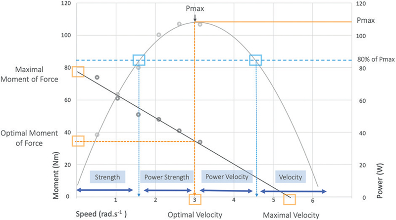
Fig. 1. Parameters obtained from the moment-velocity and power-velocity relationships and definition of the work area according to the patient’s muscle profile. 1 rad/s is equivalent to 57.69°/s. PMAX = maximum power; MOPT = optimal moment; VOPT = optimal velocity; MMAX = maximum theoretical moment; VMAX = maximum theoretical velocity. Using a methodology adapted from Jidovtseff et al. (19), the work areas (strength, power strength, power velocity, velocity) were delimited by the projection on the speed axis of the meeting point between the power-velocity curve and the predicted line of 80% of the maximum power.
The maximum power (PMAX), and optimal velocity (VOPT) were determined from this equation by:

The moment value which corresponds to a 0°/s velocity value was the maximum theoretical moment (MMAX); the velocity value, which corresponds to the 0 Nm moment value, was the maximum theoretical velocity (VMAX) (Fig. 1).
For each parameter, the paretic/non-paretic limb ratio were computed in order to quantify the balance between the 2 sides, as the muscular asymmetry is common in stroke and can affect gait. A value of 1 corresponds to a perfect balance between the 2 sides. For the values less than 1, the smaller the value, the worse is the paretic limb compared with the non-paretic.
The curves representing the power-velocity relationship and the moment-velocity relation were computed using the 6 MMAX (1 MMAX by speed) obtained from the isokinetic assessment. These curves were used to define the work area (strength, power strength, power velocity, velocity) for the individualized isokinetic strengthening. The work area was defined according to the patient’s muscle profile, using a methodology adapted from Jidovtseff et al. (19). The work areas were delimited by the projection on the speed axis of the meeting point between the power-velocity curve and the predicted line of 80% of the maximum power (Fig. 1).
Rehabilitation intervention
Conventional rehabilitation. The conventional rehabilitation comprised physiotherapy and occupational therapy. The physiotherapy sessions lasted 30 min and were performed during a period of 4–5 weeks. The content of rehabilitation was based on the deficits identified during the analytic assessment. The occupational therapy sessions also lasted 30 min and were performed during a period of 2–3 weeks during the functional recovery.
Isokinetic strength training. In addition to the conventional rehabilitation in the routine care, patients in the isokinetic group performed strength training on the isokinetic ergometer 2–3 times a week. The duration of the sessions was between 20 and 30 min and the content of session was adapted according to the level of fatigue of each patient. At the beginning of each session, fatigue was quantified by the patient between 0 (not at all tired) and 5 (extremely tired). At the end of each session, the rating of perceived exertion was assessed using the Borg scale (25), with the following question: “What was the highest perceived intensity of effort during those tasks, on a scale of 0–10, 0 being no effort and 10 being maximal intensity of effort?”.
Strengthening sessions were individualized. They were designed to prioritize the most important deficits, taking into consideration the strength and/or velocity deficit specific to the patient, the extent of muscular weakness in paretic and non-paretic limbs, and the difference between them. For example, when the muscular power of the paretic limb was very low, while that of the non-paretic limb was good (approximately 1.5 W/kg), training sessions were more focused on the paretic limb. For the first 2 weeks of the intervention, training sessions were focused on “strength” or “velocity” work according to the patient’s deficit. The choice of speed to work on “strength” or “velocity” was based on the power-velocity profile of the patient. More specifically, work areas to target the “strength”, “strength power”, “velocity” or “velocity power” were delimited by the projection on the speed axis of the meeting point between the power-velocity curve and the predicted line of 80% of the maximum power (Fig. 1). For “strength” work, the knee flexion-extensions on the isokinetic ergometer were performed at low speed in the strength area, the number of repetitions was low, and the therapist checked that the patient developed at least 90% of the maximum moment of force at that speed (identified during the first evaluation). For “velocity” work, the number of repetitions was intermediate and the knee flexion-extensions on the isokinetic ergometer were performed at high speed in the velocity area. In the following weeks, in addition to “strength” and/or “velocity” work, the programme included sessions of strength endurance and sessions aimed at improving motor control. Strength endurance was targeted with submaximal contractions and a large number of repetitions, in order to work in conditions of fatigue. The objective was to develop the capacity to maintain a level of strength over a long period despite the onset of fatigue, as required in daily living. According to the patient’s maximum moment, a minimum moment threshold was chosen, and the therapist encouraged the patient to reach this threshold throughout the series. Motor control was targeted using coordination exercises. The objective was to develop the ability to perform an intentional movement in space with precision, efficiency and speed, as well as interlimb coordination and the capacity to adapt to changing conditions as requested, notably in gait. For this, several exercises with different constraints were used, such as performing differentiated or lateralized motor actions.
Statistical analysis
For all statistical procedures, the significance level was set at p < 0.05. For the Barthel Index clinical measure, a 2-group (isokinetic, control) × 2-time (before, after intervention) repeated measures analysis of variance (ANOVA) was conducted. For muscular parameters, the data were analysed only in the isokinetic group. In samples with a non-normal distribution, the non-parametric Mann–Whitney test was used to compare the paretic and non-paretic limb, and the non-parametric Wilcoxon test was used to compare the muscular performance before and after the isokinetic intervention. In samples with a normal distribution, Student t-tests were performed. The effect size was evaluated with Cohen’s d (26). Effect sizes of 0.2, 0.5 and 0.8 Cohen’s d correspond to small, medium, and large effects, respectively. The sample size was determined by performing an a priori power analysis (α = 0.05, power = 0.95, effect size = 0.2, number of groups = 2, total sample size = 84).
RESULTS
Data from a total of 88 patients aged from 70 to 96 (mean age 79.52 ± 5.19) years were included in the analysis. Groups were equivalent for all characteristics. No significant differences were found at baseline in NIHSS score and Barthel Index between the 44 patients of the isokinetic group (age = 79.66 ± 5.47 years, BMI = 24.64 ± 4.63 kg/m2, NIHSS score = 4.41 ± 2.98, Barthel score = 61.59 ± 26.34) and the 44 patients of the conventional group (age = 79.39 ± 4.95 years, BMI = 25.44 ± 4.0 kg/m2, NIHSS score = 5.34 ± 4.79, Barthel score = 61.70 ± 26.5). Demographic and clinical characteristics are described in Table I.
Clinical scales
Functional independence: the Barthel Index. For the conventional group, the mean scores of Barthel Index increased from 61.70±26.5 to 76.93 ± 18.12. For the isokinetic group, the score increased from 61.59±26.34 to 88.18 ± 12.16 (Fig. 2). The repeated measures ANOVA revealed no significant Group effect (F(1,86) = 1.96, p = 0.17), but a significant Time effect (F(1,86) = 80.63, p < 0.001) and Time × Group interaction (F(1,86) = 5.95, p = 0.02) were found. The effect size was medium in the conventional group (d = 0.68) and large in the isokinetic group (d = 1.38). Similarly, for the Barthel subscore of mobility, no significant Group effect (F(1,85) = 1.60, p = 0.21), but a significant Time effect (F(1,85) = 53.02, p < 0.001) and Time × Group interaction (F(1,85) = 12.74, p = 0.001) were found. The Cohen’s d revealed a small effect for the conventional group (d = 0.35) and a large effect for the isokinetic group (d = 1.14).
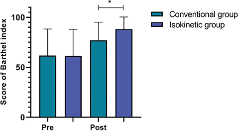
Fig. 2. Barthel Index score before and after intervention in conventional and isokinetic groups.
Spasticity: the Modified Ashworth Scale. For the spasticity, no significant Group effect (F(1,85) = 3.44, p = 0.07), Time effect (F(1,85) = 0.96, p = 0.33), or Time × Group interaction (F(1,85) = 0.99) were found (Fig. 3).
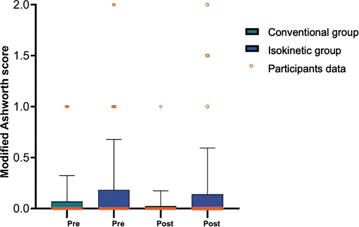
Fig. 3. Modified Ashworth Scale score before and after intervention in conventional and isokinetic groups.
Muscular parameters
Isokinetic parameters between the paretic and non-paretic side. The comparisons of isokinetic parameters between paretic limbs and non-paretic limbs are shown in Table II. At baseline, the maximum power of knee extension, the optimal moment and the maximum moment were significantly lower in the paretic limbs compared with the non-paretic limbs. These differences between the 2 limbs, in favour of the non-paretic side, were also expressed in the paretic/non-paretic limb ratio.
| Isokinetic parameters | Before intervention | After intervention | p-value | Cohen’s d |
| Paretic limbs | ||||
| Maximum power (W/kg)** | 1.22 (0.52) | 1.90 (0.83) | < 0.001* | 1.27 |
| Optimal moment (Nm/kg)** | 0.40 (0.16) | 0.46 (0.20) | 0.006* | 0.40 |
| Optimal velocity (rad/s) | 2.96 (0.77) | 3.95 (0.50) | < 0.001* | 0.82 |
| Maximum moment (Nm/kg)** | 0.87 (0.32) | 1.06 (0.38) | < 0.001* | 0.79 |
| Maximum velocity (rad/s) | 5.56 (1.40) | 7.07 (2.72) | < 0.001* | 0.74 |
| Non-paretic limbs | ||||
| Maximum power (W/kg) | 1.67 (0.55) | 2.12 (0.79) | < 0.001* | 0.89 |
| Optimal moment (Nm/kg) | 0.57 (0.19) | 0.56 (0.20) | 0.527 | 0.01 |
| Optimal velocity (rad/s) | 2.94 (0.83) | 3.62 (1.20) | < 0.001* | 0.63 |
| Maximum moment (Nm/kg) | 1.24 (0.38) | 1.29 (0.42) | 0.051 | 0.25 |
| Maximum velocity (rad/s) | 5.40 (1.20) | 6.47 (2.12) | < 0.001* | 0.58 |
| Paretic/Non-paretic limb ratio | ||||
| Maximum power | 0.72 (0.32) | 0.92 (0.27) | < 0.001* | 0.62 |
| Optimal moment | 0.73 (0.46) | 0.85 (0.36) | 0.038* | 0.36 |
| Optimal velocity | 1.05 (0.31) | 1.13 (0.38) | 0.180 | 0.23 |
| Maximum moment | 0.68 (0.29) | 0.86 (0.26) | < 0.001* | 0.61 |
| Maximum velocity | 1.08 (0.24) | 1.11 (0.27) | 0.196 | 0.23 |
| Wilcoxon signed-rank test or Student t-test was used to compare the paretic limbs and the non-paretic limbs at baseline and to compare parameters before and after intervention. | ||||
| *Significant difference from baseline, p <0.05. | ||||
| **Significant difference between the paretic limbs and the non-paretic limbs at baseline. | ||||
| A 0.2, 0.5 and 0.8 Cohen’s d correspond to small, medium and large effects. | ||||
| A ratio of 1 corresponds to a perfect balance between the 2 lower limbs. | ||||
Isokinetic parameters before and after individualized isokinetic strengthening. Comparisons of isokinetic parameters before and after isokinetic strengthening are shown in Table II and Fig. 4. In the paretic limbs, the maximum power increased significantly and the effect size was large (d = 1.27). Compared with baseline, the optimal moment and optimal velocity were significantly higher after the intervention and the effect sizes were small and large, respectively (d = 0.40, d = 0.82). For the maximum moment and maximum velocity, values were significantly higher after intervention and the effect sizes were medium (d = 0.79, d = 0.74). In the non-paretic limb, significant increases were found in the maximum power, optimal velocity and maximum velocity (d = 0.58). Finally, after intervention, significant differences were found in the paretic/non-paretic ratio for maximum power, the optimal moment and the maximum moment (P/NP). The increase in ratio values towards 1 indicated an improvement in balance between the 2 limbs.
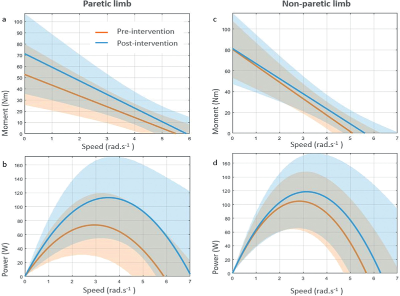
Fig. 4. Moment-velocity and power-velocity profile of paretic limb and non-paretic limb before and after intervention. (a) Mean of the moment-velocity relations of the paretic limbs before and after intervention, with standard deviation (SD). (b) Mean of the power-velocity relations of the paretic limbs before and after intervention, with SD. (c) Mean of the moment-velocity relations of the non-paretic limbs before and after intervention, with SD. (d) Mean of the power-velocity relations of the non-paretic limbs before and after intervention, with SD.
DISCUSSION
The aim of this clinical observational study was to retrospectively assess the effectiveness of an individualized isokinetic strengthening programme using the moment-velocity profile on functional recovery during post-stroke rehabilitation of older patients. As hypothesized, individualized isokinetic strengthening in addition to the conventional rehabilitation provided better recovery in basic daily tasks compared with conventional rehabilitation without added individualized isokinetic strengthening. Furthermore, all muscular parameters improved on the paretic limb in the isokinetic group, suggesting also promising effects on muscular parameters.
The global Barthel Index score and the mobility subscore increased in both conventional and isokinetic groups, but a higher increase was found in the isokinetic group. According to the Cohen’s criteria, the effect sizes of individualized isokinetic strengthening in addition to the conventional rehabilitation were large on both functional capacities and mobility, while they were medium on functional capacities and small on mobility for the conventional rehabilitation alone. Also, an increase in group-mean of 1.85 points on the 20-point Barthel Index is considered a clinically important change (27). Using this minimal clinically important difference (MCID), it would seem therefore that the effects of individualized isokinetic strengthening were clinically meaningful. Conversely, the isokinetic strengthening did not increase the spasticity score, as reported by previous studies (28, 29). Thus, taken together, these results suggest that individualized isokinetic strengthening can have additional positive and clinically relevant effects on functional recovery, and no deleterious impact on spasticity. To our knowledge, this is the first study to assess an individualized isokinetic strengthening using the moment-velocity profile in stroke rehabilitation. While a comparison with previous findings is not straightforward, the current results are consistent with others that showed the clinical pertinence of an isokinetic intervention on the upper as well as the lower limbs during stroke rehabilitation. For example, Kerimov et al. (30) found a greater improvement in the Disabilities Arm, Shoulder and Hand (DASH) score after an isokinetic intervention in comparison with strengthening with exercise bands. In the sutdy Büyükvural Şen et al., (12), conventional rehabilitation was given to all patients and further concentric isokinetic strengthening of the knees was undertaken by the isokinetic group 5 days a week for 3 weeks. In both groups, the authors found an improvement in the Functional Independence Measure (FIM) and the Rivermead Mobility Index (RMI) scores, but greater changes were reported in the isokinetic group.
It could be supposed that these functional improvements were associated with improvement in muscular parameters. However, the current retrospective study, could not compare the effects of the 2 interventions on muscular parameters, as they were not available for the conventional group. Thus, only intragroup effects in the isokinetic group, on power, force and velocity were examined.
Power is the most discriminating predictor of functional performance in older people (31) and is a determinant of walking ability in stroke patients (15). It is the product of the strength and the velocity of muscular contraction, and can be improved by increasing strength and/or velocity. The current study observed an increase in the maximum power and in both strength and velocity of muscular contraction. The whole muscular profile was improved in the current study, as seen in the power-velocity relationship and the moment-velocity relationship, which shifted upward for slow and fast speeds after intervention (Fig. 4). These results are consistent with the findings of previous studies, which showed a significant increase in the knee extensor moment at different speeds following isokinetic strengthening (10–13, 28). However, comparison with other studies is limited, because the strength indicator used in the current study is the maximum moment, whereas other studies used the moment at different speeds. For instance, in the study by Sharp et al. (28), knee extensor moments increased by 16.64% and 19.53% at 60°/s and 120°/s, but no significant increase was reported at 30°/s, and, more recently, Singhal et al. (13) found an increase of 62.23% in the moment at 60°/s after isokinetic strengthening during stroke patient rehabilitation. We report here a mean increase of 27.26% and 67.28% in maximum moment and maximum power, respectively. For both, the effects size was large according to the Cohen’s criteria, and suggests the pertinence of isokinetic strengthening in a clinical context.
Regarding the optimal moment and the optimal velocity that corresponds to the moment and the velocity at which the maximal power occurs, they are also linked to physical performance (31, 32) and both were increased after intervention on the paretic limb. This could result from improvement in the recruitment and firing of the motor unit (33). It could also result from improvement in coordination and motor learning and, more specifically, development of coordinated neuromotor patterns between the agonist and antagonist, but interpretations are limited and changes could also be due to a learning effect.
Furthermore, beyond the effect on the paretic side, improvements were also found in the non-paretic side. Although most studies focus on the paretic limb, where the motor consequences of stroke are higher (1, 5), care of the non-paretic side is also important in rehabilitation because both sides of knee extension strength are related to gait performance (34). Consistently with previous studies, the current study reported less pronounced improvement in the non-paretic side, but improvement on both sides (28). The maximum power, optimal velocity and maximum velocity increased in the non-paretic limb.
Finally, several studies found that greater strength and power asymmetry are associated with slower walking velocity and a variable gait pattern. This phenomenon is very common in patients with stroke, and can be partly explained by muscular asymmetry between the paretic and non-paretic limb (35, 36) and poor interlimb coordination (36, 37). The ratio that quantifies the muscular balance between the 2 lower limbs is therefore particularly relevant in stroke rehabilitation and, in the current study, these ratios were improved after intervention for the maximum power, maximum moment and optimal moment. In addition, this study observed that maximum power of the paretic side increased with a large effect and reached a level close to the healthy side (Fig. 4). Thus, taken together, these results suggest that individualized isokinetic strengthening could be pertinent to improve the balance between the 2 limbs.
The current study relies on clinical data from a stroke rehabilitation unit, and therefore supports the feasibility of using individualized isokinetic strengthening in a clinical context. Nevertheless, the results must be interpreted with caution and some methodological limitations should be mentioned. First, only the patients able to perform the transfer on the isokinetic ergometer were included and the results therefore do not apply to all stroke patients. Secondly, the control group was established retrospectively with patients who did not receive isokinetic strengthening because of lack of availability of the material. Even if the volunteer bias was limited and control group patients were similar in functional impairment at baseline, future studies should perform randomization. Finally, some co-morbidities or confounder factors that could have had an impact on the isokinetic strengthening. For instance, while some clinical characteristic, such as arthrosis or pain, were taken into account by the health professionals during the rehabilitation, they have been not been considered in the analysis. In addition, the duration of rehabilitation was not recorded. saved. All of this information was not available in the database and should be considered in future studies. The limitations of the current study result mostly from its retrospective design. However, individualized isokinetic strengthening appears to have promising effects, and therefore prospective studies should be conducted. In addition, it would be of interest to compare the individualized isokinetic strengthening using the moment-velocity profile with a classic non-individualized isokinetic programme that applies the same protocol to all patients.
In conclusion, this retrospective clinical study suggests that individualized isokinetic strengthening in addition to conventional rehabilitation can provide additional positive and clinically meaningful effects on functional recovery of older adults with stroke in the sub-acute phase. For muscular performance, intragroup effects of isokinetic strengthening also suggest benefits, although further studies are needed. Rehabilitation is an essential step, and the isokinetic ergometer is a promising tool that could be integrated into clinical management for muscle strengthening and optimization of functional performance. Patient management is becoming increasingly personalized, and the individualization of muscle strengthening programmes could be pertinent to obtain the greatest possible benefits. Therefore, using the patient’s moment-velocity profile to design isokinetic strengthening sessions would be a useful approach.
ACKNOWLEDGEMENTS
The authors thank all the medical staff of the post-stroke unit who participated in the rehabilitation.
REFERENCES
- Khan F, Anjamparuthikal H, Chevidikunnan MF. The comparison between isokinetic knee muscles strength in the ipsilateral and contralateral limbs and correlating with function of patients with stroke. J Neurosci Rural Pract 2019; 10: 683–689.
- Canning CG, Ada L, O’Dwyer N. Slowness to develop force contributes to weakness after stroke. Arch Phys Med Rehabil 1999; 80: 66–70.
- Hyngstrom AS, Onushko T, Heitz RP, Rutkowski A, Hunter SK, Schmit BD. Stroke-related changes in neuromuscular fatigue of the hip flexors and functional implications. Am J Phys Med Rehabil 2012; 91: 33–42.
- Kotwani S, Gadgil S, Ranade P. Comparison of neuromuscular fatigue in chronic stroke patients with healthy controls. Clin Investig 2018; 08: 1000140.
- Bohannon RW (editor). Isokinetic testing of muscle strength of older individuals post-stroke: an integrative review. Isokinet Exerc Sci 2020; 28: 303–316.
- Kim CM, Eng JJ. The relationship of lower-extremity muscle torque to locomotor performance in people with stroke. Phys Ther 2003; 83: 49–57.
- Clark DJ, Condliffe EG, Patten C. Reliability of concentric and eccentric torque during isokinetic knee extension in post-stroke hemiparesis. Clin Biomech 2006; 21: 395–404.
- Hsu AL, Tang PF, Jan MH. Test-retest reliability of isokinetic muscle strength of the lower extremities in patients with stroke. Arch Phys Med Rehabil 2002; 83: 1130–1137.
- Rabelo M, Nunes GS, da Costa Amante NM, de Noronha M, Fachin-Martins E. Reliability of muscle strength assessment in chronic post-stroke hemiparesis: a systematic review and meta-analysis. Top Stroke Rehabil 2016; 23: 26–36.
- Hammami N, Coroian FO, Julia M, Amri M, Mottet D, Hérisson C, et al. Isokinetic muscle strengthening after acquired cerebral damage: a literature review. Ann Phys Rehabil Med 2012; 55: 279–291.
- Pontes SS, de Carvalho ALR, Almeida KO, Neves MP, Ribeiro Schindler IFS, Alves IGN, et al. Effects of isokinetic muscle strengthening on muscle strength, mobility, and gait in post-stroke patients: a systematic review and meta-analysis. Clin Rehabil 2019; 33: 381–394.
- Büyükvural Şen S, Özbudak Demir S, Ekiz T, Özgirgin N. Effects of the bilateral isokinetic strengthening training on functional parameters, gait, and the quality of life in patients with stroke. Int J Clin Exp Med 2015; 8: 16871–16879.
- Singhal S, Pattnaik M, Mohanty P. Comparison of isokinetic strengthening with functional strengthening of lower limb and their effect on gait in hemiparesis due to stroke. neurology-neurorehabilitation 2017; 02: 10.35841/neurology-neurorehabilitation.2.1.25-33.
- Hill KD, Hunter SW, Batchelor FA, Cavalheri V, Burton E. Individualized home-based exercise programs for older people to reduce falls and improve physical performance: a systematic review and meta-analysis. Maturitas 2015; 82: 72–84.
- Knight RL, Saunders DH, Mead G. Maximal muscle power after stroke: a systematic review. Clin Pract 2014; 11: 183–191.
- Alcazar J, Rodriguez-Lopez C, Ara I, Alfaro-Acha A, Rodríguez-Gómez I, Navarro-Cruz R, et al. Force-velocity profiling in older adults: an adequate tool for the management of functional trajectories with aging. Exp Gerontol 2018; 108: 1–6.
- Pickering C, Kiely J. Exercise response efficiency: a novel way to enhance population health? Lifestyle Genom 2018; 11: 129–135.
- Alcazar J, Rodriguez-Lopez C, Ara I, Alfaro-Acha A, Mañas-Bote A, Guadalupe-Grau A, et al. The force-velocity relationship in older people: reliability and validity of a systematic procedure. Int J Sports Med 2017; 38: 1097–1104.
- Jidovtseff B, Quièvre J, Hanon C, Crielaard JM. Les profils musculaires inertiels permettent une définition plus précise des charges d’entraînement. Sci Sports 2009; 24: 91–96.
- Jiménez-Reyes P, Samozino P, Brughelli M, Morin JB. Effectiveness of an individualized training based on force-velocity profiling during jumping. Front Physiol 2017; 7: 677.
- Morin JB, Samozino P. Interpreting power-force-velocity profiles for individualized and specific training. Int J Sports Physiol Perform 2016; 11: 267–272.
- Goldstein LB, Samsa GP. Reliability of the National Institutes of Health Stroke Scale. Extension to non-neurologists in the context of a clinical trial. Stroke 1997; 28: 307–310.
- Mahoney FI, Barthel DW. Functional evaluation: the Barthel Index. Md State Med J 1965; 14: 61–65.
- Bohannon RW, Smith MB. Interrater reliability of a modified Ashworth scale of muscle spasticity. Phys Ther 1987; 67: 206–207.
- Borg GA. Psychophysical bases of perceived exertion. Med Sci Sports Exerc 1982; 14: 377–381.
- Cohen J. Statistical power Analysis. Current Directions in Psychological Science 2013; 1: 98–101.
- Hsieh YW, Wang CH, Wu SC, Chen PC, Sheu CF, Hsieh CL. Establishing the minimal clinically important difference of the Barthel Index in stroke patients. Neurorehabil Neural Repair 2007; 21: 233–238.
- Sharp SA, Brouwer BJ. Isokinetic strength training of the hemiparetic knee: effects on function and spasticity. Arch Phys Med Rehabil 1997; 78: 1231–1236.
- Rouleaud S, Gaujard E, Petit H, Picard D, Dehail P, Joseph PA, et al. Isocinétisme et rééducation de la marche de l’hémiplégique. Ann Réadapt Méd Phys 2000; 43: 428–436.
- Kerimov K, Coskun Benlidayi I, Ozdemir C, Gunasti O. The Effects of Upper Extremity Isokinetic Strengthening in Post-Stroke Hemiplegia: A Randomized Controlled Trial. J Stroke Cerebrovasc Dis 2021; 30: 105729.
- Reid KF, Fielding RA. Skeletal muscle power: a critical determinant of physical functioning in older adults. Exerc Sport Sci Rev 2012; 40: 4–12.
- Clémençon M, Hautier CA, Rahmani A, Cornu C, Bonnefoy M. Potential role of optimal velocity as a qualitative factor of physical functional performance in women aged 72 to 96 years. Arch Phys Med Rehabil 2008; 89: 1594–1599.
- Bohannon RW. Contribution of neural and muscular factors to the short duration tension- developing capacity of skeletal muscle. J Orthop Sports Phys Ther 1983; 5: 139–147.
- Watanabe M, Suzuki M, Sugimura Y, Kawaguchi T, Watanabe A, Shibata K, et al. The relationship between bilateral knee muscle strength and gait performance after stroke: the predictive value for gait performance. J Phys Ther Sci 2015; 27: 3227–3232.
- Laroche DP, Cook SB, Mackala K. Strength asymmetry increases gait asymmetry and variability in older women. Med Sci Sports Exerc 2012; 44: 2172–2181.
- Yen SC, Schmit BD, Wu M. Using swing resistance and assistance to improve gait symmetry in individuals post-stroke. Hum Mov Sci 2015; 42: 212–224.
- Hollands K, van Vliet P, Pelton T. Interventions for improving coordination of axial segments and lower limbs during walking following stroke: systematic review. JBI Libr Syst Rev 2011; 10: 1260–1362.