
18F-FDG-PET/CT in breast cancer imaging: Restaging and Implications for treatment decisions in a clinical practice setting
DOI:
https://doi.org/10.2340/1651-226X.2024.40003Keywords:
breast cancer, imaging, staging, PET/CT, clinical management, 18F-FDGAbstract
Background and purpose: Although the diagnostic accuracy of 18F-fluorodeoxyglucose – positron emission tomography/computed tomography (18F-FDG-PET/CT) for breast cancer (BC) has been well studied, few studies have evaluated the impact of 18F-FDG-PET/CT on BC patient care. This study aimed to investigate restaging and 18F-FDG-PET/CT-induced changes in clinical decision-making in patients with BC.
Material and methods: We retrospectively evaluated 18F-FDG-PET/CT-scans performed for BC-related indications in a prospectively collected consecutive cohort of adult patients at Skane University Hospital, Sweden. Patients with all BC stages were included and divided into three groups based on the indication for 18F-FDG-PET/CT: Group A (primary staging), Group B (response evaluation), and Group C (recurrence). The impact of 18F-FDG-PET/CT-scans on clinical management was categorized as no change, minor change (e.g. modification of treatment plans), or major change (e.g. shift from curative to palliative treatment intention).
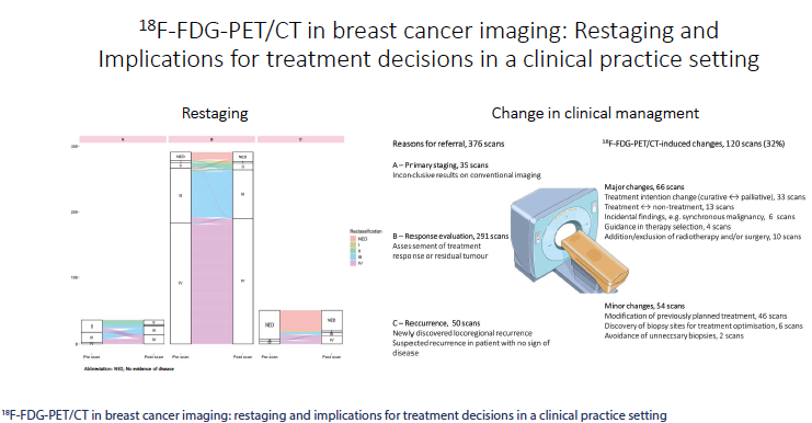
Results: A total of 376 scans (151 patients) were included: Group A 9.3% (35 of 376 scans), Group B 77.4% (291 of 376 scans), and Group C 13.3% (50 of 376 scans). Significant stage migration, predominantly upstaging, occurred in Group A (45.7%) and Group C (28.0%). Changes in clinical management were observed in 120 scans (31.9%), of which 66 were major and 54 were minor. The largest proportion of 18F-FDG-PET/CT-induced management changes were observed in Group A (57.1%), most commonly a shift from curative to palliative treatment intention due to upstaging.
Interpretation: Our study indicates the clinical utility of 18F-FDG-PET/CT in BC restaging and changes in clinical management; the latter observed in approximately one-third of all cases.
Downloads
References
Carter CL, Allen C, Henson DE. Relation of tumor size, lymph node status, and survival in 24,740 breast cancer cases. Cancer. 1989 Jan 1;63(1):181–7.
https://doi.org/10.1002/1097-0142(19890101)63:1<181::aid-cncr2820630129>3.0.co;2-h DOI: https://doi.org/10.1002/1097-0142(19890101)63:1<181::AID-CNCR2820630129>3.0.CO;2-H
Cook GJ, Azad GK, Goh V. Imaging bone metastases in breast cancer: staging and response assessment. J Nucl Med. 2016 Feb;57(Suppl 1):27S–33S.
https://doi.org/10.2967/jnumed.115.157867 DOI: https://doi.org/10.2967/jnumed.115.157867
Han S, Choi JY. Impact of 18F-FDG PET, PET/CT, and PET/MRI on staging and management as an initial staging modality in breast cancer: a systematic review and meta-analysis. Clin Nucl Med. 2021 Apr 1;46(4):271–82.
https://doi.org/10.1097/RLU.0000000000003502 DOI: https://doi.org/10.1097/RLU.0000000000003502
Groheux D, Hindie E. Breast cancer: initial workup and staging with FDG PET/CT. Clin Transl Imaging. 2021;9(3):221–31.
https://doi.org/10.1007/s40336-021-00426-z DOI: https://doi.org/10.1007/s40336-021-00426-z
Pak K, Yoon H-J, Lim W, Kim HY. Impact of 18F-FDG PET on the management of recurrent breast cancer: a meta-analysis. Clin Transl Imaging. 2021;9(3):255–63.
https://doi.org/10.1007/s40336-021-00424-1 DOI: https://doi.org/10.1007/s40336-021-00424-1
Boellaard R, Delgado-Bolton R, Oyen WJ, et al. FDG PET/CT: EANM procedure guidelines for tumour imaging: version 2.0. Eur J Nucl Med Mol Imaging. 2015;42(2):328–54.
https://doi.org/10.1007/s00259-014-2961-x DOI: https://doi.org/10.1007/s00259-014-2961-x
Hanahan D, Weinberg RA. Hallmarks of cancer: the next generation. Cell. 2011;144(5):646–74.
https://doi.org/10.1016/j.cell.2011.02.013 DOI: https://doi.org/10.1016/j.cell.2011.02.013
Miles KA, Williams RE. Warburg revisited: imaging tumour blood flow and metabolism. Cancer Imaging. 2008;8(1):81–6.
https://doi.org/10.1102/1470-7330.2008.0011 DOI: https://doi.org/10.1102/1470-7330.2008.0011
Hong S, Li J, Wang S. 18FDG PET-CT for diagnosis of distant metastases in breast cancer patients. A meta-analysis. Surg Oncol. 2013;22(2):139–43.
https://doi.org/10.1016/j.suronc.2013.03.001 DOI: https://doi.org/10.1016/j.suronc.2013.03.001
Koolen BB, Valdes Olmos RA, Elkhuizen PH, et al. Locoregional lymph node involvement on 18F-FDG PET/CT in breast cancer patients scheduled for neoadjuvant chemotherapy. Breast Cancer Res Treat. 2012;135(1):231–40.
https://doi.org/10.1007/s10549-012-2179-1 DOI: https://doi.org/10.1007/s10549-012-2179-1
Hogan MP, Goldman DA, Dashevsky B, et al. Comparison of 18F-FDG PET/CT for systemic staging of newly diagnosed invasive lobular carcinoma versus invasive ductal carcinoma. J Nucl Med. 2015;56(11):1674–80.
https://doi.org/10.2967/jnumed.115.161455 DOI: https://doi.org/10.2967/jnumed.115.161455
Koo HR, Park JS, Kang KW, et al. 18F-FDG uptake in breast cancer correlates with immunohistochemically defined subtypes. Eur Radiol. 2014;24(3):610–18.
https://doi.org/10.1007/s00330-013-3037-1 DOI: https://doi.org/10.1007/s00330-013-3037-1
NCCN Clinical Practice Guidelines in Oncology (NCCN Guidelines®) for Guideline Breast Cancer V.3.2024. © National Comprehensive Cancer Network, Inc. 2024. All rights reserved. [Cited date: June 23, 2024].
American College of Radiology. ACR appropriateness criteria® [Internet]. Reston, VA: American College of Radiology. [Cited date: January 5, 2024] Available from: https://www.acr.org/Clinical-Resources/ACR-Appropriateness-Criteria
Gradishar WJ, Moran MS, Abraham J, et al. Breast cancer, version 3.2022, NCCN clinical practice guidelines in oncology. J Natl Compr Canc Netw. 2022;20(6):691–722.
https://doi.org/10.6004/jnccn.2022.0030 DOI: https://doi.org/10.6004/jnccn.2022.0030
Regionala cancercentrum i samverkan, Nationellt vårdprogram för bröstcancer version 4.3 [Internet]. Stockholm; 2023. [Cited date: November 22, 2023] Available from: https://kunskapsbanken.cancercentrum.se/diagnoser/brostcancer/vardprogram/
Cardoso F, Kyriakides S, Ohno S, et al. Early breast cancer: ESMO clinical practice guidelines for diagnosis, treatment and follow-updagger. Ann Oncol. 2019;30(8):1194–220.
https://doi.org/10.1093/annonc/mdz173 DOI: https://doi.org/10.1093/annonc/mdz173
Gennari A, Andre F, Barrios CH, et al. ESMO clinical practice guideline for the diagnosis, staging and treatment of patients with metastatic breast cancer. Ann Oncol. 2021;32(12):1475–95.
https://doi.org/10.1016/j.annonc.2021.09.019 DOI: https://doi.org/10.1016/j.annonc.2021.09.019
Bossuyt PM, Reitsma JB, Linnet K, et al. Beyond diagnostic accuracy: the clinical utility of diagnostic tests. Clin Chem. 2012;58(12):1636–43.
https://doi.org/10.1373/clinchem.2012.182576 DOI: https://doi.org/10.1373/clinchem.2012.182576
von Elm E, Altman DG, Egger M, et al. The strengthening the reporting of observational studies in epidemiology (STROBE) statement: guidelines for reporting observational studies. J Clin Epidemiol. 2008;61(4):344–9.
https://doi.org/10.1016/j.jclinepi.2007.11.008 DOI: https://doi.org/10.1016/j.jclinepi.2007.11.008
Giuliano AE, Edge SB, Hortobagyi GN. Eighth edition of the AJCC cancer staging manual: breast cancer. Ann Surg Oncol. 2018;25(7):1783–5.
https://doi.org/10.1245/s10434-018-6486-6 DOI: https://doi.org/10.1245/s10434-018-6486-6
Groheux D, Giacchetti S, Espie M, et al. The yield of 18F-FDG PET/CT in patients with clinical stage IIA, IIB, or IIIA breast cancer: a prospective study. J Nucl Med. 2011;52(10):1526–34.
https://doi.org/10.2967/jnumed.111.093864 DOI: https://doi.org/10.2967/jnumed.111.093864
Yararbas U, Avci NC, Yeniay L, et al. The value of 18F-FDG PET/CT imaging in breast cancer staging. Bosn J Basic Med Sci. 2018;18(1):72–9.
https://doi.org/10.17305/bjbms.2017.2179 DOI: https://doi.org/10.17305/bjbms.2017.2179
Vogsen M, Jensen JD, Christensen IY, et al. FDG-PET/CT in high-risk primary breast cancer-a prospective study of stage migration and clinical impact. Breast Cancer Res Treat. 2021;185(1):145–53.
https://doi.org/10.1007/s10549-020-05929-3 DOI: https://doi.org/10.1007/s10549-020-05929-3
Naghavi-Behzad M, Oltmann HR, Alamdari TA, et al. Clinical impact of FDG-PET/CT compared with CE-CT in response monitoring of metastatic breast cancer. Cancers (Basel). 2021;13(16):4080.
https://doi.org/10.3390/cancers13164080 DOI: https://doi.org/10.3390/cancers13164080
Choi JH, Kim HA, Kim W, et al. Early prediction of neoadjuvant chemotherapy response for advanced breast cancer using PET/MRI image deep learning. Sci Rep. 2020;10(1):21149.
https://doi.org/10.1038/s41598-020-77875-5 DOI: https://doi.org/10.1038/s41598-020-77875-5
Rousseau C, Devillers A, Sagan C, et al. Monitoring of early response to neoadjuvant chemotherapy in stage II and III breast cancer by [18F]fluorodeoxyglucose positron emission tomography. J Clin Oncol. 2006;24(34):5366–72.
https://doi.org/10.1200/JCO.2006.05.7406 DOI: https://doi.org/10.1200/JCO.2006.05.7406
Tian F, Shen G, Deng Y, et al. The accuracy of (18)F-FDG PET/CT in predicting the pathological response to neoadjuvant chemotherapy in patients with breast cancer: a meta-analysis and systematic review. Eur Radiol. 2017;27(11):4786–96.
https://doi.org/10.1007/s00330-017-4831-y DOI: https://doi.org/10.1007/s00330-017-4831-y
Vogsen M, Jensen JD, Gerke O, et al. Benefits and harms of implementing [(18)F]FDG-PET/CT for diagnosing recurrent breast cancer: a prospective clinical study. EJNMMI Res. 2021;11(1):93.
https://doi.org/10.1186/s13550-021-00833-3 DOI: https://doi.org/10.1186/s13550-021-00833-3
Hadebe B, Harry L, Ebrahim T, et al. The role of PET/CT in breast cancer. Diagnostics (Basel). 2023;13(4):597.
https://doi.org/10.3390/diagnostics13040597 DOI: https://doi.org/10.3390/diagnostics13040597
Hildebrandt MG, Naghavi-Behzad M, Vogsen M. A role of FDG-PET/CT for response evaluation in metastatic breast cancer? Semin Nucl Med. 2022;52(5):520–30.
https://doi.org/10.1053/j.semnuclmed.2022.03.004 DOI: https://doi.org/10.1053/j.semnuclmed.2022.03.004
Vogsen M, Naghavi-Behzad M, Harbo FG, et al. 2-[(18)F]FDG-PET/CT is a better predictor of survival than conventional CT: a prospective study of response monitoring in metastatic breast cancer. Sci Rep. 2023;13(1):5552.
https://doi.org/10.1038/s41598-023-32727-w DOI: https://doi.org/10.1038/s41598-023-32727-w
Segaert I, Mottaghy F, Ceyssens S, et al. Additional value of PET-CT in staging of clinical stage IIB and III breast cancer. Breast J. 2010;16(6):617–24.
https://doi.org/10.1111/j.1524-4741.2010.00987.x DOI: https://doi.org/10.1111/j.1524-4741.2010.00987.x
Additional Files
Published
How to Cite
Issue
Section
Categories
License
Copyright (c) 2023 Ida Skarping

This work is licensed under a Creative Commons Attribution 4.0 International License.

