
Time efficiency, geometric accuracy, and clinical impact of AI-assisted contouring of organs at risk in head and neck cancer radiotherapy
DOI:
https://doi.org/10.2340/1651-226X.2025.44015Keywords:
Radiotherapy, artificial intelligence, contouring, Auto-segmentation, Organs at risk, Head and neck cancerAbstract
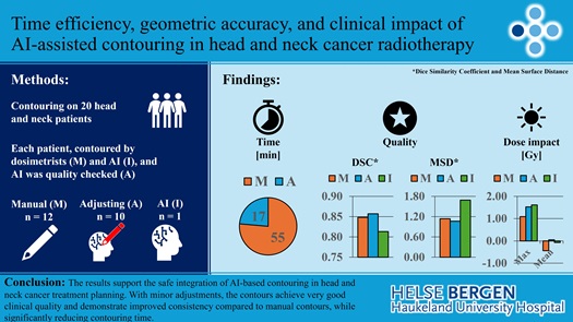
Background and purpose: Ensuring the reliability and accuracy of artificial intelligence (AI)-generated contours is paramount, as discrepancies could lead to inadequate protection of healthy tissues. With increasing clinical workload, the aim of this study was to assess the time-saving potential of AI-assisted organs at risk (OAR) contouring in head and neck cancer (HNC) treatment planning, while also evaluating geometric accuracy, variability, and dosimetric impact.
Patient/material and methods: Twenty patients had 12 OAR contoured by 11 certified dosimetrists and ARTplan (Therapanacea), including the brainstem, cochleas, larynx, mandible, oral cavity, parotid glands, pharynx constrictor muscles, spinal cord, right submandibular gland and thyroid gland. Comparisons were made using geometrical metrics, including Mean Surface Distance, Dice Similarity Coefficient (DSC), Hausdorff Distance, Volume Difference, and Centre of Mass Difference, as well as relevant dose-volume metrics, and total contouring time.
Results: Median manual contouring time of the OARs was 55 (range: 17–151) minutes per patient, while adjusted AI-based structures required 17 (7–42), resulting in 69% time saved. For manual, adjusted and AI-contours, the mean DSC were generally high, averaging 0.85, 0.86, and 0.81 respectively across the evaluated structures. Notably, variability was lowest for the AI and adjusted contours. Average mean and max dose differences were acceptably low (<3.2 Gy) for all OARs.
Interpretation: The results support the integration of AI-based contouring in HNC treatment planning. With minor adjustments, the contours achieve very good clinical quality and demonstrate improved consistency compared to manual contours, while significantly reducing contouring time.
Downloads
References
Harari PM, Song S, Tomé WA. Emphasizing conformal avoidance versus target definition for IMRT planning in head-and-neck cancer. Int J Radiat Oncol Biol Phys. 2010;77(3):950–8.
https://doi.org/10.1016/j.ijrobp.2009.09.062 DOI: https://doi.org/10.1016/j.ijrobp.2009.09.062
Brouwer CL, Steenbakkers RJ, Bourhis J, Budach W, Grau C, Grégoire V, et al. CT-based delineation of organs at risk in the head and neck region: DAHANCA, EORTC, GORTEC, HKNPCSG, NCIC CTG, NCRI, NRG Oncology and TROG consensus guidelines. Radiother Oncol. 2015;117(1):83–90.
https://doi.org/10.1016/j.radonc.2015.07.041 DOI: https://doi.org/10.1016/j.radonc.2015.07.041
Boero IJ, Paravati AJ, Xu B, Cohen EE, Mell LK, Le Q-T, et al. Importance of radiation oncologist experience among patients with head-and-neck cancer treated with intensity-modulated radiation therapy. J Clin Oncol. 2016;34(7):684–90 https://doi.org/10.1200/JCO.2015.63.9898 DOI: https://doi.org/10.1200/JCO.2015.63.9898
Vrtovec T, Močnik D, Strojan P, Pernuš F, Ibragimov B. Auto‐segmentation of organs at risk for head and neck radiotherapy planning: from atlas‐based to deep learning methods. Med Phys. 2020;47(9):e929–50 https://doi.org/10.1002/mp.14320 DOI: https://doi.org/10.1002/mp.14320
Harrison K, Pullen H, Welsh C, Oktay O, Alvarez-Valle J, Jena R. Machine learning for auto-segmentation in radiotherapy planning. Clin Oncol. 2022;34(2):74–88.
https://doi.org/10.1016/j.clon.2021.12.003 DOI: https://doi.org/10.1016/j.clon.2021.12.003
Cardenas CE, Yang J, Anderson BM, Court LE, Brock KB. Advances in auto-segmentation. Semin Radiat Oncol. 2019;29(3):185–97.
https://doi.org/10.1016/j.semradonc.2019.02.001 DOI: https://doi.org/10.1016/j.semradonc.2019.02.001
Claessens M, Oria CS, Brouwer CL, Ziemer BP, Scholey JE, Lin H, et al. Quality assurance for AI-based applications in radiation therapy. Semin Radiat Oncol. 2022;32(4):421–31.
https://doi.org/10.1016/j.semradonc.2022.06.011 DOI: https://doi.org/10.1016/j.semradonc.2022.06.011
Kosmin M, Ledsam J, Romera-Paredes B, Mendes R, Moinuddin S,
De Souza D, et al. Rapid advances in auto-segmentation of organs at risk and target volumes in head and neck cancer. Radiother Oncol. 2019;135:130–40.
https://doi.org/10.1016/j.radonc.2019.03.004 DOI: https://doi.org/10.1016/j.radonc.2019.03.004
Walker GV, Awan M, Tao R, Koay EJ, Boehling NS, Grant JD, et al. Prospective randomized double-blind study of atlas-based organ-at-risk autosegmentation-assisted radiation planning in head and neck cancer. Radiother Oncol. 2014;112(3):321–5.
https://doi.org/10.1016/j.radonc.2014.08.028 DOI: https://doi.org/10.1016/j.radonc.2014.08.028
Tao C-J, Yi J-L, Chen N-Y, Ren W, Cheng J, Tung S, et al. Multi-subject atlas-based auto-segmentation reduces interobserver variation and improves dosimetric parameter consistency for organs at risk in nasopharyngeal carcinoma: a multi-institution clinical study. Radi-other Oncol. 2015;115(3):407–11.
https://doi.org/10.1016/j.radonc.2015.05.012 DOI: https://doi.org/10.1016/j.radonc.2015.05.012
Peters LJ, O’Sullivan B, Giralt J, Fitzgerald TJ, Trotti A, Bernier J, et al. Critical impact of radiotherapy protocol compliance and quality in the treatment of advanced head and neck cancer: results from TROG 02.02. J Clin Oncol. 2010;28(18):2996–3001.
https://doi.org/10.1200/JCO.2009.27.4498 DOI: https://doi.org/10.1200/JCO.2009.27.4498
Mackay K, Bernstein D, Glocker B, Kamnitsas K, Taylor A. A review of the metrics used to assess auto-contouring systems in radiotherapy. Clin Oncol. 2023;35(6):354–69.
https://doi.org/10.1016/j.clon.2023.01.016 DOI: https://doi.org/10.1016/j.clon.2023.01.016
Doolan PJ, Charalambous S, Roussakis Y, Leczynski A, Peratikou M, Benjamin M, et al. A clinical evaluation of the performance of five com-mercial artificial intelligence contouring systems for radiotherapy. Front Oncol. 2023;13:1213068 https://doi.org/10.3389/fonc.2023.1213068 DOI: https://doi.org/10.3389/fonc.2023.1213068
Guo H, Wang J, Xia X, Zhong Y, Peng J, Zhang Z, et al. The dosimetric impact of deep learning-based auto-segmentation of organs at risk on nasopharyngeal and rectal cancer. Radiat Oncol. 2021;16(1):113.
https://doi.org/10.1186/s13014-021-01837-y DOI: https://doi.org/10.1186/s13014-021-01837-y
Smine Z, Poeta S, De Caluwé A, Desmet A, Garibaldi C, Boni KB, et al. Automated segmentation in planning-CT for breast cancer radiother-apy: a review of recent advances. Radiother Oncol. 2025;202:110615.
https://doi.org/10.1016/j.radonc.2024.110615 DOI: https://doi.org/10.1016/j.radonc.2024.110615
Sarria G, Kugel F, Layer J, Dejonckheere C, Scafa D, Roehner F, et al. AI-based auto-segmentation: advantages in delineation, absorbed dose-distribution and logistics. Adv Radiat Oncol. 2023;9(3):101394.
https://doi.org/10.1016/j.adro.2023.101394 DOI: https://doi.org/10.1016/j.adro.2023.101394
Mir R, Kelly SM, Xiao Y, Moore A, Clark CH, Clementel E, et al. Organ at risk delineation for radiation therapy clinical trials: Global Harmo-nization Group consensus guidelines. Radiother Oncol. 2020;150:30–9.
https://doi.org/10.1016/j.radonc.2020.05.038 DOI: https://doi.org/10.1016/j.radonc.2020.05.038
Bisello S, Cilla S, Benini A, Cardano R, Nguyen NP, Deodato F, et al. Dose–volume constraints fOr oRganS At risk In Radiotherapy (CORSAIR): an ‘all-in-one’ multicenter–multidisciplinary practical summary. Curr Oncol. 2022;29(10):7021–50.
https://doi.org/10.3390/curroncol29100552 DOI: https://doi.org/10.3390/curroncol29100552
Jensen K, Friborg J, Hansen CR, Samsøe E, Johansen J, Andersen M, et al. The Danish head and neck cancer group (DAHANCA) 2020 radio-therapy guidelines. Radiother Oncol. 2020;151:149–51.
https://doi.org/10.1016/j.radonc.2020.07.037 DOI: https://doi.org/10.1016/j.radonc.2020.07.037
Ogrinc G, Davies L, Goodman D, Batalden P, Davidoff F, Stevens D. SQUIRE 2.0 (S tandards for QU ality I mprovement R eporting E xcel-lence): revised publication guidelines from a detailed consensus process. J Contin Educ Nurs. 2015;46(11):501–7.
https://doi.org/10.3928/00220124-20151020-02 DOI: https://doi.org/10.3928/00220124-20151020-02
Pang EPP, Tan HQ, Wang F, Niemelä J, Bolard G, Ramadan S, et al. Multicentre evaluation of deep learning CT autosegmentation of the head and neck region for radiotherapy. NPJ Digit Med. 2025;8(1):312.
https://doi.org/10.1038/s41746-025-01624-z DOI: https://doi.org/10.1038/s41746-025-01624-z
Fritscher KD, Peroni M, Zaffino P, Spadea MF, Schubert R, Sharp G. Automatic segmentation of head and neck CT images for radiotherapy treatment planning using multiple atlases, statistical appearance models, and geodesic active contours. Med Phys. 2014;41(5):051910.
https://doi.org/10.1118/1.4871623 DOI: https://doi.org/10.1118/1.4871623
Ibragimov B, Xing L. Segmentation of organs‐at‐risks in head and neck CT images using convolutional neural networks. Med Phys. 2017;44(2):547–57.
https://doi.org/10.1002/mp.12045 DOI: https://doi.org/10.1002/mp.12045
Teguh DN, Levendag PC, Voet PW, Al-Mamgani A, Han X, Wolf TK, et al. Clinical validation of atlas-based auto-segmentation of multiple target volumes and normal tissue (swallowing/mastication) structures in the head and neck. Int J Radiat Oncol Biol Phys. 2011;81(4):950–7.
https://doi.org/10.1016/j.ijrobp.2010.07.009 DOI: https://doi.org/10.1016/j.ijrobp.2010.07.009
Van Dijk LV, Van den Bosch L, Aljabar P, Peressutti D, Both S, Steenbakkers RJ, et al. Improving automatic delineation for head and neck organs at risk by deep learning contouring. Radiother Oncol. 2020;142:115–23.
https://doi.org/10.1016/j.radonc.2019.09.022 DOI: https://doi.org/10.1016/j.radonc.2019.09.022
Fritscher K, Raudaschl P, Zaffino P, Spadea MF, Sharp GC, Schubert R, editors. Deep neural networks for fast segmentation of 3D medical images. Medical Image Computing and Computer-Assisted Intervention–MICCAI 2016: 19th International Conference; 2016 October 17–21; Athens, Proceedings, Part II 19. Springer; 2016.
https://doi.org/10.1007/978-3-319-46723-8_19 DOI: https://doi.org/10.1007/978-3-319-46723-8_19
Additional Files
Published
How to Cite
Issue
Section
Categories
License
Copyright (c) 2025 Johan M Søbstad, Turid H Sulen, Helge E S Pettersen, Grete May Engeseth, Lukas A Hirschi, Camilla H Stokkevåg

This work is licensed under a Creative Commons Attribution 4.0 International License.

