
The Diagnostic Accuracy of Ex Vivo Confocal Laser Scanning Microscopy for Basal Cell Carcinoma: A Systematic Review and Meta-analysis
DOI:
https://doi.org/10.2340/actadv.v106.adv-2026-0393Keywords:
basal cell carcinoma, confocal laser scanning microscopy, diagnostic accuracy, dermatologic surgical procedures, Mohs surgery, meta-analysisAbstract
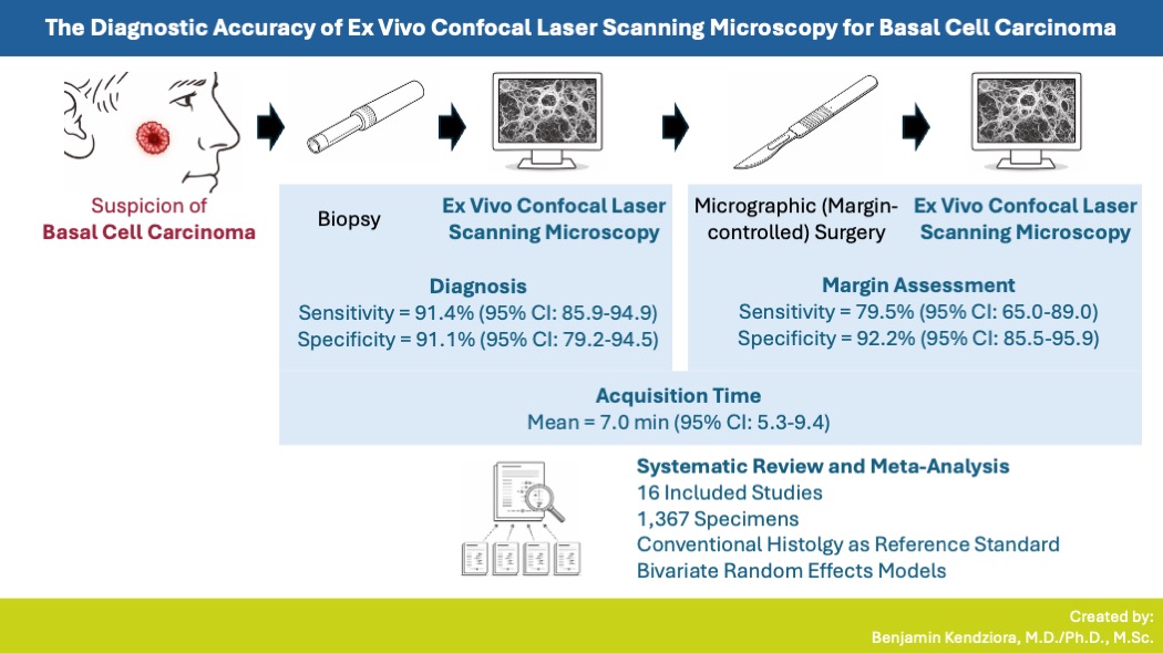
This systematic review and meta-analysis evaluated available data on the diagnostic accuracy of ex vivo confocal laser scanning microscopy (EVCM) for the diagnosis of basal cell carcinoma (BCC) in tissue specimens and for margin assessment in micrographic surgery, using conventional histopathological analysis as reference standard. MEDLINE and Embase were systematically searched in November 2024 in accordance with PRISMA guidelines to identify diagnostic accuracy studies evaluating EVCM for BCC. Pooled sensitivity and specificity were estimated using bivariate random effects models. Sixteen studies comprising 1,387 specimens were included. The pooled mean EVCM acquisition time was 7.0 min (95% confidence interval [CI]: 5.3–9.4). For BCC diagnosis in tissue specimens, pooled sensitivity was 91.4% (95% CI: 85.9–94.9) and pooled specificity was 91.1% (95% CI: 79.2–94.5). Regarding margin assessment, pooled sensitivity and specificity were 79.5% (95% CI: 65.0–89.0) and 92.2% (95% CI: 85.5–95.9). In conclusion, EVCM shows high sensitivity and specificity for BCC diagnosis in tissue specimens and may selectively replace conventional histology when rapid diagnosis is desired. For margin assessment, EVCM has high specificity but moderate sensitivity. Therefore, conventional histology should still be considered as a subsequent step, particularly after the final excision stage of stepwise micrographic surgery.
Downloads
References
Lomas A, Leonardi-Bee J, Bath-Hextall F. A systematic review of worldwide incidence of nonmelanoma skin cancer. Br J Dermatol 2012; 166: 1069–1080. DOI: https://doi.org/10.1111/j.1365-2133.2012.10830.x
Tang S, Thompson S, Smee R. Metastatic basal cell carcinoma: case series and review of the literature. Australas J Dermatol 2017; 58: e40–e43. DOI: https://doi.org/10.1111/ajd.12459
Basset-Seguin N, Herms F. Update in the management of basal cell carcinoma. Acta Derm Venereol 2020; 100: adv00140. DOI: https://doi.org/10.2340/00015555-3495
Brown AC, Brindley L, Hunt WTN, Earp EM, Veitch D, Mortimer NJ, et al. A review of the evidence for Mohs micrographic surgery. Part 2: basal cell carcinoma. Clin Exp Dermatol 2022; 47: 1794–1804. DOI: https://doi.org/10.1111/ced.15266
Vladimirova G, Ruini C, Kapp F, Kendziora B, Ergün EZ, Bağcı IS, et al. Ex vivo confocal laser scanning microscopy: a diagnostic technique for easy real-time evaluation of benign and malignant skin tumours. J Biophotonics 2022; 15: e202100372. DOI: https://doi.org/10.1002/jbio.202100372
Ruini C, Schuh S, Gust C, Kendziora B, Frommherz L, French LE, et al. Line-field optical coherence tomography: in vivo diagnosis of basal cell carcinoma subtypes compared with histopathology. Clin Exp Dermatol 2021; 46: 1471–1481. DOI: https://doi.org/10.1111/ced.14762
Ruini C, Vladimirova G, Kendziora B, Salzer S, Ergun E, Sattler E, et al. Ex-vivo fluorescence confocal microscopy with digital staining for characterizing basal cell carcinoma on frozen sections: a A comparison with histology. J Biophotonics 2021; 14: e202100094. DOI: https://doi.org/10.1002/jbio.202100094
McInnes MDF, Moher D, Thombs BD, McGrath TA, Bossuyt PM, Clifford T, et al. Preferred reporting items for a systematic review and meta-analysis of diagnostic test accuracy studies: The PRISMA-DTA Statement. JAMA 2018; 319: 388–396. DOI: https://doi.org/10.1001/jama.2017.19163
Connolly SM, Baker DR, Coldiron BM, Fazio MJ, Storrs PA, Vidimos AT, et al. AAD/ACMS/ASDSA/ASMS 2012 appropriate use criteria for mohs micrographic surgery: a report of the American Academy of Dermatology, American College of Mohs Surgery, American Society for Dermatologic Surgery Association, and the American Society for Mohs Surgery. J Am Acad Dermatol 2012; 67: 531–550. DOI: https://doi.org/10.1016/j.jaad.2012.06.009
Bittner GC, Cerci FB, Kubo EM, Tolkachjov SN. Mohs micrographic surgery: a review of indications, technique, outcomes, and considerations. An Bras Dermatol 2021; 96: 263–277. DOI: https://doi.org/10.1016/j.abd.2020.10.004
Ziefle S, Schüle D, Breuninger H, Schippert W, Moehrle M. Confocal laser scanning microscopy vs 3-dimensional histologic imaging in basal cell carcinoma. Arch Dermatol 2010; 146: 843–847. DOI: https://doi.org/10.1001/archdermatol.2010.191
meta SG. meta: An R package for meta-analysis. 2007; 7: 40–45.
Doebler P. Meta-analysis of diagnostic accuracy with mada. R Packages. Preprinted posted online 2015.
Reitsma JB, Glas AS, Rutjes AWS, Scholten RJPM, Bossuyt PM, Zwinderman AH. Bivariate analysis of sensitivity and specificity produces informative summary measures in diagnostic reviews. J Clin Epidemiol 2005; 58: 982–990 DOI: https://doi.org/10.1016/j.jclinepi.2005.02.022
Whiting PF, Rutjes AWS, Westwood ME, Mallett S, Deeks JJ, Reitsma JB, et al. QUADAS-2: a revised tool for the quality assessment of diagnostic accuracy studies. Ann Intern Med 2011; 155: 529–536. DOI: https://doi.org/10.7326/0003-4819-155-8-201110180-00009
Harbord RM, Egger M, Sterne JAC. A modified test for small-study effects in meta-analyses of controlled trials with binary endpoints. Stat Med 2006; 25: 3443–3457. DOI: https://doi.org/10.1002/sim.2380
Prasad M. Introduction to the GRADE tool for rating certainty in evidence and recommendations. Clin Epidemiol Glob Health 2024; 25: 101484. DOI: https://doi.org/10.1016/j.cegh.2023.101484
Bennàssar A, Vilata A, Puig S, Malvehy J. Ex vivo fluorescence confocal microscopy for fast evaluation of tumour margins during Mohs surgery. Br J Dermatol 2014; 170: 360–365. DOI: https://doi.org/10.1111/bjd.12671
Bergeret B, Masset F, Bekoy YD, Roger P, Habib F, Ovtchinnikoff B, et al. Diagnostic accuracy of digital staining ex vivo confocal microscopy for diagnosing and subtyping basal cell carcinoma in fresh pretherapeutic punch biopsies: a monocentric prospective study. Dermatology (Basel) 2022: 1–7. DOI: https://doi.org/10.1159/000524349
Espinasse M, Cinotti E, Grivet D, Labeille B, Prade V, Douchet C, et al. “En face” ex vivo reflectance confocal microscopy to help the surgery of basal cell carcinoma of the eyelid. Clin Exp Ophthalmol 2017; 45: 442–447. DOI: https://doi.org/10.1111/ceo.12904
Gellrich FF, Laske J, Steininger J, Eberl N, Meier F, Beissert S, et al. Ex vivo confocal microscopy speeds up surgical margin control of re-excised skin tumors and greatly shortens in-hospital stay. Cancers (Basel) 2024; 16: 3209. DOI: https://doi.org/10.3390/cancers16183209
Grizzetti L, Kuonen F. Ex vivo confocal microscopy for surgical margin assessment: aA histology-compared study on 109 specimens. Skin Health Dis 2022; 2: e91. DOI: https://doi.org/10.1002/ski2.91
Grupp M, Illes M, Mentzel J, Simon JC, Paasch U, Grunewald S. Routine application of ex vivo confocal laser scanning microscopy with digital staining for examination of surgical margins in basal cell carcinomas. J Dtsch Dermatol Ges 2021; 19: 685–692. DOI: https://doi.org/10.1111/ddg.14374
Hartmann D, Krammer S, Bachmann MR, Mathemeier L, Ruzicka T, von Braunmühl T. Simple 3-criteria-based ex vivo confocal diagnosis of basal cell carcinoma. J Biophotonics 2018; 11: e201800062. DOI: https://doi.org/10.1002/jbio.201800062
Karen JK, Gareau DS, Dusza SW, Tudisco M, Rajadhyaksha M, Nehal KS. Detection of basal cell carcinomas in Mohs excisions with fluorescence confocal mosaicing microscopy. Br J Dermatol 2009; 160: 1242–1250. DOI: https://doi.org/10.1111/j.1365-2133.2009.09141.x
Larson B, Abeytunge S, Seltzer E, Rajadhyaksha M, Nehal K. Detection of skin cancer margins in Mohs excisions with high-speed strip mosaicing confocal microscopy: a feasibility study. Br J Dermatol 2013; 169: 922–926. DOI: https://doi.org/10.1111/bjd.12444
Longo C, Pampena R, Bombonato C, Gardini S, Piana S, Mirra M, et al. Diagnostic accuracy of ex vivo fluorescence confocal microscopy in Mohs surgery of basal cell carcinomas: a prospective study on 753 margins. Br J Dermatol 2019; 180: 1473–1480. DOI: https://doi.org/10.1111/bjd.17507
Messner L, Deußing M, Maurer M, Buttgereit L, Stärr L, French LE, et al. Ex vivo confocal laser scanning microscopy in rare skin diseases. Cancers (Basel) 2024; 16: 1713. DOI: https://doi.org/10.3390/cancers16091713
Mu EW, Lewin JM, Stevenson ML, Meehan SA, Carucci JA, Gareau DS. Use of digitally stained multimodal confocal mosaic images to screen for nonmelanoma skin cancer. JAMA Dermatol 2016; 152: 1335–1341. DOI: https://doi.org/10.1001/jamadermatol.2016.2997
Peters N, Schubert M, Metzler G, Geppert JP, Moehrle M. Diagnostic accuracy of a new ex vivo confocal laser scanning microscope compared to H&E-stained paraffin slides for micrographic surgery of basal cell carcinoma. J Eur Acad Dermatol Venereol 2019; 33: 298–304. DOI: https://doi.org/10.1111/jdv.15243
Peris K, Fargnoli MC, Kaufmann R, Arenberger P, Bastholt L, Seguin NB, et al. European consensus-based interdisciplinary guideline for diagnosis and treatment of basal cell carcinoma-update 2023. Eur J Cancer 2023; 192: 113254. DOI: https://doi.org/10.1016/j.ejca.2023.113254
Longo C, Ragazzi M, Rajadhyaksha M, Nehal K, Bennassar A, Pellacani G, et al. In vivo and ex vivo confocal microscopy for dermatologic and mohs surgeons. Dermatol Clin 2016; 34: 497–504. DOI: https://doi.org/10.1016/j.det.2016.05.012
Malvehy J, Pérez-Anker J, Toll A, Pigem R, Garcia A, Alos LL, et al. Ex vivo confocal microscopy: revolution in fast pathology in dermatology. Br J Dermatol 2020; 183: 1011–1025. DOI: https://doi.org/10.1111/bjd.19017
Ragazzi M, Longo C, Piana S. Ex Vivo (Fluorescence) confocal microscopy in surgical pathology: state of the art. Adv Anat Pathol 2016; 23: 159–169. DOI: https://doi.org/10.1097/PAP.0000000000000114
Mohs. Aust J Dermatology 2023; 64: 97–100. DOI: https://doi.org/10.1111/ajd.14046
Kadouch DJ, Schram ME, Leeflang MM, Limpens J, Spuls PI, de Rie MA. In vivo confocal microscopy of basal cell carcinoma: a systematic review of diagnostic accuracy. J Eur Acad Dermatol Venereol 2015; 29: 1890–1897. DOI: https://doi.org/10.1111/jdv.13224
Lupu M, Popa IM, Voiculescu VM, Caruntu A, Caruntu C. A systematic review and meta-analysis of the accuracy of in vivoreflectance confocal microscopy for the diagnosis of primary basal cell carcinoma. J Clin Med 2019; 8: 8. DOI: https://doi.org/10.3390/jcm8091462
Perino F, Suarez R, Perez-Anker J, Carrera C, Rezze GG, Primiero CA, et al. Concordance of in vivo reflectance confocal microscopy and horizontal-sectioning histology in skin tumours. J Eur Acad Dermatol Venereol 2024; 38: 124–135. DOI: https://doi.org/10.1111/jdv.19491
Sendín-Martín M, Lara-Caro M, Harris U, Moronta M, Rossi A, Lee E, et al. Classification of basal cell carcinoma in ex vivo confocal microscopy images from freshly excised tissues using a deep learning algorithm. J Invest Dermatol 2022; 142: 1291–1299. DOI: https://doi.org/10.1016/j.jid.2021.09.029
Additional Files
Published
How to Cite
License

This work is licensed under a Creative Commons Attribution-NonCommercial 4.0 International License.
All digitalized ActaDV contents is available freely online. The Society for Publication of Acta Dermato-Venereologica owns the copyright for all material published until volume 88 (2008) and as from volume 89 (2009) the journal has been published fully Open Access, meaning the authors retain copyright to their work.
Unless otherwise specified, all Open Access articles are published under CC-BY-NC licences, allowing third parties to copy and redistribute the material in any medium or format and to remix, transform, and build upon the material for non-commercial purposes, provided proper attribution to the original work.


