
Comprehensive Transcriptomic Profiling of Nonatopic Prurigo Nodularis in Korean Patients Reveals Il-22–driven Epidermal Stress and Neuro-immuno-fibrotic Remodelling
DOI:
https://doi.org/10.2340/actadv.v106.adv-2025-0169Keywords:
prurigo nodularis, RNA sequencing, IL-22, IL-31RA, JAK–STAT, fibrosis, senescence, Korean, chronic itchAbstract
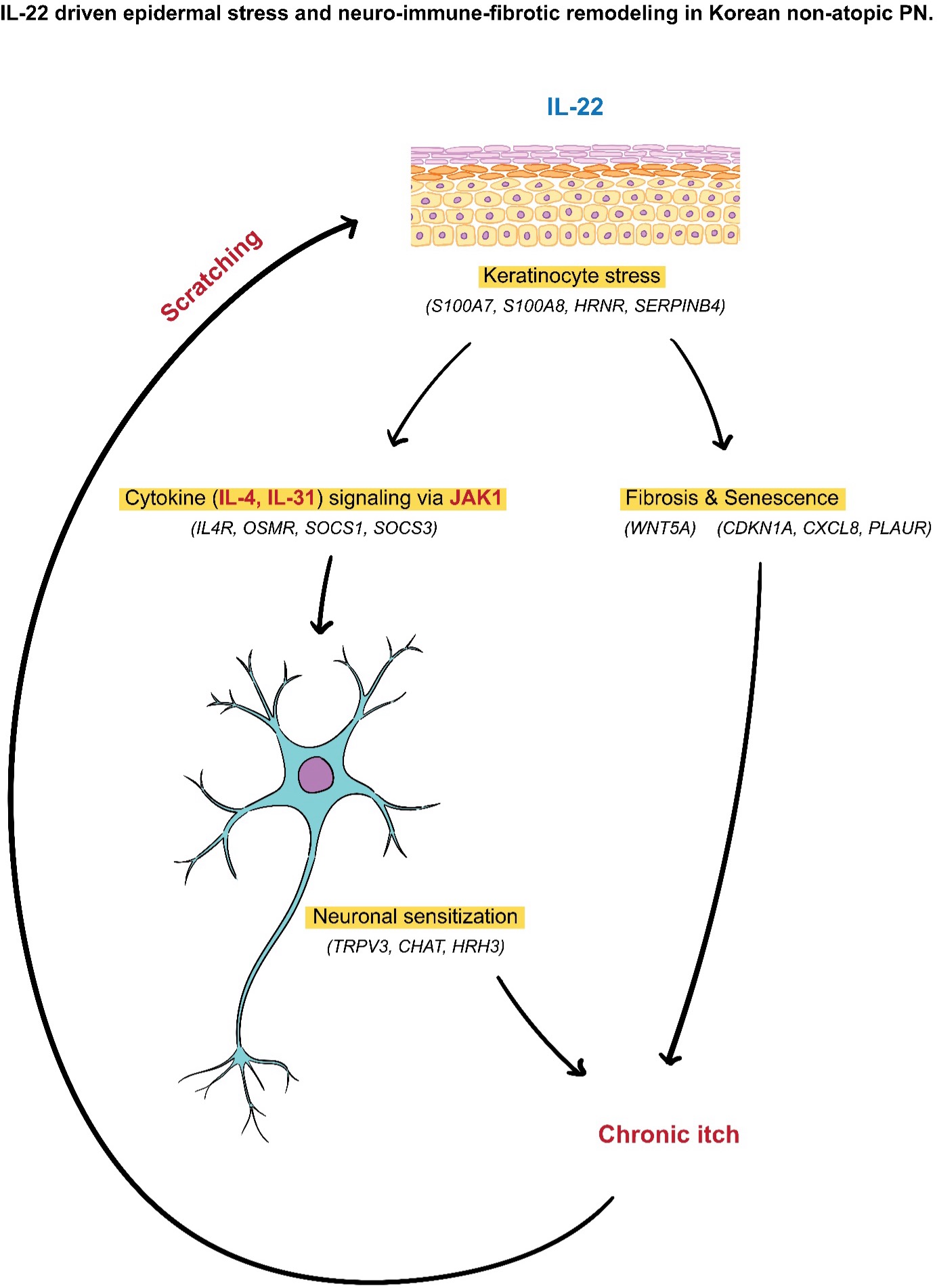
Prurigo nodularis (PN) is a chronic pruritic dermatosis with incompletely defined pathogenesis, and molecular data from East Asian populations are limited. We characterized the transcriptomic signatures of non-atopic PN in Korean patients to identify pathways linked to chronic itch and lesion persistence. RNA sequencing was performed on lesional and non-lesional skin from 17 PN patients and normal skin from 11 controls, followed by differential expression, functional enrichment, and correlation analyses. Lesional PN skin showed distinct transcriptional signatures with upregulation of Th22/IL-22–related genes and IL-22–inducible epidermal stress markers (S100A7/A8/A9, SERPINB4, HRNR), along with keratinization genes (KRT6C, KRT16, KRT17). Itch severity correlated strongly (Spearman’s ρ>0.7) with IL-22–inducible stress genes, IL4R, profibrotic mediators (WNT5A), JAK–STAT regulators (JAK3, SOCS1/3), neuromodulatory/epidermal–neural genes (TRPV3), and senescence markers (CDKN1A, CXCL8, PLAUR). Non-lesional skin showed intermediate expression patterns, consistent with subclinical inflammation. Despite the modest sample size and single-ethnicity design, these findings indicate that non-atopic PN in Korean patients is characterized by IL-22–driven epidermal stress, fibroblast remodelling, neuroimmune signalling, and senescence programsprogrammes, highlighting therapeutic targets including IL-31RA, IL-4Rα/JAK1, antifibrotic and senescence-directed pathways.
Downloads
References
Lotti T, Buggiani G, Prignano F. Prurigo nodularis and lichen simplex chronicus. Dermatol Ther 2008; 21: 42–46. DOI: https://doi.org/10.1111/j.1529-8019.2008.00168.x
Brooks SG, Coscarella G, Yosipovitch G. Prurigo nodularis: a historic perspective. Clin Dermatol 2025; 43: 606–609. DOI: https://doi.org/10.1016/j.clindermatol.2025.03.006
Singam V, Patel KR, Silverberg JI. Association of prurigo nodularis and lichen simplex chronicus with hospitalization for mental health disorders in US adults. Arch Dermatol Res 2020; 312: 587–593. DOI: https://doi.org/10.1007/s00403-020-02046-5
Pereira MP, Steinke S, Zeidler C, Forner C, Riepe C, Augustin M, et al. European academy of dermatology and venereology European prurigo project: expert consensus on the definition, classification and terminology of chronic prurigo. J Eur Acad Dermatol Venereol 2018; 32: 1059–1065. DOI: https://doi.org/10.1111/jdv.14570
Williams KA, Huang AH, Belzberg M, Kwatra SG. Prurigo nodularis: pathogenesis and management. J Am Acad Dermatol 2020; 83: 1567–1575. DOI: https://doi.org/10.1016/j.jaad.2020.04.182
Shao Y, Wang D, Zhu Y, Xiao Z, Jin T, Peng L, et al. Molecular mechanisms of pruritus in prurigo nodularis. Front Immunol 2023; 14: 1301817. DOI: https://doi.org/10.3389/fimmu.2023.1301817
Rinaldi G. The itch-scratch cycle: a review of the mechanisms. Dermatol Pract Concept 2019; 9: 90–97. DOI: https://doi.org/10.5826/dpc.0902a03
Kwatra SG, Pereira MP, Misery L, Mollanazar NK, Shah P, Wiggins S. Prurigo nodularis: disease burden, clinical features and approach to management. Br J Dermatol 2025; 193: 642–652. DOI: https://doi.org/10.1093/bjd/ljaf213
Li W, Pi Y, Xu J. Association between atopic dermatitis and prurigo nodularis: a systematic review and meta-analysis. Int J Dermatol 2025; 64: 282–286. DOI: https://doi.org/10.1111/ijd.17493
Shao Y, Zhu Y, Xiao Z, Shen Y, Dai B, Tang H, et al. RNA sequencing reveals the transcriptome profile of the atopic prurigo nodularis with severe itching. Exp Dermatol 2023; 32: 30–40. DOI: https://doi.org/10.1111/exd.14678
Kim HS, Keum HL, Chung IY, Nattkemper L, Head CR, Koh A, et al. Characterization of a perturbed skin microbiome in prurigo nodularis and lichen simplex chronicus. J Invest Dermatol 2023; 143: 2082–2085. DOI: https://doi.org/10.1016/j.jid.2023.03.1669
Sutaria N, Alphonse MP, Marani M, Parthasarathy V, Deng J, Wongvibulsin S, et al. Cluster analysis of circulating plasma biomarkers in prurigo nodularis reveals a distinct systemic inflammatory signature in African Americans. J Invest Dermatol 2022; 142: 1300–1308. DOI: https://doi.org/10.1016/j.jid.2021.10.011
Parthasarathy V, Cravero K, Xu L, Deng J, Sun Z, Engle SM, et al. The blood proteomic signature of prurigo nodularis reveals distinct inflammatory and neuropathic endotypes: a cluster analysis. J Am Acad Dermatol 2023; 88: 1101–1109. DOI: https://doi.org/10.1016/j.jaad.2023.01.042
Ständer S, Guttman-Yassky E, Yosipovitch G, Wiegmann H, Metze D, Kim M, et al. Th2 mRNA gene expression analysis separates prurigo nodularis into two immune signature groups. J Eur Acad Dermatol Venereol 2025; 39: 1750–1759. DOI: https://doi.org/10.1111/jdv.20812
Cornman HL, Manjunath J, Reddy SV, Adams J, Rajeh A, Samuel C, et al. Comprehensive plasma cytokine and chemokine profiling in prurigo nodularis reveals endotypes in Type 2 inflammation. Sci Rep 2024; 14: 8098. DOI: https://doi.org/10.1038/s41598-024-58013-x
Ma F, Gharaee-Kermani M, Tsoi LC, Plazyo O, Chaskar P, Harms P, et al. Single-cell profiling of prurigo nodularis demonstrates immune-stromal crosstalk driving profibrotic responses and reversal with nemolizumab. J Allergy Clin Immunol 2024; 153: 146–160. DOI: https://doi.org/10.1016/j.jaci.2023.07.005
Tsoi LC, Hacini-Rachinel F, Fogel P, Rousseau F, Xing X, Patrick MT, et al. Transcriptomic characterization of prurigo nodularis and the therapeutic response to nemolizumab. J Allergy Clin Immunol 2022; 149: 1329–1339. DOI: https://doi.org/10.1016/j.jaci.2021.10.004
Belzberg M, Alphonse MP, Brown I, Williams KA, Khanna R, Ho B, et al. Prurigo nodularis is characterized by systemic and cutaneous T helper 22 immune polarization. J Invest Dermatol 2021; 141: 2208–2218.e2214. DOI: https://doi.org/10.1016/j.jid.2021.02.749
Deng J, Parthasarathy V, Marani M, Bordeaux Z, Lee K, Trinh C, et al. Extracellular matrix and dermal nerve growth factor dysregulation in prurigo nodularis compared to atopic dermatitis. Front Med 2022; 9: 1022889. DOI: https://doi.org/10.3389/fmed.2022.1022889
Brunner PM, Emerson RO, Tipton C, Garcet S, Khattri S, Coats I, et al. Nonlesional atopic dermatitis skin shares similar T-cell clones with lesional tissues. Allergy 2017; 72: 2017–2025. DOI: https://doi.org/10.1111/all.13223
Moon S, Stasikowska-Kanicka O, Wągrowska-Danilewicz M, Hawro M, Metz M, Maurer M, et al. Clinically uninvolved but not healthy-the skin of patients with atopic dermatitis is primed for itch and inflammation. J Eur Acad Dermatol Venereol 2024; 38: 1089–1100. DOI: https://doi.org/10.1111/jdv.19694
Sutaria N, Alphonse MP, Roh YS, Choi J, Parthasarathy V, Deng J, et al. Cutaneous transcriptomics identifies fibroproliferative and neurovascular gene dysregulation in prurigo nodularis compared with psoriasis and atopic dermatitis. J Invest Dermatol 2022; 142: 2537–2540. DOI: https://doi.org/10.1016/j.jid.2022.02.010
Gao Z, Dou Y, Zhao P, Wang CJ, Zhang J. Successful treatment of prurigo nodularis by dupilumab: report of 24 patients. Dermatology 2023; 239: 658–663. DOI: https://doi.org/10.1159/000529965
Patel JR, Joel MZ, Lee KK, Kambala A, Cornman H, Oladipo O, et al. Single-cell RNA sequencing reveals dysregulated POSTN+WNT5A+ fibroblast subclusters in prurigo nodularis. J Invest Dermatol 2024; 144: 1568–1578. DOI: https://doi.org/10.1016/j.jid.2023.12.021
O’Reilly S, Tsou PS, Varga J. Senescence and tissue fibrosis: opportunities for therapeutic targeting. Trends Mol Med 2024; 30: 1113–1125. DOI: https://doi.org/10.1016/j.molmed.2024.05.012
Lee YI, Shim JE, Kim J, Lee WJ, Kim JW, Nam KH, et al. WNT5A drives interleukin-6-dependent epithelial-mesenchymal transition via the JAK/STAT pathway in keloid pathogenesis. Burns Trauma 2022; 10: tkac023. DOI: https://doi.org/10.1093/burnst/tkac023
Le TTT, Karmouty-Quintana H, Melicoff E, Le TTT, Weng T, Chen NY, et al. Blockade of IL-6 trans signaling attenuates pulmonary fibrosis. J Immunol 2014; 193: 3755–3768. DOI: https://doi.org/10.4049/jimmunol.1302470
Hashimoto T, Nattkemper LA, Kim HS, Kursewicz CD, Fowler E, Shah SM, et al. Itch intensity in prurigo nodularis is closely related to dermal interleukin-31, oncostatin M, IL-31 receptor alpha and oncostatin M receptor beta. Exp Dermatol 2021; 30: 804–810. DOI: https://doi.org/10.1111/exd.14279
Chen W, Li Y, Steinhoff M, Zhang W, Buddenkotte J, Buhl T, et al. The PLAUR signaling promotes chronic pruritus. FASEB J 2022; 36: e22368.Chen W, Li Y, Steinhoff M, Zhang W, Buddenkotte J, Buhl T, et al. The PLAUR signaling promotes chronic pruritus. FASEB J 2022; 36: e22368. DOI: https://doi.org/10.1096/fj.202200079R
Rukwied R, Weinkauf B, Main M, Obreja O, Schmelz M. Inflammation meets sensitization--an explanation for spontaneous nociceptor activity? Pain 2013; 154: 2707–2714. DOI: https://doi.org/10.1016/j.pain.2013.07.054
Zhong W, Wu X, Zhang W, Zhang J, Chen X, Chen S, et al. Aberrant expression of histamine-independent pruritogenic mediators in keratinocytes may be involved in the pathogenesis of prurigo nodularis. Acta Derm Venereol 2019; 99: 579–586. DOI: https://doi.org/10.2340/00015555-3150
Steinhoff M, Bíró T. A TR(I)P to pruritus research: role of TRPV3 in inflammation and itch. J Invest Dermatol 2009; 129: 531–535. DOI: https://doi.org/10.1038/jid.2008.440
Cowan A, Kehner GB, Inan S. Targeting itch with ligands selective for κ opioid receptors. Handb Exp Pharmacol 2015; 226: 291–314. DOI: https://doi.org/10.1007/978-3-662-44605-8_16
Nattkemper LA, Tey HL, Valdes-Rodriguez R, Lee H, Mollanazar NK, Albornoz C, et al. The genetics of chronic itch: gene expression in the skin of patients with atopic dermatitis and psoriasis with severe itch. J Invest Dermatol 2018; 138: 1311–1317. DOI: https://doi.org/10.1016/j.jid.2017.12.029
Hu X, Li J, Fu M, Zhao X, Wang W. The JAK/STAT signaling pathway: from bench to clinic. Signal Transduct Target Ther 2021; 6: 402. DOI: https://doi.org/10.1038/s41392-021-00791-1
Kershaw NJ, Murphy JM, Liau NPD, Varghese LN, Laktyushin A, Whitlock EL, et al. SOCS3 binds specific receptor-JAK complexes to control cytokine signaling by direct kinase inhibition. Nat Struct Mol Biol 2013; 20: 469–476. DOI: https://doi.org/10.1038/nsmb.2519
Tamiya T, Kashiwagi I, Takahashi R, Yasukawa H, Yoshimura A. Suppressors of cytokine signaling (SOCS) proteins and JAK/STAT pathways: regulation of T-cell inflammation by SOCS1 and SOCS3. Arterioscler Thromb Vasc Biol 2011; 31: 980–985. DOI: https://doi.org/10.1161/ATVBAHA.110.207464
O’Shea JJ, Schwartz DM, Villarino AV, Gadina M, McInnes IB, Laurence A. The JAK-STAT pathway: impact on human disease and therapeutic intervention. Annu Rev Med 2015; 66: 311–328. DOI: https://doi.org/10.1146/annurev-med-051113-024537
Weisshaar E, Szepietowski JC, Bernhard JD, Hait H, Legat FJ, Nattkemper L, et al. Efficacy and safety of oral nalbuphine extended release in prurigo nodularis: results of a phase 2 randomized controlled trial with an open-label extension phase. J Eur Acad Dermatol Venereol 2022; 36: 453–461. DOI: https://doi.org/10.1111/jdv.17816
Additional Files
Published
How to Cite
License

This work is licensed under a Creative Commons Attribution-NonCommercial 4.0 International License.
All digitalized ActaDV contents is available freely online. The Society for Publication of Acta Dermato-Venereologica owns the copyright for all material published until volume 88 (2008) and as from volume 89 (2009) the journal has been published fully Open Access, meaning the authors retain copyright to their work.
Unless otherwise specified, all Open Access articles are published under CC-BY-NC licences, allowing third parties to copy and redistribute the material in any medium or format and to remix, transform, and build upon the material for non-commercial purposes, provided proper attribution to the original work.


