
Distinctive Reactivity to the C-terminal Epitope of BP180 Characterizes Immune Checkpoint Inhibitor–associated Bullous Pemphigoid, and an ELISA Based on the BP180 Ectodomain Enables Prompt Diagnosis in a Subset of Patients
DOI:
https://doi.org/10.2340/actadv.v106.adv-2026-0336Keywords:
autoantibodies, BP180, bullous pemphigoid, epitope profile, humoral response, immune checkpoint inhibitorsAbstract
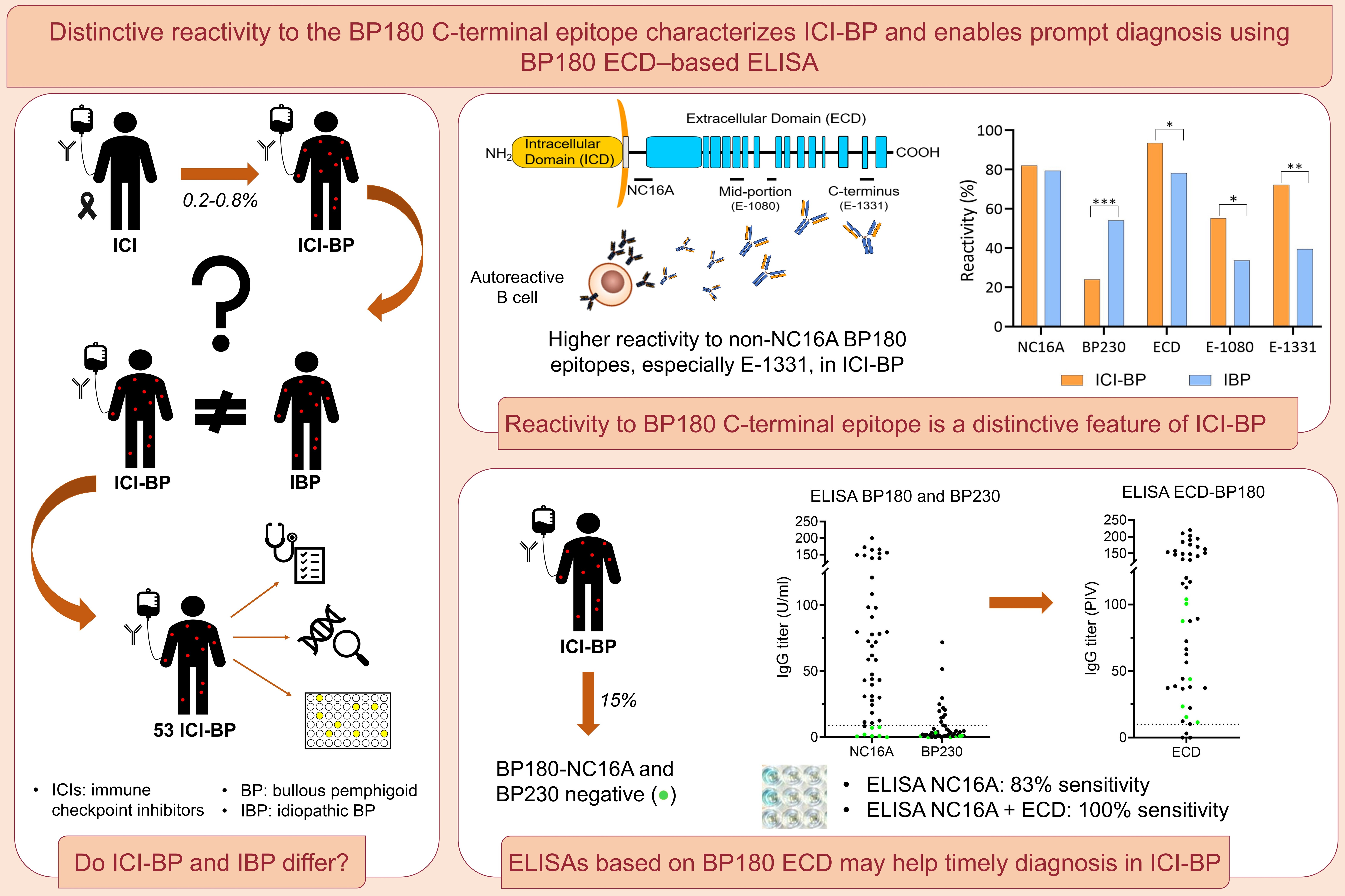
Growing evidence has linked bullous pemphigoid (BP) to immune checkpoint inhibitor (ICI) therapy in cancer treatment. However, the immunological features of ICI-associated BP (ICI-BP) are not yet fully elucidated. In order to characterize the humoral response in ICI BP patients and investigate whether their epitope profile differs from idiopathic BP (IBP), 53 ICI-BP patients were enrolled, immunologically characterized and compared with 59 IBP patients. ICI-BP had a distinctive IgG humoral profile, with reduced reactivity toward BP230 and recognition of multiple BP180 epitopes beyond the immunodominant extracellular noncollagenous 16A domain (NC16A). Specifically, reactivity to BP180 ectodomain was present in 94% of ICI-BP and 78% of IBP (p=0.044). Moreover, BP180 C-terminal epitope was more frequently targeted in ICI-BP than IBP (72% vs 41%, p=0.002). Notably, the combined use of an in-house BP180 ectodomain ELISA and the commercial BP180 test increased diagnostic sensitivity from 83% to 100%. Enhanced IgG reactivity toward nonimmunodominant epitopes, and especially C-terminal epitope recognition, characterize the humoral immune response in ICI-BP. Our data suggest that combining NC16A and fulllength BP180 ectodomain ELISAs may help reduce diagnostic delay in ICI-BP patients, in whom a timely diagnosis is crucial to appropriately manage the disease and ultimately avoid discontinuation of
cancer therapy.
Downloads
References
Di Zenzo G, Della Torre R, Zambruno G, Borradori L. Bullous pemphigoid: Ffrom the clinic to the bench. Clin Dermatol 2012; 30: 3–16. DOI: https://doi.org/10.1016/j.clindermatol.2011.03.005
Genovese G, Di Zenzo G, Cozzani E, Berti E, Cugno M, Marzano AV. New insights into the pathogenesis of bullous pemphigoid: 2019 Update. Front Immunol 2019; 10: 1506. DOI: https://doi.org/10.3389/fimmu.2019.01506
Swiderski M, Vinogradova Y, Knaggs RD, Harman K, Harwood RH, Prasad V, et al. Association between drugs and vaccines commonly prescribed to older people and bullous pemphigoid: a case-control study. Br J Dermatol 2025; 192: 440–449. DOI: https://doi.org/10.1093/bjd/ljae416
Merli M, Accorinti M, Romagnuolo M, Marzano A, Di Zenzo G, Moro F, et al. Autoimmune bullous dermatoses in cancer patients treated by immunotherapy: a literature review and Italian multicentric experience. Front Med (Lausanne) 2023; 10: 1208418. DOI: https://doi.org/10.3389/fmed.2023.1208418
Siegel J, Totonchy M, Damsky W, Berk-Krauss J, Castiglione F Jr, Sznol M, et al. Bullous disorders associated with anti-PD-1 and anti-PD-L1 therapy: A retrospective analysis evaluating the clinical and histopathologic features, frequency, and impact on cancer therapy. J Am Acad Dermatol 2018; 79: 1081–1088. DOI: https://doi.org/10.1016/j.jaad.2018.07.008
Kawsar A, Edwards C, Patel P, Heywood RM, Gupta A, Mann J, et al. Checkpoint inhibitor-associated bullous cutaneous immune-related adverse events: Aa multicentre observational study. Br J Dermatol 2022; 187: 981–987. DOI: https://doi.org/10.1111/bjd.21836
Said JT, Liu M, Talia J, Singer SB, Semenov YR, Wei EX, et al. Risk factors for the development of bullous pemphigoid in US patients receiving immune checkpoint inhibitors. JAMA Dermatol 2022; 158: 552–557. DOI: https://doi.org/10.1001/jamadermatol.2022.0354
Andreani M, Mariotti F, Pira A, Locatelli F, Testa G, Battarra M, et al. HLA alleles associated to susceptibility to gliptin-associated bullous pemphigoid in Italian patients. HLA 2024; 104: e15616. DOI: https://doi.org/10.1111/tan.15616
Gandarillas S, Berger A, Stephenson R, Mehregan D, Dasgeb B. Enriched class II HLA inherence in patients with checkpoint inhibitor-associated bullous pemphigoid. Int J Dermatol 2025; 64: 399–401. DOI: https://doi.org/10.1111/ijd.17563
Gandarillas S, Newland ES, Toppmeyer D, Stephenson R, Denzin L, Dasgeb B. HLA inherence as a potential parameter in checkpoint inhibitor-associated autoimmune adverse event assessment. Front Med 2024; 10: 1288844. DOI: https://doi.org/10.3389/fmed.2023.1288844
Mariotti F, Pira A, De Luca N, Giampetruzzi AR, Russo F, Cerri A, et al. Bullous pemphigoid and mucous membrane pemphigoid humoral responses differ in reactivity towards BP180 midportion and BP230. Front Immunol 2024; 15: 1494294. DOI: https://doi.org/10.3389/fimmu.2024.1494294
Salemme A, Fania L, Scarabello A, Caproni M, Marzano AV, Cozzani E, et al. Gliptin-associated bullous pemphigoid shows peculiar features of anti-BP180 and -BP230 humoral response: Results of a multicenter study. J Am Acad Dermatol 2022; 87: 56–63. DOI: https://doi.org/10.1016/j.jaad.2022.02.036
Mariotti F, Grosso F, Terracina M, Ruffelli M, Cordiali-Fei P, Sera F, et al. Development of a novel ELISA system for detection of anti-BP180 IgG and characterization of autoantibody profile in bullous pemphigoid patients. Br J Dermatol 2004; 151: 1004–1010. DOI: https://doi.org/10.1111/j.1365-2133.2004.06245.x
Thoma-Uszynski S, Uter W, Schwietzke S, Schuler G, Borradori L, Hertl M. Autoreactive T and B cells from bullous pemphigoid (BP) patients recognize epitopes clustered in distinct regions of BP180 and BP230. J Immunol 2006; 176: 2015–2023. DOI: https://doi.org/10.4049/jimmunol.176.3.2015
Saffuri N, Boyango I, Cohen I, Ali-Saleh Z, Dawood M, Khamaysi Z, et al. The immunophenotype of immune checkpoint-induced bullous pemphigoid: Aa cohort study. Cancer Immunol Immunother 2025; 74: 360. DOI: https://doi.org/10.1007/s00262-025-04172-3
Borradori L, Van Beek N, Feliciani C, Tedbirt B, Antiga E, Bergman R, et al. Updated S2 K guidelines for the management of bullous pemphigoid initiated by the European Academy of Dermatology and Venereology (EADV). J Eur Acad Dermatol Venereol 2022; 36: 1689–1704. DOI: https://doi.org/10.1111/jdv.18220
Di Zenzo G, Thoma-Uszynski S, Fontao L, Calabresi V, Hofmann SC, Hellmark T, et al. Multicenter prospective study of the humoral autoimmune response in bullous pemphigoid. Clin Immunol 2008; 128: 415–426. DOI: https://doi.org/10.1016/j.clim.2008.04.012
Gammon WR, Briggaman RA, Inman AO III, Queen LL, Wheeler CE. Differentiating anti-lamina lucida and anti-sublamina densa anti-BMZ antibodies by indirect immunofluorescence on 1.0 M sodium chloride-separated skin. J Invest Dermatol 1984; 82: 139–144. DOI: https://doi.org/10.1111/1523-1747.ep12259692
Wang J, Hu X, Jiang W, Zhou W, Tang M, Wu C, et al. Analysis of the clinical characteristics of pembrolizumab-induced bullous pemphigoid. Front Oncol 2023; 13: 1095694. DOI: https://doi.org/10.3389/fonc.2023.1095694
Lopez AT, Khanna T, Antonov N, Audrey-Bayan C, Geskin L. A review of bullous pemphigoid associated with PD-1 and PD-L1 inhibitors. Int J Dermatol 2018; 57: 664–669. DOI: https://doi.org/10.1111/ijd.13984
Moro F, Fania L, Sinagra JLM, Salemme A, Di Zenzo G. Bullous pemphigoid: Trigger and predisposing factors. Biomolecules 2020; 10: 1432. DOI: https://doi.org/10.3390/biom10101432
de Groot PM, Wu CC, Carter BW, Munden RF. The epidemiology of lung cancer. Transl Lung Cancer Res 2018; 7: 220–233. DOI: https://doi.org/10.21037/tlcr.2018.05.06
Olsen CM, Thompson JF, Pandeya N, Whiteman DC. Evaluation of sex-specific incidence of melanoma. JAMA Dermatol 2020; 156: 553–560. DOI: https://doi.org/10.1001/jamadermatol.2020.0470
Stelkovics E, Korom I, Marczinovits I, Molnar J, Rasky K, Raso E, et al. Collagen XVII/BP180 protein expression in squamous cell carcinoma of the skin detected with novel monoclonal antibodies in archived tissues using tissue microarrays and digital microscopy. Appl Immunohistochem Mol Morphol 2008; 16: 433–441. DOI: https://doi.org/10.1097/PAI.0b013e318162f8aa
Krenacs T, Kiszner G, Stelkovics E, Balla P, Teleki I, Nemeth I, et al. Collagen XVII is expressed in malignant but not in benign melanocytic tumors and it can mediate antibody induced melanoma apoptosis. Histochem Cell Biol 2012; 138: 653–667. DOI: https://doi.org/10.1007/s00418-012-0981-9
Russo F, Pira A, Mariotti F, Papaccio F, Giampetruzzi AR, Bellei B, et al. The possible and intriguing relationship between bullous pemphigoid and melanoma: Sspeculations on significance and clinical relevance. Front Immunol 2024; 15: 1416473. DOI: https://doi.org/10.3389/fimmu.2024.1416473
Koga H, Tsutsumi M, Teye K, Shirahama T, Ishii N, Azuma K, et al. Epitope spreading in immune checkpoint inhibitor-associated bullous pemphigoid. JAMA Dermatol 2025; 161: 557–559. DOI: https://doi.org/10.1001/jamadermatol.2024.6665
Di Zenzo G, Thoma-Uszynski S, Calabresi V, Fontao L, Hofmann SC, Lacour JP, et al. Demonstration of epitope-spreading phenomena in bullous pemphigoid: Rresults of a prospective multicenter study. J Invest Dermatol 2011; 131: 2271–2280. DOI: https://doi.org/10.1038/jid.2011.180
Jensen-Jarolim E, Achatz G, Turner MC, Karagiannis S, Legrand F, Capron M, et al. AllergoOncology: the role of IgE-mediated allergy in cancer. Allergy 2008; 63: 1255–1266. DOI: https://doi.org/10.1111/j.1398-9995.2008.01768.x
Brahmer JR, Lacchetti C, Schneider BJ, Atkins MB, Brassil KJ, Caterino JM, et al. Management of immune-related adverse events in patients treated with immune checkpoint inhibitor therapy: American Society of Clinical Oncology Clinical Practice Guideline. JCO 2018; 36: 1714–1768. DOI: https://doi.org/10.1200/JCO.2017.77.6385
Barrios DM, Phillips GS, Geisler AN, Trelles SR, Markova A, Noor SJ, et al. IgE blockade with omalizumab reduces pruritus related to immune checkpoint inhibitors and anti-HER2 therapies. Ann Oncol 2021; 32: 736–745. DOI: https://doi.org/10.1016/j.annonc.2021.02.016
Avallone G, Maronese CA, Zussino M, Muratori S, Ferrucci SM, Quaglino P, et al. Effectiveness of dupilumab and omalizumab in bullous pemphigoid: A nationwide retrospective cohort study. J Dermatol 2025; 52: 983–1000. DOI: https://doi.org/10.1111/1346-8138.17742
Chen J, Xu D, He Z, Ma S, Liu J, Dai X, et al. Successful treatment of immune checkpoint inhibitor-induced bullous pemphigoid with omalizumab: A case report and review of the literature. Clin Cosmet Investig Dermatol 2024; 17: 2865–2874. DOI: https://doi.org/10.2147/CCID.S487711
Dousset L, Pacaud A, Barnetche T, Kostine M, Dutriaux C, Pham-Ledard A, et al. Analysis of tumor response and clinical factors associated with vitiligo in patients receiving anti-programmed cell death-1 therapies for melanoma: A cross-sectional study. JAAD Int 2021; 5: 112–120. DOI: https://doi.org/10.1016/j.jdin.2021.09.002
Nelson CA, Singer S, Chen T, Puleo AE, Lian CG, Wei EX, et al. Bullous pemphigoid after anti-programmed death-1 therapy: A retrospective case-control study evaluating impact on tumor response and survival outcomes. J Am Acad Dermatol 2022; 87: 1400–1402. DOI: https://doi.org/10.1016/j.jaad.2019.12.068
Asdourian MS, Shah N, Jacoby TV, Reynolds KL, Chen ST. Association of bullous pemphigoid with immune checkpoint inhibitor therapy in patients with cancer: A systematic review. JAMA Dermatol 2022; 158: 933–941. DOI: https://doi.org/10.1001/jamadermatol.2022.1624
Du Y, Wu W, Chen M, Dong Z, Wang F. Cutaneous adverse events and cancer survival prognosis with immune checkpoint inhibitor treatment: A systematic review and meta-analysis. JAMA Dermatol 2023; 159: 1093–1101. DOI: https://doi.org/10.1001/jamadermatol.2023.3003
Grimaux X, Delva R, Jadaud E, Croue A. Nivolumab-induced bullous pemphigoid after radiotherapy and abscopal effect. Australas J Dermatol 2019; 60: e235–e236. DOI: https://doi.org/10.1111/ajd.12987
Zhang X, Sui D, Wang D, Zhang L, Wang R. Case rReport: A rare case of pembrolizumab-induced bullous pemphiRare Case of Pembrolizumab-Induced Bullous Pemphigoid. Front Immunol 2021; 12: 731774. DOI: https://doi.org/10.3389/fimmu.2021.731774
Sowerby L, Dewan AK, Granter S, Gandhi L, LeBoeuf NR. Rituximab treatment of nivolumab-induced bullous pemphigoid. JAMA Dermatol 2017; 153: 603–605. DOI: https://doi.org/10.1001/jamadermatol.2017.0091
Additional Files
Published
How to Cite
License

This work is licensed under a Creative Commons Attribution-NonCommercial 4.0 International License.
All digitalized ActaDV contents is available freely online. The Society for Publication of Acta Dermato-Venereologica owns the copyright for all material published until volume 88 (2008) and as from volume 89 (2009) the journal has been published fully Open Access, meaning the authors retain copyright to their work.
Unless otherwise specified, all Open Access articles are published under CC-BY-NC licences, allowing third parties to copy and redistribute the material in any medium or format and to remix, transform, and build upon the material for non-commercial purposes, provided proper attribution to the original work.


