
The Role of RCM and LC-OCT in Genodermatoses Associated with Skin Cancer: A Narrative Review
DOI:
https://doi.org/10.2340/actadv.v106.adv-2026-0385Keywords:
dermoscopy, microscopy, confocal, skin diseases, skin neoplasms, tomography, optical coherence, diagnosisAbstract
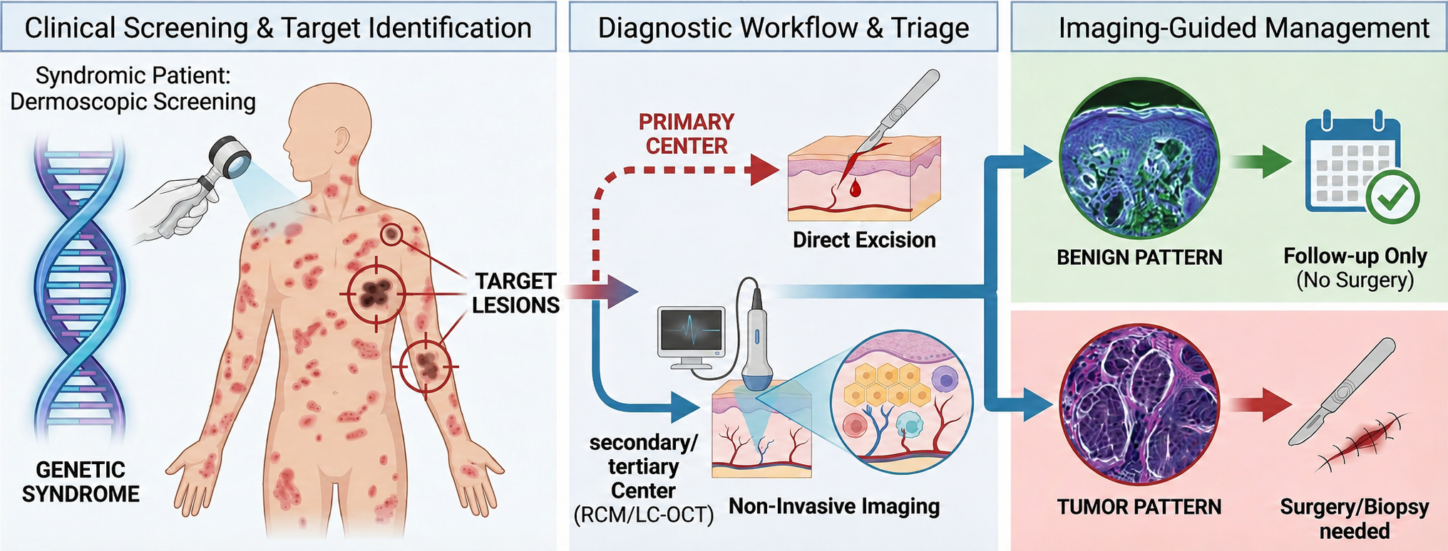
Patients with genodermatoses, such as nevoid basal cell carcinoma syndrome, recessive dystrophic epidermolysis bullosa, familial atypical multiple mole and melanoma syndrome, xeroderma pigmentosum and ichthyosis with confetti, face a high risk of skin malignancies. While dermoscopy significantly improves diagnostic accuracy compared to clinical examination, its specificity remains limited when evaluating ambiguous lesions in these complex patients. This narrative review examines the role of reflectance confocal microscopy and line-field confocal optical coherence tomography in high-risk populations. By enabling the in vivo visualization of histologic-like features at cellular resolution, these noninvasive imaging technologies overcome the limitations of dermoscopy. The analysis of illustrative cases demonstrates that integrating these tools enhances both sensitivity and specificity, allowing for a precise differentiation between true malignancies and benign mimics. While reflectance confocal microscopy remains the standard for melanocytic criteria, line-field confocal optical coherence tomography complements it by overcoming depth limitations. Consequently, the use of combined noninvasive imaging supports a more conservative management strategy, potentially reducing the number of unnecessary excisions while ensuring the timely detection of skin cancer in patients with rare genetic syndromes.
Downloads
References
Juan HY, Zhou AE, Hoegler KM, Khachemoune A. Overview of familial syndromes with increased skin malignancies. Arch Dermatol Res 2023; 315: 707–727. DOI: https://doi.org/10.1007/s00403-022-02447-8
Schierbeck J, Vestergaard T, Bygum A. Skin cancer associated genodermatoses: a literature review. Acta Derm Venereol 2019; 99: 360–369. DOI: https://doi.org/10.2340/00015555-3123
Smith MJ, Beetz C, Williams SG, Bhaskar SS, O’Sullivan J, Anderson B, et al. Germline mutations in SUFU cause Gorlin syndrome-associated childhood medulloblastoma and redefine the risk associated with PTCH1 mutations. J Clin Oncol 2014; 32: 4155–4161. DOI: https://doi.org/10.1200/JCO.2014.58.2569
Has C, Bauer JW, Bodemer C, Bolling MC, Bruckner-Tuderman L, Diem A, et al. Consensus reclassification of inherited epidermolysis bullosa and other disorders with skin fragility. Br J Dermatol 2020; 183: 614–627. DOI: https://doi.org/10.1111/bjd.18921
Christiano AM, Fine JD, Uitto J. Genetic basis of dominantly inherited transient bullous dermolysis of the newborn: a splice site mutation in the type VII collagen gene. J Invest Dermatol 1997; 109: 811–814. DOI: https://doi.org/10.1111/1523-1747.ep12341013
Soura E, Eliades PJ, Shannon K, Stratigos AJ, Tsao H. Hereditary melanoma: update on syndromes and management: genetics of familial atypical multiple mole melanoma syndrome. J Am Acad Dermatol 2016; 74: 395–407. DOI: https://doi.org/10.1016/j.jaad.2015.08.038
Black JO. Xeroderma pigmentosum. Head Neck Pathol 2016; 10: 139–144. DOI: https://doi.org/10.1007/s12105-016-0707-8
Rossi M, Pellegrini C, Cardelli L, Ciciarelli V, Di Nardo L, Fargnoli MC. Familial melanoma: diagnostic and management implications. Dermatol Pract Concept 2019; 9: 10–16. DOI: https://doi.org/10.5826/dpc.0901a03
Piscazzi F, Ardigò M, Suppa M, Malvehy J, Puig S, Perez-Anker J. Applying RCM-based criteria to LC-OCT for classifying mucosal melanoses: a proof-of-concept study. Dermatol Pract Concept 2026; 16: 6544. DOI: https://doi.org/10.5826/dpc.1602a6544
Valenti M, Piscazzi F, Franceschini C, Ferrari A, Frascione P, Ardigò M. Characterizing pigmented mucosal lesions: new insights from reflectance confocal microscopy. J Eur Acad Dermatol Venereol 2025; 39: e638–e641. DOI: https://doi.org/10.1111/jdv.20656
Ahlgrimm-Siess V, Laimer M, Rabinovitz HS, Oliviero M, Hofmann-Wellenhof R, Marghoob AA, et al. Confocal microscopy in skin cancer. Curr Dermatol Rep 2018; 7: 105–118. DOI: https://doi.org/10.1007/s13671-018-0218-9
Mozaffari M, Tavernier C, Ogien J, Godet P, Fünfer K, Wirsching H, et al. Co-localized dermoscopy and LC-OCT for AI-assisted margin assessment of basal cell carcinoma: development of a “BCC-One-Stop-Shop” workflow. Diagnostics 2026; 16: 750. DOI: https://doi.org/10.3390/diagnostics16050750
Wojtowicz I, Żychowska M. Dermoscopy of basal cell carcinoma part 1: dermoscopic findings and diagnostic accuracy—a systematic literature review. Cancers 2025; 17: 493. DOI: https://doi.org/10.3390/cancers17030493
Orte Cano C, Sanak D, Lenoir C, Diet G, Fontaine M, Boussingault L, et al. Diagnostic performance of line-field confocal optical coherence tomography for basal cell carcinoma: a prospective study. Cancers 2026; 18: 153. DOI: https://doi.org/10.3390/cancers18010153
Verkouteren BJA, Cosgun B, Reinders MGHC, Kessler PAWK, Vermeulen RJ, Klaassens M, et al. A guideline for the clinical management of basal cell naevus syndrome (Gorlin-Goltz syndrome). Br J Dermatol 2022; 186: 215–226. DOI: https://doi.org/10.1111/bjd.20700
Federico S, Cassalia F, Mazza M, Del Fiore P, Ferrera N, Malvehy J, et al. Advancements in diagnosis of neoplastic and inflammatory skin diseases: old and emerging approaches. Diagnostics 2025; 15: 2100. DOI: https://doi.org/10.3390/diagnostics15162100
Piscazzi F, Ardigò M, Carrera C, Suppa M, Cinotti E, Perrot JL, et al. Line-field confocal optical coherence tomography in diagnosing keratinizing pearls, multiple aggregated yellow–white globules and milia-like cysts in squamous cell carcinomas, basal cell carcinomas and seborrhoeic keratoses. Clin Exp Dermatol 2025; 51: 107–109. DOI: https://doi.org/10.1093/ced/llaf346
Rodeck U, Uitto J. Recessive dystrophic epidermolysis bullosa-associated squamous-cell carcinoma: an enigmatic entity with complex pathogenesis. J Invest Dermatol 2007; 127: 2295–2296. DOI: https://doi.org/10.1038/sj.jid.5700939
Jacobsen K, Ortner VK, Wiegell SR, Philipsen PA, Haedersdal M. Skin lesion triage in organ transplant recipients using line‐field confocal optical coherence tomography: a retrospective classification study. J Deutsche Derma Gesell 2026. DOI: https://doi.org/10.1111/ddg.15974
Jacobsen K, Haedersdal M. Line-field confocal optical coherence tomography assessment of dermal-epidermal junction in actinic keratosis and skin cancer. Int J Dermatol 2026. DOI: https://doi.org/10.1111/ijd.70415
Michelini S, Scribani Rossi P, Gemma G, Persechino F, Vespasiani G, Conforti C, et al. Assessing the dermal epidermal junction of hyperkeratotic actinic keratoses via line-field confocal optical coherence tomography. Int J Dermatol 2026. DOI: https://doi.org/10.1111/ijd.70328
Gallardo F, Toll A, Malvehy J, Mascaró-Galy JM, Lloreta J, Barranco C, et al. Large atypical melanocytic nevi in recessive dystrophic epidermolysis bullosa: clinicopathological, ultrastructural, and dermoscopic study. Pediatr Dermatol 2005; 22: 338–343. DOI: https://doi.org/10.1111/j.1525-1470.2005.22412.x
Pérez-Anker J, Soglia S, Puig S, García A, Albero R, Alejo B, et al. Melanocytic clefting is associated with melanoma on LC-OCT. J Eur Acad Dermatol Venereol 2025; 39: 1923–1933. DOI: https://doi.org/10.1111/jdv.20761
Begg CB, Orlow I, Hummer AJ, Armstrong BK, Kricker A, Marrett LD, et al. Lifetime risk of melanoma in CDKN2A mutation carriers in a population-based sample. J Natl Cancer Inst 2005; 97: 1507–1515. DOI: https://doi.org/10.1093/jnci/dji312
Woltsche N, Schwab C, Deinlein T, Hofmann-Wellenhof R, Zalaudek I. Dermoscopy in the era of dermato-oncology: from bed to bench side and retour. Expert Rev Anticancer Ther 2016; 16: 531–541. DOI: https://doi.org/10.1586/14737140.2016.1168700
Dinnes J, Deeks JJ, Saleh D, Chuchu N, Bayliss SE, Patel L, et al. Reflectance confocal microscopy for diagnosing cutaneous melanoma in adults. Cochrane Database Syst Rev 2018; 12: CD013190. DOI: https://doi.org/10.1002/14651858.CD013191
Pellacani G, Farnetani F, Ciardo S, Chester J, Kaleci S, Mazzoni L, et al. Effect of reflectance confocal microscopy for suspect lesions on diagnostic accuracy in melanoma: a randomized clinical trial. JAMA Dermatol 2022; 158: 754–761. DOI: https://doi.org/10.1001/jamadermatol.2022.1570
Malvehy J, Puig S, Marti-Laborda RM. Dermoscopy of skin lesions in two patients with xeroderma pigmentosum. Br J Dermatol 2005; 152: 271–278. DOI: https://doi.org/10.1111/j.1365-2133.2004.06332.x
Stern JB, Peck GL, Haupt HM, Hollingsworth HC, Beckerman T. Malignant melanoma in xeroderma pigmentosum: search for a precursor lesion. J Am Acad Dermatol 1993; 28: 591–594. DOI: https://doi.org/10.1016/0190-9622(93)70079-9
Choate KA, Lu Y, Zhou J, Choi M, Elias PM, Farhi A, et al. Mitotic recombination in patients with ichthyosis causes reversion of dominant mutations in KRT10. Science 2010; 330: 94–97. DOI: https://doi.org/10.1126/science.1192280
Lim YH, Qiu J, Saraceni C, Burrall BA, Choate KA. Genetic reversion via mitotic recombination in ichthyosis with confetti due to a KRT10 polyalanine frameshift mutation. J Invest Dermatol 2016; 136: 1725–1728. DOI: https://doi.org/10.1016/j.jid.2016.04.023
Spoerri I, Brena M, De Mesmaeker J, Schlipf N, Fischer J, Tadini G, et al. The phenotypic and genotypic spectra of ichthyosis with confetti plus novel genetic variation in the 3’ end of KRT10: from disease to a syndrome. JAMA Dermatol 2015; 151: 64–69. DOI: https://doi.org/10.1001/jamadermatol.2014.2526
Ito Y, Takeichi T, Nakagawa K, Tanahashi K, Muro Y, Ogi T, et al. Case of ichthyosis with confetti caused by KRT10 mutation, complicated with multiple malignant melanomas. J Dermatol 2022; 49: e228–e229. DOI: https://doi.org/10.1111/1346-8138.16348
Aromolo IF, Moltrasio C, Cozzaglio L, Colavito D, Leon A, Cavalli R, et al. Multiple melanomas in ichthyosis with confetti: one more piece of evidence. Australas J Dermatol 2023; 64: 576–578. DOI: https://doi.org/10.1111/ajd.14143
Wu X, Marchetti MA, Marghoob AA. Dermoscopy: not just for dermatologists. Melanoma Manag 2015; 2: 63–73. DOI: https://doi.org/10.2217/mmt.14.32
Reiter O, Mimouni I, Gdalevich M, Marghoob AA, Levi A, Hodak E, et al. The diagnostic accuracy of dermoscopy for basal cell carcinoma: a systematic review and meta-analysis. J Am Acad Dermatol 2019; 80: 1380–1388. DOI: https://doi.org/10.1016/j.jaad.2018.12.026
Boussingault L, Lenoir C, Stefani AD, Cappilli S, Fontaine M, Diet G, et al. Line-field confocal optical coherence tomography of basal cell carcinoma: systematic correlation with histopathology. Diagnostics 2025; 15: 3059. DOI: https://doi.org/10.3390/diagnostics15233059
Longo C, Guida S, Mirra M, Pampena R, Ciardo S, Bassoli S, et al. Dermatoscopy and reflectance confocal microscopy for basal cell carcinoma diagnosis and diagnosis prediction score: a prospective and multicenter study on 1005 lesions. J Am Acad Dermatol 2024; 90: 994–1001. DOI: https://doi.org/10.1016/j.jaad.2024.01.035
Zalaudek I, Giacomel J, Schmid K, Bondino S, Rosendahl C, Cavicchini S, et al. Dermatoscopy of facial actinic keratosis, intraepidermal carcinoma, and invasive squamous cell carcinoma: a progression model. J Am Acad Dermatol 2012; 66: 589–597. DOI: https://doi.org/10.1016/j.jaad.2011.02.011
Thamm JR, Daxenberger F, Viel T, Gust C, Eijkenboom Q, French LE, et al. Artificial intelligence‐based PRO score assessment in actinic keratoses from LC‐OCT imaging using Convolutional Neural Networks. J Deutsche Derma Gesell 2023; 21: 1359–1366. DOI: https://doi.org/10.1111/ddg.15194
Thomas L, Puig S. Dermoscopy, digital dermoscopy and other diagnostic tools in the early detection of melanoma and follow-up of high-risk skin cancer patients. Acta Derm Venereol 2017; Suppl 218: 14–21. DOI: https://doi.org/10.2340/00015555-2719
Segura S, Puig S, Carrera C, Palou J, Malvehy J. Development of a two-step method for the diagnosis of melanoma by reflectance confocal microscopy. J Am Acad Dermatol 2009; 61: 216–229. DOI: https://doi.org/10.1016/j.jaad.2009.02.014
Perez-Anker J, Soglia S, Lenoir C, Albero R, Alos L, García A, et al. Criteria for melanocytic lesions in LC-OCT. J Eur Acad Dermatol Venereol 2024; 38: 2005–2016. DOI: https://doi.org/10.1111/jdv.20079
Published
How to Cite
License

This work is licensed under a Creative Commons Attribution-NonCommercial 4.0 International License.
All digitalized ActaDV contents is available freely online. The Society for Publication of Acta Dermato-Venereologica owns the copyright for all material published until volume 88 (2008) and as from volume 89 (2009) the journal has been published fully Open Access, meaning the authors retain copyright to their work.
Unless otherwise specified, all Open Access articles are published under CC-BY-NC licences, allowing third parties to copy and redistribute the material in any medium or format and to remix, transform, and build upon the material for non-commercial purposes, provided proper attribution to the original work.






