
Antibiofilm and antibacterial effects of Nigella sativa extract against Streptococcus mutans predicting the role of fatty acids composition: in vitro, CLSM and in silico studies
DOI:
https://doi.org/10.2340/biid.v13.45299Keywords:
Nigella sativa, Streptococcus mutans, oral biofilm, CLSM, fatty acids, MurA enzyme, molecular dockingAbstract
Purpose: To investigate the antibiofilm and antibacterial properties of Nigella sativa seed aqueous extract (NSE) against Streptococcus (S.) mutans.
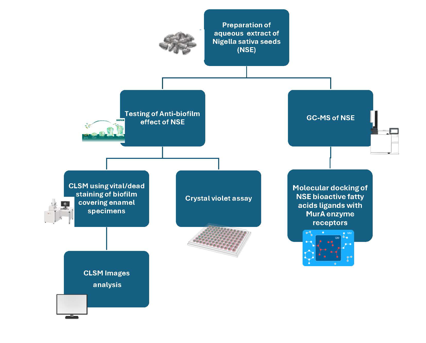
Methods: Chemical analysis of aqueous extract powder of Nigella sativa seeds was carried out using gas chromatography–mass spectrometry (GC-MS). S. mutans biofilm removal by NSE and chlorhexidine (CHX) mouthwash was evaluated using crystal violet (CV) assay. Ten extracted human teeth were sectioned to obtain 12 enamel specimens. Enamel specimens were coated by artificial saliva with bovine serum albumin, and then inoculated by S. mutans for biofilm formation. Enamel specimens were divided into four groups [positive biofilm control (without treatment), negative biofilm control (without bacteria), NSE and CHX biofilm treatment groups]. Acridine orange/propidium iodide assay was used for vital/dead biofilm staining. Confocal laser scanning microscopy (CLSM) biofilm images were digitally analyzed to calculate vital/dead stains relative fluorescence (RF) and estimate biofilm thicknesses. Molecular docking (MD) of interaction between long chain fatty acids (FAs) of NSE and MurA enzyme (PDB: 1UAE) was performed.
Results: GC-MS revealed high concentration of long chain FAs (linoleic, oleic and palmitic acids). NSE and CHX groups showed significantly the highest red (dead) RF. NSE caused the highest reduction in biofilm thickness. NSE and CHX presented the minimum absorbance in CV assay. MD predicted a high binding affinity between FAs ligands and MurA receptors (Arg371, Arg331, Asn23, Lys22), with maximum affinity to linoleic acid (–98.07 kcal/mol).
Conclusion: NSE showed antibacterial effect against S. mutans biofilm comparable to that of CHX, with a higher biofilm removal effect. NSE is expected to have a MurA enzyme inhibitory effect.
Downloads
References
Kay JG, Kramer JM, Visser MB. Danger signals in oral cavity-related diseases. J Leukoc Biol. 2019;106(1):193–200. https://doi.org/10.1002/JLB.4MIR1118-439R DOI: https://doi.org/10.1002/JLB.4MIR1118-439R
Voidarou, C., Antoniadou, M., Rozos, G., Alexopoulos, A., Giorgi, E., Tzora, A., Skoufos, I., Varzakas, T., & Bezirtzoglou, E. An In Vitro Study of Different Types of Greek Honey as Potential Natural Antimicrobials against Dental Caries and Other Oral Pathogenic Microorganisms. Case Study Simulation of Oral Cavity Conditions. Applied Sciences, 2021;11(14):6318. https://doi.org/10.3390/app11146318 DOI: https://doi.org/10.3390/app11146318
Javed S, Zakirulla M, Baig RU, Asif SM, Meer AB. Development of artificial neural network model for prediction of post-streptococcus mutans in dental caries. Comput Methods Programs Biomed. 2020;186:105198. https://doi.org/10.1016/j.cmpb.2019.105198 DOI: https://doi.org/10.1016/j.cmpb.2019.105198
Poppolo Deus F, Ouanounou A. Chlorhexidine in dentistry: pharmacology, uses, and adverse effects. Int Dent J. 2022;72(3):269–77. https://doi.org/10.1016/j.identj.2022.01.005 DOI: https://doi.org/10.1016/j.identj.2022.01.005
Cieplik F, Jakubovics NS, Buchalla W, Maisch T, Hellwig E, Al-Ahmad A. Resistance toward chlorhexidine in oral bacteria – is there cause for concern? Front Microbiol. 2019;10:587. https://doi.org/10.3389/fmicb.2019.00587 DOI: https://doi.org/10.3389/fmicb.2019.00587
McCoy LC, Wehler CJ, Rich SE, Garcia RI, Miller DR, Jones JA. Adverse events associated with chlorhexidine use: results from the Depart-ment of Veterans Affairs Dental Diabetes Study. J Am Dent Assoc. 2008;139(2):178–83. https://doi.org/10.14219/jada.archive.2008.0134 DOI: https://doi.org/10.14219/jada.archive.2008.0134
R. Copin, W.E. Sause, Y. Fulmer, D. Balasubramanian, S. Dyzenhaus, J.M. Ahmed, K. Kumar, J. Lees, A. Stachel, J.C. Fisher, K. Drlica, M. Phillips, J.N. Weiser, P.J. Planet, A. Uhlemann, D.R. Altman, R. Sebra, H. van Bakel, J. Lighter, V.J. Torres, & B. Shopsin. Sequential evolu-tion of virulence and resistance during clonal spread of community-acquired methicillin-resistant Staphylococcus aureus. Proc Natl Acad Sci U S A. 2019;116(5):1745–54. https://doi.org/10.1073/pnas.1814265116 DOI: https://doi.org/10.1073/pnas.1814265116
Vinod KS, Sunil KS, Sethi P, Bandla RC, Singh S, Patel D. A novel herbal formulation versus chlorhexidine mouthwash in efficacy against oral microflora. J Int Soc Prev Community Dent. 2018;8(2):184–90. https://doi.org/10.4103/jispcd.JISPCD_59_18 DOI: https://doi.org/10.4103/jispcd.JISPCD_59_18
Yagoub SO. Chapter four – Black cumin: morphology, physiology, growth, and agricultural yield. In: Mariod AA, editor. Biochemistry, nutrition, and therapeutics of Black cumin seed. Academic Press, Massachusetts, USA; 2023. p. 19–25. https://doi.org/10.1016/B978-0-323-90788-0.00016-0 DOI: https://doi.org/10.1016/B978-0-323-90788-0.00016-0
Yimer EM, Tuem KB, Karim A, Ur-Rehman N, Anwar F. Nigella sativa L. (Black Cumin): a promising natural remedy for wide range of ill-nesses. Evid Based Complement Alternat Med. 2019;2019:1528635. https://doi.org/10.1155/2019/1528635 DOI: https://doi.org/10.1155/2019/1528635
Kurnia D, Padilah R, Apriyanti E, Dharsono HAD. Phytochemical analysis and anti-biofilm potential that cause dental caries from black cumin seeds (Nigella sativa Linn.). Drug Des Devel Ther. 2024;18:1917–32. https://doi.org/10.2147/DDDT.S454217 DOI: https://doi.org/10.2147/DDDT.S454217
Bourgou, S., Bettaieb Rebey, I., Ben Kaab, S., Hammami, M., Dakhlaoui, S., Sawsen, S., Msaada, K., Isoda, H., Ksouri, R., & Fauconnier, M.-L. Green solvent to substitute hexane for bioactive lipids extraction from black cumin and basil seeds. Foods. 2021;10(7):1493. https://doi.org/10.3390/foods10071493 DOI: https://doi.org/10.3390/foods10071493
Bourgou S, Pichette A, Marzouk B, Legault J. Bioactivities of black cumin essential oil and its main terpenes from Tunisia. South Afr J Bot. 2010;76(2):210–16. https://doi.org/10.1016/j.sajb.2009.10.009 DOI: https://doi.org/10.1016/j.sajb.2009.10.009
Stiefel P, Rosenberg U, Schneider J, Mauerhofer S, Maniura-Weber K, Ren Q. Is biofilm removal properly assessed? Comparison of different quantification methods in a 96-well plate system. Appl Microbiol Biotechnol. 2016;100(9):4135–45. https://doi.org/10.1007/s00253-016-7396-9 DOI: https://doi.org/10.1007/s00253-016-7396-9
Radwan AA, Darwesh OM, Emam MT, Mohamed KA, Shady HMA. A combined treatment of proteinase K and biosynthesized ZnO-NPs for eradication of dairy biofilm of sporeformers. AIMS Microbiol. 2022;8(4):507–27. https://doi.org/10.3934/microbiol.2022033 DOI: https://doi.org/10.3934/microbiol.2022033
Mountcastle, S.E., Vyas, N., Villapun, V.M. et al Biofilm viability checker: an open-source tool for automated biofilm viability analysis from confocal microscopy images. npj Biofilms Microbiomes. 2021;7(1):44. https://doi.org/10.1038/s41522-021-00214-7 DOI: https://doi.org/10.1038/s41522-021-00214-7
Herdiyati Y, Astrid Y, Shadrina AAN, Wiani I, Satari MH, Kurnia D. Potential fatty acid as antibacterial agent against oral bacteria of Streptococcus mutans and Streptococcus sanguinis from Basil (Ocimum americanum): in vitro and in silico studies. Curr Drug Discov Tech-nol. 2021;18(4):532–41. https://doi.org/10.2174/1570163817666200712171652 DOI: https://doi.org/10.2174/1570163817666200712171652
Barros LS, Denucci GC, Amoral FL, Franga FM, Basting RT, Turssi CP. The potential of salivary albumin to degrade composite resin. Acta Odontol Latinoam. 2023;36(1):34–9. https://doi.org/10.54589/aol.36/1/34 DOI: https://doi.org/10.54589/aol.36/1/34
Cieplik F, Aparicio C, Kreth J, Schmalz G. Development of standard protocols for biofilm-biomaterial interface testing. JADA Found Sci. 2022;3(1):100035. https://doi.org/10.1016/j.jfscie.2022.100008 DOI: https://doi.org/10.1016/j.jfscie.2022.100008
Zayed SM, Aboulwafa MM, Hashem AM, Saleh SE. Biofilm formation by Streptococcus mutans and its inhibition by green tea extracts. AMB Express. 2021;11(1):73. https://doi.org/10.1186/s13568-021-01232-6 DOI: https://doi.org/10.1186/s13568-021-01232-6
Zhou H, Weir MD, Antonucci JM, Schumacher GE, Zhou X-D, Xu HHK. Evaluation of three-dimensional biofilms on antibacterial bonding agents containing novel quaternary ammonium methacrylates. Int J Oral Sci. 2014;6(2):77–86. https://doi.org/10.1038/ijos.2014.18 DOI: https://doi.org/10.1038/ijos.2014.18
Abd El-Kareem M, Rabbih M, Selim E, Elsherbiny EA, El-Khateeb A. Application of GC/EIMS in combination with semi-empirical calculations for identification and investigation of some volatile components in basil essential oil. Int J Anal Mass Spectrom Chromatogr. 2016;4:14–25. https://doi.org/10.4236/ijamsc.2016.41002 DOI: https://doi.org/10.4236/ijamsc.2016.41002
Mastoor, S., Nazim, F., Rizwan-ul-Hasan, S., Ahmed, K., Khan, S., Ali, S. N., & Abidi, S. H. Analysis of the antimicrobial and anti-biofilm activity of natural compounds and their analogues against Staphylococcus aureus isolates. Molecules. 2022;27(20):6874. https://doi.org/10.3390/molecules27206874 DOI: https://doi.org/10.3390/molecules27206874
Yilmaz FN, Hacioglu M, Aldogan EH. Impact of N-acetylcysteine and antibiotics against single and dual species biofilms of Pseudomonas aeruginosa and Achromobacter xylosoxidans. Curr Microbiol. 2022;80(1):5. https://doi.org/10.1007/s00284-022-03122-x DOI: https://doi.org/10.1007/s00284-022-03122-x
Priyadharsini N, Malathi N, Tamizhchelvan H, Dineshkumar T. Dental fluorosis: a histological study using light and confocal microscopy. Indian J Dent Res Off Publ Indian Soc Dent Res. 2015;26(3):248–51. https://doi.org/10.4103/0970-9290.162896 DOI: https://doi.org/10.4103/0970-9290.162896
Naveed, Muhammad & Ali, Imran & Aziz, Tariq & Ali, Nouman & Hassan, Ali & Rahman, Shafiq & Aziz, Riffat & Alharbi, Metab. Assessment of Melia azedarach plant extracts activity against hypothetical protein of Mycobacterium tuberculosis via GC-MS analysis and in silico approaches. J Comput Biophys Chem. 2024;23(3):299–320. https://doi.org/10.1142/S2737416523500631 DOI: https://doi.org/10.1142/S2737416523500631
Ali, S. K., El-Masry, S. S., El-Adl, K., Abdel-Mawgoud, M., Okla, M. K., Abdel-Raheam, H. E. F, et al. Assessment of antimicrobial activity and GC-MS using culture filtrate of local marine Bacillus strains. J Environ Sci Heal B Pestic Food Contam Agric Wastes. 2024;59(7):399–416. https://doi.org/10.1080/03601234.2024.2357465 DOI: https://doi.org/10.1080/03601234.2024.2357465
Mhade S, Kaushik KS. Tools of the trade: image analysis programs for confocal laser-scanning microscopy studies of biofilms and considera-tions for their use by experimental researchers. ACS Omega. 2023;8(23):20163–20177. https://doi.org/10.1021/acsomega.2c07255 DOI: https://doi.org/10.1021/acsomega.2c07255
Netuschil L, Auschill TM, Sculean A, Arweiler NB. Confusion over live/dead stainings for the detection of vital microorganisms in oral bio-films – which stain is suitable? BMC Oral Health. 2014;14(1):2. https://doi.org/10.1186/1472-6831-14-2 DOI: https://doi.org/10.1186/1472-6831-14-2
Berney M, Hammes F, Bosshard F, Weilenmann H-U, Egli T. Assessment and interpretation of bacterial viability by using the LIVE/DEAD BacLight Kit in combination with flow cytometry. Appl Environ Microbiol. 2007;73(10):3283–90. https://doi.org/10.1128/AEM.02750-06 DOI: https://doi.org/10.1128/AEM.02750-06
Stiefel P, Schmidt-Emrich S, Maniura-Weber K, Ren Q. Critical aspects of using bacterial cell viability assays with the fluorophores SYTO9 and propidium iodide. BMC Microbiol. 2015;15(1):36. https://doi.org/10.1186/s12866-015-0376-x DOI: https://doi.org/10.1186/s12866-015-0376-x
Rosenberg M, Azevedo NF, Ivask A. Propidium iodide staining underestimates viability of adherent bacterial cells. Sci Rep. 2019;9(1):6483. https://doi.org/10.1038/s41598-019-42906-3 DOI: https://doi.org/10.1038/s41598-019-42906-3
Oliveira F, Lima CA, Brás S, França Â, Cerca N. Evidence for inter- and intraspecies biofilm formation variability among a small group of coagulase-negative staphylococci. FEMS Microbiol Lett. 2015;362(20):fnv175. https://doi.org/10.1093/femsle/fnv175 DOI: https://doi.org/10.1093/femsle/fnv175
Fricke K, Koban I, Tresp H, Jablonowski L, Schröder K, Kramer A, et al. Atmospheric pressure plasma: a high-performance tool for the effi-cient removal of biofilms. PLoS One. 2012;7(8):e42539. https://doi.org/10.1371/journal.pone.0042539 DOI: https://doi.org/10.1371/journal.pone.0042539
Yu P, Wang C, Zhou J, Jiang L, Xue J, Li W. Influence of surface properties on adhesion forces and attachment of Streptococcus mutans to zirconia in vitro. Biomed Res Int. 2016;2016:8901253. https://doi.org/10.1155/2016/8901253 DOI: https://doi.org/10.1155/2016/8901253
Ham S-Y, Kim H-S, Cha E, Lim T, Byun Y, Park H-D. Raffinose inhibits Streptococcus mutans biofilm formation by targeting glucosyltransfer-ase. Microbiol Spectr. 2022;10(3):e0207621. https://doi.org/10.1128/spectrum.02076-21 DOI: https://doi.org/10.1128/spectrum.02076-21
Borowicz M, Krzyżanowska DM, Jafra S. Crystal violet-based assay for the assessment of bacterial biofilm formation in medical tubing. J Microbiol Methods. 2023;204:106656. https://doi.org/10.1016/j.mimet.2022.106656 DOI: https://doi.org/10.1016/j.mimet.2022.106656
Alamoudi RA, Alamoudi SA, Alamoudi RA. Biological potential of the main component, thymoquinone, of Nigella sativa in pulp therapy – in vitro study. Life. 2022;12(9):1434. https://doi.org/10.3390/life12091434 DOI: https://doi.org/10.3390/life12091434
Al-Khalifa KS, AlSheikh R, Al-Hariri MT, El-Sayyad H, Alqurashi MS, Ali S, Bugshan AS. Evaluation of the antimicrobial effect of thymoqui-none against different dental pathogens: an in vitro study. Molecules. 2021;26(21):6451. https://doi.org/10.3390/molecules26216451 DOI: https://doi.org/10.3390/molecules26216451
Rostinawati T, Karipaya S, Iskandar Y. Antibacterial activity of ethanol extract of Nigella sativa L. seed against Streptococcus mutans. IOP Conf Ser Earth Environ Sci. 2019;334(1):12050. https://doi.org/10.1088/1755-1315/334/1/012050 DOI: https://doi.org/10.1088/1755-1315/334/1/012050
Ilangovan S, Rajasekar A. Evaluation of efficacy of 20% Nigella sativa on gingival health. J Pharm Res Int. 2021;33(61A):354–63. https://doi.org/10.9734/jpri/2021/v33i61A35595 DOI: https://doi.org/10.9734/jpri/2021/v33i61A35595
Dalli M, Azizi S, Kandsi F, Gseyra N. Evaluation of the in vitro antioxidant activity of different extracts of Nigella sativa L. seeds, and the quantification of their bioactive compounds. Mater Today Proc. 2021;45(part 8):7259–63. https://doi.org/10.1016/j.matpr.2020.12.743 DOI: https://doi.org/10.1016/j.matpr.2020.12.743
Abdel-Razek AG, Hassanein MMM, Moawad S, Farouk A, Badr AN, Shehata MG, Siger A, Grygier A, Rudzińska M. Assessment of the quality, bioactive compounds, and antimicrobial activity of Egyptian, Ethiopian, and Syrian Black cumin oils. Molecules. 2024;29(21):4985. https://doi.org/10.3390/molecules29214985 DOI: https://doi.org/10.3390/molecules29214985
Albakry Z, Karrar E, Ahmed IAM, Oz E, Proestos C, El Sheikha AF, Oz F, Wu G, Wang X. Nutritional composition and volatile compounds of black cumin (Nigella sativa L.) seed, fatty acid composition and tocopherols, polyphenols, and antioxidant activity of its essential oil. Hor-ticulturae. 2022;8(7):575. https://doi.org/10.3390/horticulturae8070575 DOI: https://doi.org/10.3390/horticulturae8070575
Khoddami A, Ghazali HM, Yassoralipour A, Ramakrishnan Y, Ganjloo A. Physicochemical characteristics of Nigella seed (Nigella sativa L.) oil as affected by different extraction methods. J Am Oil Chem Soc. 2011;88(4):533–40. https://doi.org/10.1007/s11746-010-1687-6 DOI: https://doi.org/10.1007/s11746-010-1687-6
Alrashidi M, Derawi D, Salimon J, Firdaus Yusoff M. An investigation of physicochemical properties of Nigella sativa L. seed oil from Al-Qassim by different extraction methods. J King Saud Univ Sci. 2020;32(8):3337–42. https://doi.org/10.1016/j.jksus.2020.09.019 DOI: https://doi.org/10.1016/j.jksus.2020.09.019
Chamlagain M, Hu J, Sionov RV, Steinberg D. Anti-bacterial and anti-biofilm activities of arachidonic acid against the cariogenic bacterium Streptococcus mutans. Front Microbiol. 2024;15:1333274. https://doi.org/10.3389/fmicb.2024.1333274 DOI: https://doi.org/10.3389/fmicb.2024.1333274
Giancarlo Casillas-Vargas, Carlimar Ocasio-Malavé, Solymar Medina, Christian Morales-Guzmán, René García Del Valle, Néstor M. Carbal-leira, David J. Sanabria-Río. Antibacterial fatty acids: an update of possible mechanisms of action and implications in the development of the next-generation of antibacterial agents. Prog Lipid Res. 2021;82:101093. https://doi.org/10.1016/j.plipres.2021.101093 DOI: https://doi.org/10.1016/j.plipres.2021.101093
Dilika F, Bremner PD, Meyer JJM. Antibacterial activity of linoleic and oleic acids isolated from Helichrysum pedunculatum: a plant used during circumcision rites. Fitoterapia. 2000;71(4):450–2. https://doi.org/10.1016/S0367-326X(00)00150-7 DOI: https://doi.org/10.1016/S0367-326X(00)00150-7
Abdel-Aziz MM, Emam TM, Raafat MM. Hindering of cariogenic Streptococcus mutans biofilm by fatty acid array derived from an endo-phytic arthrographis kalrae strain. Biomolecules. 2020;10(5):811. https://doi.org/10.3390/biom10050811 DOI: https://doi.org/10.3390/biom10050811
Harvey V, Brady D, Kealey C. Effect of antibacterial lipids on biofilm formation by Streptococcus mutans. Access Microbiol. 2019;1(1A):po0563. https://doi.org/10.1099/acmi.ac2019.po0563 DOI: https://doi.org/10.1099/acmi.ac2019.po0563
Bruss ML. Lipids and ketones. In: Kaneko JJ, Harvey JW, Bruss ML, editors. Clinical biochemistry of domestic animals. 6th ed. San Diego, CA: Academic Press; 2008. p. 81–115. https://doi.org/10.1016/B978-0-12-370491-7.00004-0 DOI: https://doi.org/10.1016/B978-0-12-370491-7.00004-0
Jung J-E, Santosh P, Jeon J-G. Identification of linoleic acid, a main component of the n-hexane fraction from Dryopteris crassirhizoma, as an anti-Streptococcus mutans biofilm agent. Biofouling. 2014;30(7):789–98. https://doi.org/10.1080/08927014.2014.930446 DOI: https://doi.org/10.1080/08927014.2014.930446
Won S-R, Hong M-J, Kim Y-M, Li CY, Kim J-W, Rhee H-I. Oleic acid: an efficient inhibitor of glucosyltransferase. FEBS Lett. 2007;581(25):4999–5002. https://doi.org/10.1016/j.febslet.2007.09.045 DOI: https://doi.org/10.1016/j.febslet.2007.09.045
Hara, T., Sonoi, A., Handa, T. et al. Unsaturated fatty acid salts remove biofilms on dentures. Sci Rep. 2021;11(1):12524. https://doi.org/10.1038/s41598-021-92044-y DOI: https://doi.org/10.1038/s41598-021-92044-y
Aline Silva Braga, Leticia Lobo de Melo Simas, Juliana Gonçalves Pires, Beatriz Martines Souza, Fernanda Pereira de Souza Rosa de Melo, Luiz Leonardo Saldanha, Anne Ligia Dokkedal, Ana Carolina Magalhães. Antibiofilm and anti-caries effects of an experimental mouth rinse containing Matricaria chamomilla L. extract under microcosm biofilm on enamel. J Dent. 2020;99:103415. https://doi.org/10.1016/j.jdent.2020.103415 DOI: https://doi.org/10.1016/j.jdent.2020.103415
Giacaman RA, Jobet-Vila P, Muñoz-Sandoval C. Fatty acid effect on sucrose-induced enamel demineralization and cariogenicity of an ex-perimental biofilm-caries model. Odontology. 2015;103(2):169–76. https://doi.org/10.1007/s10266-014-0154-5 DOI: https://doi.org/10.1007/s10266-014-0154-5
Marimuthu SCV, Murugesan J, Babkiewicz E, Maszczyk P, Sankaranarayanan M, Thangamariappan E, Rosy JC, Ram Kumar Pandian S, Kunjiappan S, Balakrishnan V, et al. Pharmacoinformatics-based approach for uncovering the quorum-quenching activity of phytocom-pounds against the oral pathogen, Streptococcus mutans. Molecules. 2023;28(14):5514. https://doi.org/10.3390/molecules28145514 DOI: https://doi.org/10.3390/molecules28145514
Li H, Zhou Y, Wang N, XinY, Tang L, Ma Y. Identification and characterization of a MurA, UDP-N-Acetylglucosamine enolpyruvyl transferase from cariogenic streptococcus mutans. J Hard Tissue Biol. 2012;21(1):17–24. https://doi.org/10.2485/jhtb.21.17 DOI: https://doi.org/10.2485/jhtb.21.17
El Zoeiby A, Sanschagrin F, Levesque RC. Structure and function of the Mur enzymes: development of novel inhibitors. Mol Microbiol. 2003;47(1):1–12. https://doi.org/10.1046/j.1365-2958.2003.03289.x DOI: https://doi.org/10.1046/j.1365-2958.2003.03289.x
Evangelina IA, Herdiyati Y, Laviana A, Rikmasari R, Zubaedah C, Anisah, Kurnia D. Bio-mechanism inhibitory prediction of β-sitosterol from Kemangi (Ocimum basilicum L.) as an inhibitor of MurA enzyme of oral bacteria: in vitro and in silico study. Adv Appl Bioinform Chem. 2021;14:103–15. https://doi.org/10.2147/AABC.S301488 DOI: https://doi.org/10.2147/AABC.S301488
Dai HJ, Parker CN, Bao JJ. Characterization and inhibition study of MurA enzyme by capillary electrophoresis. J Chromatogr B. 2002;766(1):123–32. https://doi.org/10.1016/S0378-4347(01)00461-3 DOI: https://doi.org/10.1016/S0378-4347(01)00461-3
Bescos, R., Ashworth, A., Clarke, C. et al. Effects of chlorhexidine mouthwash on the oral microbiome. Sci Rep. 2020;10(1):5254. https://doi.org/10.1038/s41598-020-61912-4 DOI: https://doi.org/10.1038/s41598-020-61912-4
Yazicioglu O, Ucuncu MK, Guven K. Ingredients in commercially available mouthwashes. Int Dent J. 2024;74(2):223–41. https://doi.org/10.1016/j.identj.2023.08.004 DOI: https://doi.org/10.1016/j.identj.2023.08.004
Begum NF, Gheena S, Ramani P, Rajeshkumar S, Ramalingam K, Ramasubramanian A. Assessment of antimicrobial activity and
cytotoxic effect of Nigella sativa, Syzygium aromaticum, and Allium cepa formulation for use as antimicrobial gel or mouthwash. Cureus. 2023;15(11):e48549. https://doi.org/10.7759/cureus.48549 DOI: https://doi.org/10.7759/cureus.48549
Bhavikatti, S.K., Zainuddin, S.L.A., Ramli, R.B. et al. Insights into the antioxidant, anti-inflammatory and anti-microbial potential of Nigella sativa essential oil against oral pathogens. Sci Rep. 2024;14(1):11878. https://doi.org/10.1038/s41598-024-62915-1 DOI: https://doi.org/10.1038/s41598-024-62915-1
Additional Files
Published
How to Cite
Issue
Section
License
Copyright (c) 2026 Sherif ELsayed, Khaled EL-Adl, Doaa M. Sadony, Haidy A. Gad, Abdullah Yousef, Ahmed A. Radwan

This work is licensed under a Creative Commons Attribution 4.0 International License.




 Biomaterial Investigations in Dentistry is a Diamond Open Access peer-reviewed journal, publishing research in oral biomaterials science. The publishing of articles is free for authors, thanks to the support of Acta Odontologica Scandinavica Society (AOSS), a not-for-profit society.


