
A comprehensive narrative review on the significance of hyaluronic acid for dental implantology
DOI:
https://doi.org/10.2340/biid.v13.45432Keywords:
Biomolecules, Prosthodontics, biomaterials, coatings, scaffoldAbstract
Dental implants have significantly advanced the scope of oral health care and practices, providing a stable and durable solution for replacing missing teeth. Essential maintenance practices, including regular oral hygiene and professional monitoring, are imperative to prevent complications such as peri-implant diseases, which can compromise implant integrity. Supplementary agents, including hyaluronic acid (HA), have been shown to enhance healing and integration. HA is recognised for its moisture-retaining properties, its promotion of wound healing, its reduction of inflammation, and its facilitation of tissue integration. The extensive therapeutic applications of HA in dental implant therapy are due to its biocompatibility and regulatory influences on cellular behaviour, which render HA a valuable adjunct to implant success, particularly concerning soft and hard tissue integration around the implants.
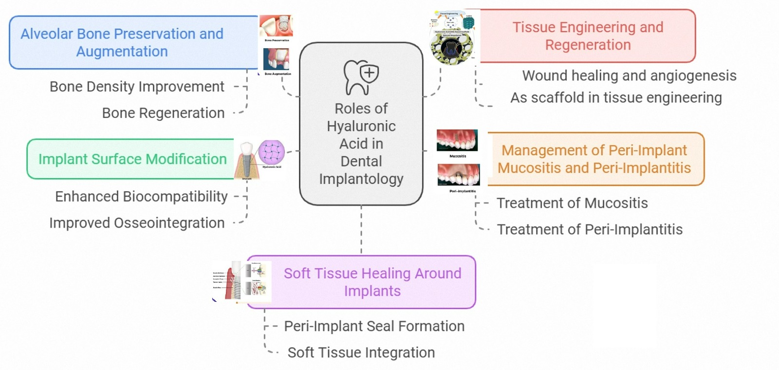
The present study aims to explore the potential applications of HA in dental implantology, including the modification of implant surfaces, the promotion of soft tissue healing around the implants, and the management of peri-implant diseases, such as mucositis and peri-implantitis. In addition, this study explores the role of HA in alveolar bone regeneration, particularly through alveolar ridge augmentation and preservation processes, as well as more advanced techniques such as tissue engineering, as the primary requirement for successful implant placement is sufficient bone width and depth. HA promotes osseointegration, increases osteogenesis, and helps treat peri-implant conditions such as mucositis and peri-implantitis. Implant biocompatibility, hydrophilicity, and bacterial adhesion resistance are all enhanced by HA coatings, which facilitate improved peri-implant bone and soft tissue healing. Stability and healing properties of HA can be increased by combining it with other biomaterials. Future studies should focus on enhancing HA’s mechanical properties, improving long-term bioactivity, and investigating synergistic biomaterial combinations; clinical trials are required to fully understand its potential in implant dentistry.
Downloads
References
Alqutaibi AY, Algabri R, Ibrahim WI, Alhajj MN, Elawady D. Dental implant planning using artificial intelligence: a systematic review and meta-analysis. J Prosthet Dent. 2024;134: 1619-1629 https://doi.org/10.1016/j.prosdent.2024.03.032 DOI: https://doi.org/10.1016/j.prosdent.2024.03.032
Marx RE. Clinical application of bone biology to mandibular and maxillary reconstruction. Clin Plastic Surg. 1994;21(3):377–92. https://doi.org/10.1016/S0094-1298(20)31014-2 DOI: https://doi.org/10.1016/S0094-1298(20)31014-2
Alqutaibi AY, Alghauli MA, Aboalrejal A, Mulla AK, Almohammadi AA, Aljayyar AW, et al. Quantitative and qualitative 3D analysis of mandibular lingual concavities: implications for dental implant planning in the posterior mandible. Clin Exp Dent Res. 2024;10(1):e858. https://doi.org/10.1002/cre2.858 DOI: https://doi.org/10.1002/cre2.858
Alqutaibi AY. CAD-CAM surgically-guided oral implant site expansion and implant placement in severely atrophic maxilla. J Taibah Univ Med Sci. 2020;15(2):153–9. https://doi.org/10.1016/j.jtumed.2020.02.006 DOI: https://doi.org/10.1016/j.jtumed.2020.02.006
Rotundo R, Pagliaro U, Bendinelli E, Esposito M, Buti J. Long‐term outcomes of soft tissue augmentation around dental implants on soft and hard tissue stability: a systematic review. Clin Oral Implants Res. 2015;26:123–38. https://doi.org/10.1111/clr.12629 DOI: https://doi.org/10.1111/clr.12629
Begam H, Nandi SK, Kundu B, Chanda A. Strategies for delivering bone morphogenetic protein for bone healing. Mater Sci Eng C Mater Biol Appl. 2017;70:856–69. https://doi.org/10.1016/j.msec.2016.09.074 DOI: https://doi.org/10.1016/j.msec.2016.09.074
Abdalla RIB, Alqutaibi AY, Kaddah A. Does the adjunctive use of platelet-rich plasma to bone graft during sinus augmentation reduce im-plant failure and complication? Systematic review and meta-analysis. Quintessence Int. 2018;49(2):139-146.
Maekawa S, Cho YD, Kauffmann F, Yao Y, Sugai JV, Zhong X, et al. BMP gene‐immobilization to dental implants enhances bone regenera-tion. Adv Mater Interfaces. 2022;9(22):2200531. DOI: https://doi.org/10.1002/admi.202200531
Choi JU, Lee SW, Pangeni R, Byun Y, Yoon I-S, Park JW. Preparation and in vivo evaluation of cationic elastic liposomes comprising highly skin-permeable growth factors combined with hyaluronic acid for enhanced diabetic wound-healing therapy. Acta Biomater. 2017;57:197–215. https://doi.org/10.1016/j.actbio.2017.04.034 DOI: https://doi.org/10.1016/j.actbio.2017.04.034
Gravante G, Sorge R, Merone A, Tamisani AM, Di Lonardo A, Scalise A, et al. Hyalomatrix PA in burn care practice: results from a national retrospective survey, 2005 to 2006. Ann Plast Surg. 2010;64(1):69–79. https://doi.org/10.1097/SAP.0b013e31819b3d59 DOI: https://doi.org/10.1097/SAP.0b013e31819b3d59
Kloss FR, Kau T, Heimes D, Kämmerer PW, Kloss-Brandstätter A. Enhanced alveolar ridge preservation with hyaluronic acid-enriched allo-grafts: a comparative study of granular allografts with and without hyaluronic acid addition. Int J Implant Dent. 2024;10(1):42. https://doi.org/10.1186/s40729-024-00559-6 DOI: https://doi.org/10.1186/s40729-024-00559-6
Yazan M, Kocyigit I, Atil F, Tekin U, Gonen Z, Onder M. Effect of hyaluronic acid on the osseointegration of dental implants. Br J Oral Max-illofac Surg. 2019;57(1):53–7. DOI: https://doi.org/10.1016/j.bjoms.2018.08.014
Moseley R, Waddington RJ, Embery G. Hyaluronan and its potential role in periodontal healing. Dent Update. 2002;29(3):144–8. DOI: https://doi.org/10.12968/denu.2002.29.3.144
Hosny Elhadidi M, Mohammed Said Ahmed W, Abdelsalam Yossef E. Effect of topical application of hyaluronic acid on stability of immedi-ate loading dental implant in posterior maxilla. Mansoura J Dent. 2020;7(3):11–2. https://doi.org/10.21608/mjd.2020.198722 DOI: https://doi.org/10.21608/mjd.2020.198722
Salih ARC, Farooqi HMU, Amin H, Karn PR, Meghani N, Nagendran S. Hyaluronic acid: comprehensive review of a multifunctional biopoly-mer. Futur J Pharm Sci. 2024;10(1):63. https://doi.org/10.1186/s43094-024-00636-y DOI: https://doi.org/10.1186/s43094-024-00636-y
Boeriu CG, Springer J, Kooy FK, van den Broek LA, Eggink G. Production methods for hyaluronan. Int J Carbohydr Chem. 2013;2013(1):624967. https://doi.org/10.1155/2013/624967 DOI: https://doi.org/10.1155/2013/624967
Boas NF. Isolation of hyaluronic acid from the cock’s comb. J Biol Chem. 1949;181(2):573–5. DOI: https://doi.org/10.1016/S0021-9258(18)56578-9
Blumberg B, Ogston A, Lowther D, Rogers H. Physicochemical properties of hyaluronic acid formed by Streptococcus haemolyticus. Biochem J. 1958;70(1):1. https://doi.org/10.1042/bj0700001 DOI: https://doi.org/10.1042/bj0700001
Fletcher E, JH J, RL M. Viscosity studies on hyaluronic acid of synovial fluid in rheumatoid arthritis and osteoarthritis. Clin Sci. 1955;14(4):653–60.
Kendall FE, Heidelberger M, Dawson MH. A serologieally inactive polysaccharide elaborated by mucoid strains of group A hemolytie streptococcus. 1937;118(1):61-69. https://doi.org/10.1016/S0021-9258(18)74517-1 DOI: https://doi.org/10.1016/S0021-9258(18)74517-1
Weissmann B, Meyer K. The structure of hyalobiuronic acid and of hyaluronic acid from umbilical Cord1, 2. J Am Chem Soc. 1954;76(7):1753–7. https://doi.org/10.1021/ja01636a010 DOI: https://doi.org/10.1021/ja01636a010
Necas J, Bartosikova L, Brauner P, Kolar J. Hyaluronic acid (hyaluronan): a review. Vet Med. 2008;53(8):397–411. https://doi.org/10.17221/1930-VETMED DOI: https://doi.org/10.17221/1930-VETMED
Balazs EA. Ultrapure hyaluronic acid and the use thereof. Google Patents; 1979. United States https://patents.google.com/patent/US4141973A
Miller D, Stegmann R. Use of Na-hyaluronate in anterior segment eye surgery. J Cataract Refract Surg. 1980;6(1):13–5. https://doi.org/10.1016/S0146-2776(80)80097-8 DOI: https://doi.org/10.1016/S0146-2776(80)80097-8
Regnault F, Bregeat P. Treatment of severe cases of retinal detachment with highly viscous hyaluronic acid. Mod Probl Ophthalmol. 1974;12:378–83.
Jones AC, Pattrick M, Doherty S, Doherty M. Intra-articular hyaluronic acid compared to intra-articular triamcinolone hexacetonide in inflammatory knee osteoarthritis. Osteoarthritis Cartilage. 1995;3(4):269–73. https://doi.org/10.1016/S1063-4584(05)80018-4 DOI: https://doi.org/10.1016/S1063-4584(05)80018-4
Pavicic T, Gauglitz GG, Lersch P, Schwach-Abdellaoui K, Malle B, Korting HC, et al. Efficacy of cream-based novel formulations of hyaluronic acid of different molecular weights in anti-wrinkle treatment. J Drugs Dermatol. 2011;10(9):990–1000.
Hellström S, Laurent C. Hyaluronan and healing of tympanic membrane perforations. An experimental study. Acta Oto-Laryngol Suppl. 1987;104(sup442):54–61. DOI: https://doi.org/10.3109/00016488709102840
Duranti F, Salti G, Bovani B, Calandra M, Rosati ML. Injectable hyaluronic acid gel for soft tissue augmentation: a clinical and histological study. Dermatol Surg. 1998;24(12):1317–25. https://doi.org/10.1111/j.1524-4725.1998.tb00007.x DOI: https://doi.org/10.1111/j.1524-4725.1998.tb00007.x
Camber O, Edman P. Sodium hyaluronate as an ophthalmic vehicle: some factors governing its effect on the ocular absorption of pilocar-pine. Curr Eye Res. 1989;8(6):563–7. DOI: https://doi.org/10.3109/02713688908995755
Benedetti L, Topp E, Stella V. Microspheres of hyaluronic acid esters – fabrication methods and in vitro hydrocortisone release. J Control Release. 1990;13(1):33–41. https://doi.org/10.1016/0168-3659(90)90072-2 DOI: https://doi.org/10.1016/0168-3659(90)90072-2
Pagnacco A, Vangelisti R, Erra C, Poma A. Double-blind clinical trial vs. placebo of a new sodium-hyaluronate-based gingival gel. Attualita Terapeutica Internazionale. 1997;15:1–7.
Miglani A, Vishnani R, Reche A, Buldeo J, Wadher B. Hyaluronic acid: exploring its versatile applications in dentistry. Cureus. 2023;15(10): e46349. DOI: https://doi.org/10.7759/cureus.46349
Sapna N. Clinical evaluation of hyaluronan gel (Gengigel) as a toipical application in the treatment of gingivitis. Rajiv Gandhi University of Health Sciences. Journal of Investigative and Clinical Dentistry Wiley-Blackwell: Hoboken, New Jersey, USA; 2006.
Petersen S, Kaule S, Teske M, Minrath I, Schmitz K-P, Sternberg K. Development and in vitro characterization of hyaluronic acid‐based coatings for implant‐associated local drug delivery systems. J Chem. 2013;2013(1):587875. https://doi.org/10.1155/2013/587875 DOI: https://doi.org/10.1155/2013/587875
Chircov C, Grumezescu AM, Bejenaru LE. Hyaluronic acid-based scaffolds for tissue engineering. Rom J Morphol Embryol. 2018;59(1):71–6.
Yun J, Lee J, Ha CW, Park SJ, Kim S, Koo KT, et al. The effect of 3‐D printed polylactic acid scaffold with and without hyaluronic acid on bone regeneration. J Periodontol. 2022;93(7):1072–82. https://doi.org/10.1002/JPER.21-0428 DOI: https://doi.org/10.1002/JPER.21-0428
Lee JH, Kim J, Baek H-R, Lee KM, Seo J-H, Lee H-K, et al. Fabrication of an rhBMP-2 loaded porous β-TCP microsphere-hyaluronic acid-based powder gel composite and evaluation of implant osseointegration. J Mater Sci Mater Med. 2014;25:2141–51. DOI: https://doi.org/10.1007/s10856-014-5250-0
Ibraheem W, Jedaiba W, Alnami A, Hussain Baiti L, Ali Manqari S, Bhati A, et al. Efficacy of hyaluronic acid gel and spray in healing of extraction wound: a randomized controlled study. Eur Rev Med Pharmacol Sci. 2022;26(10):3444–9.
Koray M, Ofluoglu D, Onal E, Ozgul M, Ersev H, Yaltirik M, et al. Efficacy of hyaluronic acid spray on swelling, pain, and trismus after surgi-cal extraction of impacted mandibular third molars. Int J Oral Maxillofac Surg. 2014;43(11):1399–403. https://doi.org/10.1016/j.ijom.2014.05.003 DOI: https://doi.org/10.1016/j.ijom.2014.05.003
Sánchez-Fernández E, Magán-Fernández A, O’Valle F, Bravo M, Mesa F. Hyaluronic acid reduces inflammation and crevicular fluid IL-1β concentrations in peri-implantitis: a randomized controlled clinical trial. J Periodontal Implant Sci. 2020;51(1):63. https://doi.org/10.5051/jpis.1903660183 DOI: https://doi.org/10.5051/jpis.1903660183
Genovesi A, Barone A, Toti P, Covani U. The efficacy of 0.12% chlorhexidine versus 0.12% chlorhexidine plus hyaluronic acid mouthwash on healing of submerged single implant insertion areas: a short‐term randomized controlled clinical trial. Int J Dent Hyg. 2017;15(1):65–72. DOI: https://doi.org/10.1111/idh.12158
Avila-Ortiz G, Elangovan S, Kramer K, Blanchette D, Dawson D. Effect of alveolar ridge preservation after tooth extraction: a systematic review and meta-analysis. J Dent Res. 2014;93(10):950–8. https://doi.org/10.1177/0022034514541127 DOI: https://doi.org/10.1177/0022034514541127
Iasella JM, Greenwell H, Miller RL, Hill M, Drisko C, Bohra AA, et al. Ridge preservation with freeze‐dried bone allograft and a collagen membrane compared to extraction alone for implant site development: a clinical and histologic study in humans. J Periodontol. 2003;74(7):990–9. DOI: https://doi.org/10.1902/jop.2003.74.7.990
Kloss FR, Offermanns V, Kloss‐Brandstätter A. Comparison of allogeneic and autogenous bone grafts for augmentation of alveolar ridge defects – a 12‐month retrospective radiographic evaluation. Clin Oral Implants Res. 2018;29(11):1163–75. https://doi.org/10.1111/clr.13380 DOI: https://doi.org/10.1111/clr.13380
Kämmerer PW, Tunkel J, Götz W, Würdinger R, Kloss F, Pabst A. The allogeneic shell technique for alveolar ridge augmentation: a multi-center case series and experiences of more than 300 cases. Int J Implant Dent. 2022;8(1):48. DOI: https://doi.org/10.1186/s40729-022-00446-y
Majzoub J, Ravida A, Starch-Jensen T, Tattan M, Del Amo FS-L. The influence of different grafting materials on alveolar ridge preserva-tion: a systematic review. J Oral Maxillofac Res. 2019;10(3): e6. https://doi.org/10.5037/jomr.2019.10306 DOI: https://doi.org/10.5037/jomr.2019.10306
Majidinia M, Sadeghpour A, Yousefi B. The roles of signaling pathways in bone repair and regeneration. J Cell Physiol. 2018;233(4):2937–48. DOI: https://doi.org/10.1002/jcp.26042
Zhai P, Peng X, Li B, Liu Y, Sun H, Li X. The application of hyaluronic acid in bone regeneration. Int J Biol Macromol. 2020;151:1224–39. https://doi.org/10.1016/j.ijbiomac.2019.10.169 DOI: https://doi.org/10.1016/j.ijbiomac.2019.10.169
Rao NV. Hyaluronic acid–based hydrogels for tissue engineering. In: Biomaterials for organ and tissue regeneration. Hindawi Publishing Corporation. Elsevier: Cairo, Egypt. 2020;551–65. DOI: https://doi.org/10.1016/B978-0-08-102906-0.00014-3
Husseini B, Friedmann A, Wak R, Ghosn N, Khoury G, Ghoul TE, et al. Clinical and radiographic assessment of cross-linked hyaluronic acid addition in demineralized bovine bone based alveolar ridge preservation: a human randomized split-mouth pilot study. J Stomatol Oral Maxillofac Surg. 2023;124(4):101426. https://doi.org/10.1016/j.jormas.2023.101426 DOI: https://doi.org/10.1016/j.jormas.2023.101426
Aldeen Abdelzaher KM. Clinical and radiographic assessment of xenogenic bone graft with or without hyaluronic acid for post extraction socket preservation (randomized clinical trial). Future. 2022;8(2):101–8. DOI: https://doi.org/10.54623/fdj.8024
Abdullah AAB, Ali HE-DM, Al-Ashmawy MMM, Mwafey IM. Volumetric and histological evaluation of Osteon II collagen with hyaluronic acid versus sticky bone graft in three dimensional socket preservation. Egypt Dent J. 2020;66(3-ِJuly (Oral Surgery)):1483–94. https://doi.org/10.21608/edj.2020.26333.1081 DOI: https://doi.org/10.21608/edj.2020.26333.1081
Ronsivalle V, Santonocito S, Giudice R, Bocchieri S, Didomenico S, Cicciù M. The role of hyaluronic acid in alveolar ridge preservation: a systematic review of its biological and regenerative potential according to PRISMA guidelines and the Cochrane handbook. Biomedicines. 2025;13(2):451. https://doi.org/10.3390/biomedicines13020451 DOI: https://doi.org/10.3390/biomedicines13020451
Helal MH, Sheta MS, Alsherif AA, Hassan MA, Aboushelib MN, Ghouraba RF. The effectiveness of hyaluronic acid on prefabricated CAD CAM bone blocks for ridge augmentation: a split mouth study. Clin Implant Dent Relat Res. 2025;27(2):e70035. https://doi.org/10.1111/cid.70035 DOI: https://doi.org/10.1111/cid.70035
Abaza G, Abdel Gaber HK, Afifi NS, Adel‐Khattab D. Injectable platelet rich fibrin versus hyaluronic acid with bovine derived xenograft for alveolar ridge preservation. A randomized controlled clinical trial with histomorphometric analysis. Clin Implant Dent Relat Res. 2024;26(1):88–102. DOI: https://doi.org/10.1111/cid.13289
Lee JB, Chu S, Ben Amara H, Song HY, Son MJ, Lee J, et al. Effects of hyaluronic acid and deproteinized bovine bone mineral with 10% collagen for ridge preservation in compromised extraction sockets. J Periodontol. 2021;92(11):1564–75. https://doi.org/10.1002/JPER.20-0832 DOI: https://doi.org/10.1002/JPER.20-0832
Kang H-J, Park S-S, Saleh T, Ahn K-M, Lee B-T. In vitro and in vivo evaluation of Ca/P-hyaluronic acid/gelatin based novel dental plugs for one-step socket preservation. Mater Design. 2020;194:108891. DOI: https://doi.org/10.1016/j.matdes.2020.108891
Kloss FR, Kämmerer PW, Kloss-Brandstätter A. First clinical case report of a xenograft–allograft combination for alveolar ridge augmenta-tion using a bovine bone substitute material with hyaluronate (Cerabone® Plus) combined with allogeneic bone granules (Maxgraft®). J Clin Med. 2023;12(19):6214. https://doi.org/10.3390/jcm12196214 DOI: https://doi.org/10.3390/jcm12196214
El Halawani GN, Khalil MM. Evaluation of melatonin and hyaluronic acid in maxillary sinus augmentation (A randomized controlled clinical trial). Alex Dent J. 2022;47(3):80–7.
Zhao N, Qin L, Liu Y, Zhai M, Li D. Improved new bone formation capacity of hyaluronic acid-bone substitute compound in rat calvarial critical size defect. BMC Oral Health. 2024;24(1):994. https://doi.org/10.1186/s12903-024-04679-8 DOI: https://doi.org/10.1186/s12903-024-04679-8
Darby I. Risk factors for periodontitis & peri‐implantitis. Periodontology 2000. 2022;90(1):9–12. DOI: https://doi.org/10.1111/prd.12447
Pontoriero R, Tonelli M, Carnevale G, Mombelli A, Nyman SR, Lang NP. Experimentally induced peri‐implant mucositis. A clinical study in humans. Clin Oral Implants Res. 1994;5(4):254–9. https://doi.org/10.1034/j.1600-0501.1994.050409.x DOI: https://doi.org/10.1034/j.1600-0501.1994.050409.x
Schimmel M, Mombelli A. Experimental mucositis and experi-mental gingivitis in persons aged 70 or over. Clinical and biological responses. Clinical Oral Implants Research . 2016;28(8):1005–1012. https://doi.org/10.1111/clr.12912 DOI: https://doi.org/10.1111/clr.12912
Berglundh T, Armitage G, Araujo MG, Avila‐Ortiz G, Blanco J, Camargo PM, et al. Peri‐implant diseases and conditions: consensus report of workgroup 4 of the 2017 world workshop on the classification of periodontal and peri‐implant diseases and conditions. J Periodontol. 2018;89:S313–8. https://doi.org/10.1002/JPER.17-0739 DOI: https://doi.org/10.1002/JPER.17-0739
Schwarz F, Derks J, Monje A, Wang HL. Peri‐implantitis. J Clin Periodontol. 2018;45:S246–66. DOI: https://doi.org/10.1111/jcpe.12954
Herrera D, Berglundh T, Schwarz F, Chapple I, Jepsen S, Sculean A, et al. Prevention and treatment of peri‐implant diseases – the EFP S3 level clinical practice guideline. J Clin Periodontol. 2023;50:4–76. https://doi.org/10.1111/jcpe.13823 DOI: https://doi.org/10.1111/jcpe.13823
Monje A, Salvi GE. Diagnostic methods/parameters to monitor peri‐implant conditions. Periodontology 2000. 2024;95: 20-39 DOI: https://doi.org/10.1111/prd.12584
Sahrmann P, Kühl S, Dagassan‐Berndt D, Bornstein MM, Zitzmann NU. Radiographic assessment of the peri‐implant site. Periodontology 2000. 2024;95: 70-86. https://doi.org/10.1111/prd.12577 DOI: https://doi.org/10.1111/prd.12577
Salvi GE, Ramseier CA. Efficacy of patient‐administered mechanical and/or chemical plaque control protocols in the management of peri‐implant mucositis. A systematic review. J Clin Periodontol. 2015;42:S187–201. DOI: https://doi.org/10.1111/jcpe.12321
Scully MF, Kakkar VV, Goodwin CA, O’Regan M. Inhibition of fibrinolytic activity by hyaluronan and its alcohol ester derivatives. Thromb Res. 1995;78(3):255–8. https://doi.org/10.1016/0049-3848(95)90876-H DOI: https://doi.org/10.1016/0049-3848(95)90876-H
Shirakata Y, Nakamura T, Kawakami Y, Imafuji T, Shinohara Y, Noguchi K, et al. Healing of buccal gingival recessions following treatment with coronally advanced flap alone or combined with a cross‐linked hyaluronic acid gel. An experimental study in dogs. J Clin Periodontol. 2021;48(4):570–80. https://doi.org/10.1111/jcpe.13433 DOI: https://doi.org/10.1111/jcpe.13433
Shirakata Y, Imafuji T, Nakamura T, Shinohara Y, Iwata M, Setoguchi F, et al. Cross‐linked hyaluronic acid gel with or without a collagen matrix in the treatment of class III furcation defects: a histologic and histomorphometric study in dogs. J Clin Periodontol. 2022;49(10):1079–89. DOI: https://doi.org/10.1111/jcpe.13694
Iorio-Siciliano V, Marasca D, Mauriello L, Vaia E, Stratul S-I, Ramaglia L. Treatment of peri-implant mucositis using spermidine and calcium chloride as local adjunctive delivery to non-surgical mechanical debridement: a double-blind randomized controlled clinical trial. Clin Oral Investig. 2024;28(10):1–11. https://doi.org/10.1007/s00784-024-05924-8 DOI: https://doi.org/10.1007/s00784-024-05924-8
Lopez M, Manzulli N, D’Angelo A, Candotto V, Casale M, Lauritano D. The use of hyaluronic acid as an adjuvant in the management of mucositis. J Biol Regul Homeost Agents. 2017;31(4 Suppl 2):115–8.
Parfenova LV, Galimshina ZR, Gil’fanova GU, Alibaeva EI, Danilko KV, Pashkova TM, et al. Hyaluronic acid bisphosphonates as antifouling antimicrobial coatings for PEO-modified titanium implants. Surf Interfaces. 2022;28:101678. https://doi.org/10.1016/j.surfin.2021.101678 DOI: https://doi.org/10.1016/j.surfin.2021.101678
Derks J, Tomasi C. Peri‐implant health and disease. A systematic review of current epidemiology. J Clin Periodontol. 2015;42:S158–71. https://doi.org/10.1111/jcpe.12334 DOI: https://doi.org/10.1111/jcpe.12334
Bohara S, Suthakorn J. Surface coating of orthopedic implant to enhance the osseointegration and reduction of bacterial colonization: a review. Biomater Res. 2022;26(1):26. DOI: https://doi.org/10.1186/s40824-022-00269-3
Sanz M, Chapple IL, Periodontology* WGotVEWo. Clinical research on peri‐implant diseases: consensus report of W orking G roup 4. J Clin Periodontol. 2012;39:202–6. https://doi.org/10.1111/j.1600-051X.2011.01837.x DOI: https://doi.org/10.1111/j.1600-051X.2011.01837.x
Sutherland IW. Novel and established applications of microbial polysaccharides. Trends Biotechnol. 1998;16(1):41–6. https://doi.org/10.1016/S0167-7799(97)01139-6 DOI: https://doi.org/10.1016/S0167-7799(97)01139-6
Moreland LW. Intra-articular hyaluronan (hyaluronic acid) and hylans for the treatment of osteoarthritis: mechanisms of action. Arthritis Res Ther. 2003;5:1–14. https://doi.org/10.1186/ar623 DOI: https://doi.org/10.1186/ar623
Chen WJ. Functions of hyaluronan in wound repair. Hyaluronan. 2002:147–56. DOI: https://doi.org/10.1533/9781845693121.147
Manzanares D, Monzon M-E, Savani RC, Salathe M. Apical oxidative hyaluronan degradation stimulates airway ciliary beating via RHAMM and RON. Am J Respir Cell Mol Biol. 2007;37(2):160–8. https://doi.org/10.1165/rcmb.2006-0413OC DOI: https://doi.org/10.1165/rcmb.2006-0413OC
Soriano‐Lerma A, Magán‐Fernández A, Gijón J, Sánchez‐Fernández E, Soriano M, García‐Salcedo JA, et al. Short‐term effects of hyaluronic acid on the subgingival microbiome in peri‐implantitis: a randomized controlled clinical trial. J Periodontol. 2020;91(6):734–45. DOI: https://doi.org/10.1002/JPER.19-0184
Zhou Z, Zhang Q, Wang Y. Preparation and characterization of antibacterial and anti-inflammatory hyaluronic acid-chitosan-dexamethasone hydrogels for peri-implantitis repair. J Biomater Appl. 2022;36(7):1141–50. https://doi.org/10.1177/08853282211047939 DOI: https://doi.org/10.1177/08853282211047939
Rakašević D, Šćepanović M, Mijailović I, Mišić T, Janjić B, Soldatović I, et al. Reconstructive peri-implantitis therapy by using bovine bone substitute with or without hyaluronic acid: a randomized clinical controlled pilot study. J Funct Biomater. 2023;14(3):149. DOI: https://doi.org/10.3390/jfb14030149
López-Valverde N, López-Valverde A, Blanco Rueda JA. Role of hyaluronic acid in the treatment of peri-implant diseases: results of a meta-analysis. Front Oral Health. 2025;6:1564599. https://doi.org/10.3389/froh.2025.1564599 DOI: https://doi.org/10.3389/froh.2025.1564599
Friedmann A, Jung R, Bilhan H, Ghawi-Begovic HA, Kauffmann F, Diehl D. Reconstructive surgical therapy of peri-implant defects with ribose cross-linked collagen matrix and crosslinked hyaluronic acid–a prospective case series. Clin Oral Investig. 2024;28(10):536. DOI: https://doi.org/10.1007/s00784-024-05942-6
Alharbi MS, Alshehri FA. High molecular weight hyaluronic acid reduces the expression of virulence genes fima, mfa1, haga, rgpa, and kgp in the oral pathogen Porphyromonas gingivalis. Pharmaceutics. 2022;14(8):1628. https://doi.org/10.3390/pharmaceutics14081628 DOI: https://doi.org/10.3390/pharmaceutics14081628
Lopez M, Manzulli N, D’Angelo A, Lauritano D, Papalia R, Candotto V. The use of hyaluronic acid as an adjuvant in the management of peri-implantitis. J Biol Regul Homeost Agents. 2017;31(4 Suppl 2):123–7.
Yumashev A, Karapetyan A, Garnova N, Berestova A. Characteristics of biocompatible coatings on dental implants. J Global Pharma Tech-nol. 2020;12(1):30.
Schulz MC, Korn P, Stadlinger B, Range U, Möller S, Becher J, et al. Coating with artificial matrices from collagen and sulfated hyaluronan influences the osseointegration of dental implants. J Mater Sci Mater Med. 2014;25:247–58. https://doi.org/10.1007/s10856-013-5066-3 DOI: https://doi.org/10.1007/s10856-013-5066-3
Dreifke MB, Ebraheim NA, Jayasuriya AC. Investigation of potential injectable polymeric biomaterials for bone regeneration. J Biomed Mater Res Part A. 2013;101(8):2436–47. DOI: https://doi.org/10.1002/jbm.a.34521
Schanté CE, Zuber G, Herlin C, Vandamme TF. Chemical modifications of hyaluronic acid for the synthesis of derivatives for a broad range of biomedical applications. Carbohydr Polym. 2011;85(3):469–89. https://doi.org/10.1016/j.carbpol.2011.03.019 DOI: https://doi.org/10.1016/j.carbpol.2011.03.019
Mendes RM, Silva GA, Lima MF, Calliari MV, Almeida AP, Alves JB, et al. Sodium hyaluronate accelerates the healing process in tooth sockets of rats. Archiv Oral Biol. 2008;53(12):1155–62. https://doi.org/10.1016/j.archoralbio.2008.07.001 DOI: https://doi.org/10.1016/j.archoralbio.2008.07.001
Hamdy M, Nasr S, Mater N, ElSayed O. Assessment of osseointegration properties of hyaluronic acid coated titanium implant versus sand-blasted large thread acid etched implants in posterior maxilla. A randomized clinical trial. MSA Dent J. 2024;3(4):1–7. DOI: https://doi.org/10.21608/msadj.2024.301280.1040
Abdulghafor MA, Amin ZM. The impact of hyaluronic acid coating on polyether ether ketone dental implant surface: an in vitro analysis. Saudi Dent J. 2024;36(10):1326–32. https://doi.org/10.1016/j.sdentj.2024.07.012 DOI: https://doi.org/10.1016/j.sdentj.2024.07.012
Ganesh S, Gheena S, Madhu K. Commercially pure titanium implants with selenium and hyaluronic acid coating for dental applications. Cureus. 2024;16(1):e52984. DOI: https://doi.org/10.7759/cureus.52984
Del Olmo JA, Alonso JM, Martínez VS, Ruiz-Rubio L, González RP, Vilas-Vilela JL, et al. Biocompatible hyaluronic acid-divinyl sulfone inject-able hydrogels for sustained drug release with enhanced antibacterial properties against Staphylococcus aureus. Mater Sci Eng C. 2021;125:112102. https://doi.org/10.1016/j.msec.2021.112102 DOI: https://doi.org/10.1016/j.msec.2021.112102
Jin H, Wang J, Tian L, Gao M, Zhao J, Ren L. Recent advances in emerging integrated antifouling and anticorrosion coatings. Mater Des. 2022;213:110307. https://doi.org/10.1016/j.matdes.2021.110307 DOI: https://doi.org/10.1016/j.matdes.2021.110307
Zamboni F, Okoroafor C, Ryan MP, Pembroke JT, Strozyk M, Culebras M, et al. On the bacteriostatic activity of hyaluronic acid composite films. Carbohydr Polym. 2021;260:117803. https://doi.org/10.1016/j.carbpol.2021.117803 DOI: https://doi.org/10.1016/j.carbpol.2021.117803
Sánchez-Bodón J, Andrade del Olmo J, Alonso JM, Moreno-Benítez I, Vilas-Vilela JL, Pérez-Álvarez L. Bioactive coatings on titanium: a re-view on hydroxylation, self-assembled monolayers (SAMs) and surface modification strategies. Polymers. 2021;14(1):165. DOI: https://doi.org/10.3390/polym14010165
Del Olmo JA, Alonso JM, Sáez-Martínez V, Benito-Cid S, Pérez-González R, Vilas-Vilela JL, et al. Hyaluronic acid-based hydrogel coatings on Ti6Al4V implantable biomaterial with multifunctional antibacterial activity. Carbohydr Polym. 2023;301:120366. DOI: https://doi.org/10.1016/j.carbpol.2022.120366
Hu X, Neoh K-G, Shi Z, Kang E-T, Poh C, Wang W. An in vitro assessment of titanium functionalized with polysaccharides conjugated with vascular endothelial growth factor for enhanced osseointegration and inhibition of bacterial adhesion. Biomaterials. 2010;31(34):8854–63. https://doi.org/10.1016/j.biomaterials.2010.08.006 DOI: https://doi.org/10.1016/j.biomaterials.2010.08.006
Valverde A, Pérez-Álvarez L, Ruiz-Rubio L, Olivenza MAP, Blanco MBG, Díaz-Fuentes M, et al. Antibacterial hyaluronic acid/chitosan multilayers onto smooth and micropatterned titanium surfac-es. Carbohydr Polym. 2019;207:824–33. https://doi.org/10.1016/j.carbpol.2018.12.039 DOI: https://doi.org/10.1016/j.carbpol.2018.12.039
Zhong X, Song Y, Yang P, Wang Y, Jiang S, Zhang X, et al. Titanium surface priming with phase-transited lysozyme to establish a silver nanoparticle-loaded chitosan/hyaluronic acid antibacterial multilayer via layer-by-layer self-assembly. PLoS One. 2016;11(1):e0146957. DOI: https://doi.org/10.1371/journal.pone.0146957
Yurttutan E, Dereci Ö, Karagöz MA. Biomechanical and histologic evaluation of osseointegration of titanium dental implants modified by various combinations of sandblasting, acid-etching, hydroxyapatite, and hyaluronic acid coating techniques. Int J Oral Maxillofac Implants. 2023;38(3): 583-590. https://doi.org/10.11607/jomi.9935 DOI: https://doi.org/10.11607/jomi.9935
Shamma MM, Ayad SS, El-dibany RM, Nagui DA. Evaluation of the effect of hyaluronic acid mixed with biphasic calcium phosphate on bone healing around dental implants (experimental study). Alexandria Dent J. 2017;42(1):104–7. https://doi.org/10.21608/adjalexu.2017.57868 DOI: https://doi.org/10.21608/adjalexu.2017.57868
Ibrahim WS, Mohammed FI, Reda HM. Evaluation of the effect of hyaluronic acid gel loaded with simvastatin on the osseointegration and stability of the dental implants. Al-Azhar Dent J Girls. 2023;10(1):393–400. DOI: https://doi.org/10.58675/2974-4164.1540
Bassiouny G, Abu Brika M, Ezzat M, Hommos A. Evaluation of the efficacy of flowable leukocyte and platelet–rich fibrin versus hyaluronic acid as bioactive implant coatings (randomized controlled clinical trial). Egypt Dent J. 2024;70(2):1339–53. https://doi.org/10.21608/edj.2024.267529.2920 DOI: https://doi.org/10.21608/edj.2024.267529.2920
Chai WL, Razali M, Ngeow WC. Dimension and structures of biological seal of peri-implant tissues. Dent Implantol Biomater. 2016;39: 39-62. https://doi.org/10.5772/63950 DOI: https://doi.org/10.5772/63950
Favia G, Mariggio M, Maiorano F, Cassano A, Capodiferro S, Ribatti D. Accelerated wound healing of oral soft tissues and angiogenic effect induced by a pool of aminoacids combined to sodium hyaluronate (AMINOGAM). J Biol Regul Homeost Agents. 2008;22(2):109–16. DOI: https://doi.org/10.1177/039463200902200225
Cohen IK, Die-Gelmann RF, Lindblad WJ, Hugo NE. Wound healing: biochemical and clinical aspects. Plast Reconstr Surg. 1992;90(5):926. https://doi.org/10.1097/00006534-199211000-00034 DOI: https://doi.org/10.1097/00006534-199211000-00034
Brown LF, Yeo K, Berse B, Yeo T-K, Senger DR, Dvorak HF, et al. Expression of vascular permeability factor (vascular endothelial growth factor) by epidermal keratinocytes during wound healing. J Exp Med. 1992;176(5):1375–9. DOI: https://doi.org/10.1084/jem.176.5.1375
Kim J-W, Han Y-S, Lee H-M, Kim J-K, Kim Y-J. Effect of morphological characteristics and biomineralization of 3D-printed gelatin/hyaluronic acid/hydroxyapatite composite scaffolds on bone tissue regeneration. Int J Mol Sci. 2021;22(13):6794. https://doi.org/10.3390/ijms22136794 DOI: https://doi.org/10.3390/ijms22136794
Partsch G, Schwarzer C, Neumüller J, Dunky A, Petera P, Bröll H, et al. Modulation of the migration and chemotaxis of PMN cells by hyalu-ronic acid. Zeitschrift fur Rheumatologie. 1989;48(3):123–8.
Ouasti S, Donno R, Cellesi F, Sherratt MJ, Terenghi G, Tirelli N. Network connectivity, mechanical properties and cell adhesion for hyalu-ronic acid/PEG hydrogels. Biomaterials. 2011;32(27):6456–70. DOI: https://doi.org/10.1016/j.biomaterials.2011.05.044
Fallacara A, Baldini E, Manfredini S, Vertuani S. Hyaluronic acid in the third millennium. Polymers. 2018;10(7):701. https://doi.org/10.3390/polym10070701 DOI: https://doi.org/10.3390/polym10070701
Scholz N. Personalised medicine: the right treatment for the right person at the right time. European Parliamentary Research Service (EPRS), Members’ Research Service, European Parliament: 2015.
Mariggiò MA, Cassano A, Vinella A, Vincenti A, Fumarulo R, Muzio LL, et al. Enhancement of fibroblast proliferation, collagen biosynthesis and production of growth factors as a result of combining sodium hyaluronate and aminoacids. Int J Immunopathol Pharmacol. 2009;22(2):485–92. https://doi.org/10.1177/039463200902200225 DOI: https://doi.org/10.1177/039463200902200225
Tammi MI, Day AJ, Turley EA. Hyaluronan and homeostasis: a balancing act. J Biol Chem. 2002;277(7):4581–4. https://doi.org/10.1074/jbc.R100037200 DOI: https://doi.org/10.1074/jbc.R100037200
Mesa F, Aneiros J, Cabrera A, Bravo M, Caballero T, Revelles F, et al. Antiproliferative effect of topic hyaluronic acid gel. Study in gingival biopsies of patients with periodontal disease. Histol Histopathol; 2002;17: 747–753.
Litwiniuk M, Krejner A, Speyrer MS, Gauto AR, Grzela T. Hyaluronic acid in inflammation and tissue regeneration. Wounds. 2016;28(3):78–88.
S.p.A. PD. Aminogam. 2021. Available from: https://aminogam.com/ [cited 28-October-2024]
Canciani E, Sirello R, Pellegrini G, Henin D, Perrotta M, Toma M, et al. Effects of vitamin and amino acid-enriched hyaluronic acid gel on the healing of oral mucosa: in vivo and in vitro study. Medicina. 2021;57(3):285. https://doi.org/10.3390/medicina57030285 DOI: https://doi.org/10.3390/medicina57030285
Capodiferro S, Tempesta A, Bucci S, Maiorano E, Favia G, Limongelli L. Aminogam® gel allows faster wound healing after oral surgery by formation of mature connective tissue with low vascular density and reducing inflammatory infiltration. A retrospective study on 580 cases with histological and confocal laser investigation. Appl Sci. 2020;10(3):1105. DOI: https://doi.org/10.3390/app10031105
Bawankar PV, Tuli P, Kolte AP, Kolte RA. Evaluation of the efficacy of microneedling alone and in combination with injectable hyaluronic acid in augmentation of peri-implant soft tissues: a randomized controlled trial. J Indian Soc Periodontol. 2024;28(6):643–50. https://doi.org/10.4103/jisp.jisp_158_24 DOI: https://doi.org/10.4103/jisp.jisp_158_24
Pilipchuk SP, Plonka AB, Monje A, Taut AD, Lanis A, Kang B, et al. Tissue engineering for bone regeneration and osseointegration in the oral cavity. Dent Mater. 2015;31(4):317–38. DOI: https://doi.org/10.1016/j.dental.2015.01.006
Kostadinova M, Raykovska M, Simeonov R, Lolov S, Mourdjeva M. Recent advances in bone tissue engineering: enhancing the potential of mesenchymal stem cells for regenerative therapies. Curr Issues Mol Biol. 2025;47(4):287. https://doi.org/10.3390/cimb47040287 DOI: https://doi.org/10.3390/cimb47040287
Zhang C, Chen Z, Liu J, Wu M, Yang J, Zhu Y, et al. 3D-printed pre-tapped-hole scaffolds facilitate one-step surgery of predictable alveolar bone augmentation and simultaneous dental implantation. Compos Part B Eng. 2022;229:109461. DOI: https://doi.org/10.1016/j.compositesb.2021.109461
Wong SK, Yee MMF, Chin K-Y, Ima-Nirwana S. A review of the application of natural and synthetic scaffolds in bone regeneration. J Funct Biomater. 2023;14(5):286. https://doi.org/10.3390/jfb14050286 DOI: https://doi.org/10.3390/jfb14050286
Roseti L, Parisi V, Petretta M, Cavallo C, Desando G, Bartolotti I, et al. Scaffolds for bone tissue engineering: state of the art and new per-spectives. Mater Sci Eng C Mater Biol Appl. 2017;78:1246–62. DOI: https://doi.org/10.1016/j.msec.2017.05.017
D’Albis G, D’Albis V, Palma M, Plantamura M, Nizar AK. Use of hyaluronic acid for regeneration of maxillofacial bones. Genesis. 2022;60(8–9):e23497. DOI: https://doi.org/10.1002/dvg.23497
Hwang HS, Lee C-S. Recent progress in hyaluronic-acid-based hydrogels for bone tissue engineering. Gels. 2023;9(7):588. https://doi.org/10.3390/gels9070588 DOI: https://doi.org/10.3390/gels9070588
AlHowaish NA, AlSudani DI, AlMuraikhi NA. Evaluation of a hyaluronic acid hydrogel (Restylane Lyft) as a scaffold for dental pulp regen-eration in a regenerative endodontic organotype model. Odontology. 2022;110(4):726–34. DOI: https://doi.org/10.1007/s10266-022-00710-y
Seliktar D. Designing cell-compatible hydrogels for biomedical applications. Science. 2012;336(6085):1124–8. https://doi.org/10.1126/science.1214804 DOI: https://doi.org/10.1126/science.1214804
Alshehadat SA, Thu HA, Hamid SSA, Nurul AA, Rani SA, Ahmad A. Scaffolds for dental pulp tissue regeneration: a review. Int Dent Med J Adv Res. 2016;2(1):1–12. DOI: https://doi.org/10.15713/ins.idmjar.36
Hamlet SM, Vaquette C, Shah A, Hutmacher DW, Ivanovski S. 3‐Dimensional functionalized polycaprolactone‐hyaluronic acid hydrogel constructs for bone tissue engineering. J Clin Periodontol. 2017;44(4):428–37. DOI: https://doi.org/10.1111/jcpe.12686
Subramaniam S, Fang Y-H, Sivasubramanian S, Lin F-H, Lin C-p. Hydroxyapatite-calcium sulfate-hyaluronic acid composite encapsulated with collagenase as bone substitute for alveolar bone regeneration. Biomaterials. 2016;74:99–108. https://doi.org/10.1016/j.biomaterials.2015.09.044 DOI: https://doi.org/10.1016/j.biomaterials.2015.09.044
Zanchetta P, Lagarde N, Uguen A, Marcorelles P. Mixture of hyaluronic acid, chondroitin 6 sulphate and dermatan sulphate used to com-pletely regenerate bone in rat critical size defect model. J Cranio-Maxillofac Surg. 2012;40(8):783–7. https://doi.org/10.1016/j.jcms.2012.02.011 DOI: https://doi.org/10.1016/j.jcms.2012.02.011
Azhar IS, Nariswari VS, Kusumawardhani DP, Maksum MA. Combination of hDPSCs and oxysterol in Hyaluronic Acid scaffolds for Dental Implant Therapy: a narrative review. J Int Oral Health. 2022;14(5):440–6. https://doi.org/10.4103/jioh.jioh_299_21 DOI: https://doi.org/10.4103/jioh.jioh_299_21
Li S, De Wijn JR, Li J, Layrolle P, De Groot K. Macroporous biphasic calcium phosphate scaffold with high permeability/porosity ratio. Tis-sue Eng. 2003;9(3):535–48. DOI: https://doi.org/10.1089/107632703322066714
Park S, Huang NW, Wong CX, Pan J, Albakr L, Gu J, et al. Microstructured hyaluronic acid hydrogel for tooth germ bioengineering. Gels. 2021;7(3):123. DOI: https://doi.org/10.3390/gels7030123
Pan H, Han JJ, Park Y-D, Cho TH, Hwang SJ. Effect of sustained release of rhBMP-2 from dried and wet hyaluronic acid hydrogel carriers compared with direct dip coating of rhBMP-2 on peri-implant osteogenesis of dental implants in canine mandibles. J Cranio-Maxillofac Surg. 2016;44(2):116–25. DOI: https://doi.org/10.1016/j.jcms.2015.11.018
Park SH, Park JY, Ji YB, Ju HJ, Min BH, Kim MS. An injectable click-crosslinked hyaluronic acid hydrogel modified with a BMP-2 mimetic peptide as a bone tissue engineering scaffold. Acta Biomater. 2020;117:108–20. https://doi.org/10.1016/j.actbio.2020.09.013 DOI: https://doi.org/10.1016/j.actbio.2020.09.013
Miranda DG, Malmonge SM, Campos DM, Attik NG, Grosgogeat B, Gritsch K. A chitosan‐hyaluronic acid hydrogel scaffold for periodontal tissue engineering. J Biomed Mater Res Part B Appl Biomater. 2016;104(8):1691–702. DOI: https://doi.org/10.1002/jbm.b.33516
Chrepa V, Austah O, Diogenes A. Evaluation of a commercially available hyaluronic acid hydrogel (Restylane) as injectable scaffold for dental pulp regeneration: an in vitro evaluation. J Endod. 2017;43(2):257–62. https://doi.org/10.1016/j.joen.2016.10.026 DOI: https://doi.org/10.1016/j.joen.2016.10.026
Jung YS, Ye JR, Kwack KH, Lee M-H, Kweon D-K, Chae YK, et al. Bilayer cellulose-coated hyaluronic acid-based scaffold for accelerating oral wound healing. Cellulose. 2024:31;1–14. DOI: https://doi.org/10.21203/rs.3.rs-4263630/v1
Sabu A, Kaarthikeyan G, Eswaramoorthy R, Priyangha P. Development of a Cissus quadrangularis-doped extracellular matrix and a hyalu-ronic acid-incorporated scaffold for periodontal regeneration: an in vitro study. Cureus. 2024;16(3): e56507.
ElShammaa IM, Ghoraba SF, Shoukheba MY. Effect of hyaluronic acid with a collagen sponge scaffold in the treatment of intra-bony de-fects of chronic periodontitis. Tanta Dent J. 2023;20(3):224–32. https://doi.org/10.4103/tdj.tdj_10_23 DOI: https://doi.org/10.4103/tdj.tdj_10_23
Lee S-w, Kim J, Do M, Namkoong E, Lee H, Ryu JH, et al. Developmental role of hyaluronic acid and its application in salivary gland tissue engineering. Acta Biomater. 2020;115:275–87. https://doi.org/10.1016/j.actbio.2020.08.030 DOI: https://doi.org/10.1016/j.actbio.2020.08.030
Sieger D, Korzinskas T, Jung O, Stojanovic S, Wenisch S, Smeets R, et al. The addition of high doses of hyaluronic acid to a biphasic bone substitute decreases the proinflammatory tissue response. Int J Mol Sci. 2019;20(8):1969. DOI: https://doi.org/10.3390/ijms20081969
Rőszer T. Understanding the mysterious M2 macrophage through activation markers and effector mechanisms. Mediators Inflamm. 2015;2015(1):816460. DOI: https://doi.org/10.1155/2015/816460
Bertl K, Gotfredsen K, Jensen SS, Bruckmann C, Stavropoulos A. Adverse reaction after hyaluronan injection for minimally invasive papilla volume augmentation. A report on two cases. Clin Oral Implants Res. 2017;28(7):871–6. https://doi.org/10.1111/clr.12892 DOI: https://doi.org/10.1111/clr.12892
De Jong WH, Jennen D, Keizers PH, Hodemaekers HM, Vermeulen JP, Bakker F, et al. Evaluation of adverse effects of resorbable hyalu-ronic acid fillers: determination of macrophage responses. Int J Mol Sci. 2022;23(13):7275. https://doi.org/10.3390/ijms23137275 DOI: https://doi.org/10.3390/ijms23137275
Cox SE, Adigun CG. Complications of injectable fillers and neurotoxins. Dermatol Ther. 2011;24(6):524–36. DOI: https://doi.org/10.1111/j.1529-8019.2012.01455.x
Lafaille P, Benedetto A. Fillers: contraindications, side effects and precautions. J Cutan Aesthet Surg. 2010;3(1):16–9. https://doi.org/10.4103/0974-2077.63222 DOI: https://doi.org/10.4103/0974-2077.63222
Houseman ND, Taylor GI, Pan W-R. The angiosomes of the head and neck: anatomic study and clinical applications. Plast Reconstr Surg. 2000;105(7):2287–313. DOI: https://doi.org/10.1097/00006534-200006000-00001
Cohen JL. Understanding, avoiding, and managing dermal filler complications. Dermatol Surg. 2008;34:S92–9. https://doi.org/10.1097/00042728-200806001-00020 DOI: https://doi.org/10.1097/00042728-200806001-00020
Weinberg MJ, Solish N. Complications of hyaluronic acid fillers. Facial Plast Surg. 2009;25(05):324–8. https://doi.org/10.1055/s-0029-1243081 DOI: https://doi.org/10.1055/s-0029-1243081
Sadowitz B, Seymour K, Gahtan V, Maier KG. The role of hyaluronic acid in atherosclerosis and intimal hyperplasia. J Surg Res. 2012;173(2):e63–72. https://doi.org/10.1016/j.jss.2011.09.025 DOI: https://doi.org/10.1016/j.jss.2011.09.025
Brody HJ. Use of hyaluronidase in the treatment of granulomatous hyaluronic acid reactions or unwanted hyaluronic acid misplacement. Dermatol Surg. 2005;31(8):893–7. https://doi.org/10.1097/00042728-200508000-00001 DOI: https://doi.org/10.1097/00042728-200508000-00001
Menon H, Thomas M, D’silva J. Low dose of Hyaluronidase to treat over correction by HA filler – a case report. J Plast Reconstr Aesthet Surg. 2010;63(4):e416–7. https://doi.org/10.1016/j.bjps.2010.01.005 DOI: https://doi.org/10.1016/j.bjps.2010.01.005
Costerton JW, Cheng K-J, Geesey GG, Ladd TI, Nickel JC, Dasgupta M, et al. Bacterial biofilms in nature and disease. Annual Review of Microbiology. 1987;41: 435–464 https://doi.org/10.1146/annurev.mi.41.100187.002251 DOI: https://doi.org/10.1146/annurev.mi.41.100187.002251
Hoyle BD, Jass J, Costerton JW. The biofilm glycocalyx as a resistance factor. J Antimicrob Chemother. 1990;26(1):1–5. https://doi.org/10.1093/jac/26.1.1 DOI: https://doi.org/10.1093/jac/26.1.1
Sadashivaiah AB, Mysore V. Biofilms: their role in dermal fillers. J Cutan Aesthet Surg. 2010;3(1):20–2. https://doi.org/10.4103/0974-2077.63257 DOI: https://doi.org/10.4103/0974-2077.63257
Carlos-Fabuel L, Marzal-Gamarra C, Martí-Álamo S, Mancheño-Franch A. Foreign body granulomatous reactions to cosmetic fillers. J Clin Exp Dentist. 2012;4(4):e244. https://doi.org/10.4317/jced.50868 DOI: https://doi.org/10.4317/jced.50868
Reisberger E-M, Landthaler M, Wiest L, Schröder J, Stolz W. Foreign body granulomas caused by polymethylmethacrylate microspheres: successful treatment with allopurinol. Archiv Dermatol. 2003;139(1):17–20. https://doi.org/10.1001/archderm.139.1.17 DOI: https://doi.org/10.1001/archderm.139.1.17
Published
How to Cite
Issue
Section
License
Copyright (c) 2026 Ahmed Yaseen Alqutaibi, Nazrah Maher, Anum Mahmood, Faiza Amin, Ghulam Irtiza Mustafa, Naresh Kumar, Muhammad Sohail Zafar

This work is licensed under a Creative Commons Attribution 4.0 International License.


 Biomaterial Investigations in Dentistry is a Diamond Open Access peer-reviewed journal, publishing research in oral biomaterials science. The publishing of articles is free for authors, thanks to the support of Acta Odontologica Scandinavica Society (AOSS), a not-for-profit society.

