
Effects of HCl concentration and immersion time on physicochemical properties, BMP-2 detection, degradation, and osteoblast response of human demineralized tooth matrix
DOI:
https://doi.org/10.2340/biid.v13.45645Keywords:
human demineralized tooth matrix, tooth-derived graft, hydrochloric acid, demineralization parameters, degradation, BMP-2, osteoblast responseAbstract
Objective: Human demineralized tooth matrix (hDTM) is a promising tooth-derived graft material; however, demineralization protocols vary widely and may alter matrix structure, degradation behavior, and growth factor detectability. This study investigates the combined effects of hydrochloric acid (HCl) concentration (0.5 vs 1.0M) and immersion time (10 vs 20 min) on the physicochemical characteristics, Bone Morphogenetic Protein-2 (BMP-2) detection, degradation behavior and osteoblast response of hDTM.
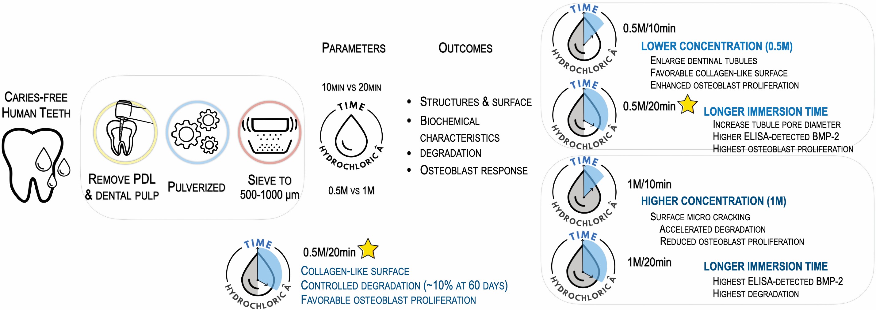
Methods: Caries-free permanent teeth (particle size 500–1000 µm) were demineralized using four HCl protocols: 0.5M/10 min, 0.5M/20 min, 1M/10 min, and 1M/20 min. Morphology, crystallinity, and surface features were characterized by scanning electron microscopy (SEM), X-ray diffraction, X-ray fluorescence, and Brunauer-Emmett-Teller analysis. Total protein and BMP-2 were quantified using Bradford and enzyme-linked immunosorbent assay (ELISA) assays. In-vitro degradation was monitored for 60 days in simulated body fluid. MC3T3-E1 osteoblast adhesion and proliferation were evaluated using the SEM and PrestoBlue® assay. Two-way analysis of variance was performed to assess main and interaction effects of concentration and time.
Results: Both HCl concentration and immersion time significantly influenced hDTM properties. Lower concentration enlarged dentinal tubules and collagen exposure, whereas higher concentration produced surface microcracking and reduced collagen exposure. Higher HCl concentration increased degradation (1M/20 min: 23.76 ± 3.23% vs. 0.5M/20 min: 9.02 ± 0.63%, p < 0.01), while longer immersion increased ELISA-detected BMP-2 levels (1M/20 min: 11.8 ± 1.9 ng/g; 0.5M/20 min: 5.3 ± 1.6 ng/g vs. control: 0.1 ng/g, p < 0.001). Significant independent and interactive effects of HCl concentration and immersion time were observed (p < 0.001). Among the test conditions, a 0.5M/20 min protocol demonstrated balanced collagen preservation, controlled degradation, and favorable osteoblast proliferation.
Conclusion: HCl concentration and immersion time independently and interactively modulate hDTM properties. Within the test conditions, 0.5M HCl for 20 minutes provides a practical solution and balances collagen preservation, controlled degradation, and osteoblast proliferation, supporting its use as a feasible approach for fabricating tooth-derived bone graft materials.
Downloads
References
Desarda HM, Shetgar SS, Chaudhari SU, Chaudhari RK, Chandrahas B, Burli VVA. Evaluation of human tooth properties to use as an autogenous graft – an in vitro study. J Pharmacy Bioallied Sci. 2025;17(Suppl 1):S878–80.
https://doi.org/10.4103/jpbs.jpbs_1781_24 DOI: https://doi.org/10.4103/jpbs.jpbs_1781_24
Tanwatana S, Kiewjurat A, Suttapreyasri S. Chemical and thermal deproteinization of human demineralized tooth matrix: physicochemical characterization and osteoblast cell biocompatibility. J Biomater Appl. 2019;34(5):651–63.
https://doi.org/10.1177/0885328219866039 DOI: https://doi.org/10.1177/0885328219866039
Yamakoshi Y. Dentinogenesis and dentin sialophosphoprotein (DSPP). J Oral Biosci. 2009;51(3):134. DOI: https://doi.org/10.2330/joralbiosci.51.134
https://doi.org/10.1016/S1349-0079(09)80021-2 DOI: https://doi.org/10.1016/S1349-0079(09)80021-2
Linde A. Dentin matrix proteins: composition and possible functions in calcification. Anatom Record. 1989;224(2):154–66.
https://doi.org/10.1002/ar.1092240206 DOI: https://doi.org/10.1002/ar.1092240206
Butler WT, Mikulski A, Urist MR, Bridges G, Uyeno S. Noncollagenous proteins of a rat dentin matrix possessing bone morphogenetic activity. J Dent Res. 1977;56(3):228–32.
https://doi.org/10.1177/00220345770560030601 DOI: https://doi.org/10.1177/00220345770560030601
Huggins CB, Urist MR. Dentin matrix transformation: rapid induction of alkaline phosphatase and cartilage. Science. 1970;167(3919):896–8.
https://doi.org/10.1126/science.167.3919.896 DOI: https://doi.org/10.1126/science.167.3919.896
Urist MR. Bone: formation by autoinduction. Science. 1965;150(698):893–9.
https://doi.org/10.1126/science.150.3698.893 DOI: https://doi.org/10.1126/science.150.3698.893
Katz JM, Nataraj C, Jaw R, Deigl E, Bursac P. Demineralized bone matrix as an osteoinductive biomaterial and in vitro predictors of its biological potential. J Biomed Mater Res. 2009;89B(1):127–34.
https://doi.org/10.1002/jbm.b.31195 DOI: https://doi.org/10.1002/jbm.b.31195
Pang S, Su FY, Green A, Salim J, McKittrick J, Jasiuk I. Comparison of different protocols for demineralization of cortical bone. Sci Rep. 2021;11(1):7012.
https://doi.org/10.1038/s41598-021-86257-4 DOI: https://doi.org/10.1038/s41598-021-86257-4
Ariffin AF, Yusof N, Mohd S, Rahman SA, Ramalingam S, Mansor A, et al. Verifying measurements of residual calcium content in demineralised cortical bone. Cell Tissue Bank. 2019;20(4):527–34.
https://doi.org/10.1007/s10561-019-09785-4 DOI: https://doi.org/10.1007/s10561-019-09785-4
Figueiredo M, Cunha S, Martins G, Freitas J, Judas F, Figueiredo H. Influence of hydrochloric acid concentration on the demineralization of cortical bone. Chem Eng Res Design. 2011;89(1):116–24.
https://doi.org/10.1016/j.cherd.2010.04.013 DOI: https://doi.org/10.1016/j.cherd.2010.04.013
Kiviranta I, Tammi M, Lappalainen R, Kuusela T, Helminen HJ. The rate of calcium extraction during EDTA decalcification from thin bone slices as assessed with atomic absorption spectrophotometry. Histochemistry. 1980;68(2):119–27.
https://doi.org/10.1007/BF00489507 DOI: https://doi.org/10.1007/BF00489507
John S, Devi P, Sharma P, Gupta S, Chandra S. Comparison of various decalcifying agents to evaluate their efficacy. Int J Basic Clin Pharmacol. 2023;12(4):522–7.
https://doi.org/10.18203/2319-2003.ijbcp20231886 DOI: https://doi.org/10.18203/2319-2003.ijbcp20231886
Cornelison JB, Isaac CV, Devota CJ, Billian J, Brown TT, deJong JL, et al. A comparison of three decalcification agents for assessments of cranial fracture histomorphology. J Forens Sci. 2022;67(3):1157–66.
https://doi.org/10.1111/1556-4029.14990 DOI: https://doi.org/10.1111/1556-4029.14990
Khurshid Z, Adanir N, Ratnayake J, Dias G, Cooper PR. Demineralized dentin matrix for bone regeneration in dentistry: a critical update. Saudi Dent J. 2024;36(3):443–50.
https://doi.org/10.1016/j.sdentj.2023.11.028 DOI: https://doi.org/10.1016/j.sdentj.2023.11.028
Inchingolo AM, Marinelli G, Inchingolo F, Giorgio RV, Colonna V, Pennacchio BFP, et al. Autologous tooth-derived biomaterials in alveolar bone regeneration: a systematic review of clinical outcomes and histological evidence. J Funct Biomater. 2025;16(10):367.
https://doi.org/10.3390/jfb16100367 DOI: https://doi.org/10.3390/jfb16100367
Minetti E, Taschieri S, Berardini M, Corbella S. New classification of autologous tooth-derived grafting materials: fundamental concepts. Int J Dent. 2025;2025:6646405.
https://doi.org/10.1155/ijod/6646405 DOI: https://doi.org/10.1155/ijod/6646405
Sultan N, Mowafey B, Ata F, El-Zekrid MH, Jayash SN. Enhanced bone regeneration using demineralized dentin matrix: a comparative study in alveolar bone repair. Int Dent J. 2025;75(4):100817.
https://doi.org/10.1016/j.identj.2025.03.026 DOI: https://doi.org/10.1016/j.identj.2025.03.026
Hesham R, Shemais N, Saleh HA, Fawzy El-Sayed K. Autogenous demineralized dentin graft with high molecular weight hyaluronic acid in ridge preservation: pilot trial. Clin Implant Dent Relat Res. 2025;27(6):e70100.
https://doi.org/10.1111/cid.70100 DOI: https://doi.org/10.1111/cid.70100
Heggendorn FL, Nascimento MBD, Lima AM, Ribeiro AA. Demineralized dentin matrix technique – a comparison of different demineralizing solutions. Braz Dent J. 2023;34(4):72–84.
https://doi.org/10.1590/0103-6440202305353 DOI: https://doi.org/10.1590/0103-6440202305353
Grawish ME, Grawish LM, Grawish HM, Grawish MM, Holiel AA, Sultan N, et al. Demineralized dentin matrix for dental and alveolar bone tissues regeneration: an innovative scope review. Tissue Eng Regener Med. 2022;19(4):687–701.
https://doi.org/10.1007/s13770-022-00438-4 DOI: https://doi.org/10.1007/s13770-022-00438-4
de Dios Teruel J, Alcolea A, Hernandez A, Ruiz AJ. Comparison of chemical composition of enamel and dentine in human, bovine, porcine and ovine teeth. Arch Oral Biol. 2015;60(5):768–75.
https://doi.org/10.1016/j.archoralbio.2015.01.014 DOI: https://doi.org/10.1016/j.archoralbio.2015.01.014
Wildemann B, Kadow-Romacker A, Pruss A, Haas NP, Schmidmaier G. Quantification of growth factors in allogenic bone grafts extracted with three different methods. Cell Tissue Bank. 2007;8(2):107–14.
https://doi.org/10.1007/s10561-006-9021-0 DOI: https://doi.org/10.1007/s10561-006-9021-0
Kokubo T, Takadama H. How useful is SBF in predicting in vivo bone bioactivity? Biomaterials. 2006;27(15):2907–15.
https://doi.org/10.1016/j.biomaterials.2006.01.017 DOI: https://doi.org/10.1016/j.biomaterials.2006.01.017
Park SM, Kim DH, Pang EK. Bone formation of demineralized human dentin block graft with different demineralization time: in vitro and in vivo study. J Craniomaxillofac Surg. 2017;45(6):903–12.
https://doi.org/10.1016/j.jcms.2017.03.007 DOI: https://doi.org/10.1016/j.jcms.2017.03.007
Kabir MA, Murata M, Akazawa T, Kusano K, Yamada K, Ito M. Evaluation of perforated demineralized dentin scaffold on bone regeneration in critical-size sheep iliac defects. Clin Oral Implants Res. 2017;28(11):e227–35.
https://doi.org/10.1111/clr.13000 DOI: https://doi.org/10.1111/clr.13000
Saebe M, Suttapreyasri S. Mini-review: dentin as bone graft substitution. Songklanakarin Dent J. 2014;2(1):40–7.
Rey C, Combes C, Drouet C, Glimcher MJ. Bone mineral: update on chemical composition and structure. Osteoporos Int. 2009;20(6):1013–21.
https://doi.org/10.1007/s00198-009-0860-y DOI: https://doi.org/10.1007/s00198-009-0860-y
Dorozhkin SV. Nanodimensional and nanocrystalline calcium orthophosphates. Am J Biomed Eng. 2012;2(3):48–97.
https://doi.org/10.5923/j.ajbe.20120203.01 DOI: https://doi.org/10.5923/j.ajbe.20120203.01
Danilchenko S, Kalinkevich A, Zhovner M, Li H, Kochenko A, Danylchenko P, et al. X-ray diffraction studies of a partially demineralized oriented cortical bone with the controlled depth of analysis. Heliyon. 2023;9(7):e17809.
https://doi.org/10.1016/j.heliyon.2023.e17809 DOI: https://doi.org/10.1016/j.heliyon.2023.e17809
Madupalli H, Pavan B, Tecklenburg MMJ. Carbonate substitution in the mineral component of bone: discriminating the structural changes, simultaneously imposed by carbonate in A and B sites of apatite. J Solid State Chem. 2017;255:27–35.
https://doi.org/10.1016/j.jssc.2017.07.025 DOI: https://doi.org/10.1016/j.jssc.2017.07.025
Wang B, Zhang Z, Pan H. Bone apatite nanocrystal: crystalline structure, chemical composition and architecture. Biominetics. 2023;8(1):90.
https://doi.org/10.3390/biomimetics8010090 DOI: https://doi.org/10.3390/biomimetics8010090
Piga G, Santos-Cubedo A, Moya Solà S, Brunetti A, Malgosa A, Enzo S. An X-ray diffraction (XRD) and X-ray fluorescence (XRF) investigation in human and animal fossil bones from Holocene to Middle Triassic. J Archaeol Sci. 2009;36(9):1857–68.
https://doi.org/10.1016/j.jas.2009.04.013 DOI: https://doi.org/10.1016/j.jas.2009.04.013
Maestracci B, Delchini S, Chateigner D, Pilliere H, Lutterotti L, Borovin E. Simultaneous combined XRF-XRD analysis of geological sample: new methodological approach for on-site analysis on New-Caledonian Ni-rich harzburgite. J Geochem Explor. 2023;252:107250.
https://doi.org/10.1016/j.gexplo.2023.107250 DOI: https://doi.org/10.1016/j.gexplo.2023.107250
Park M, Mah YJ, Kim BD, Kim ES, Park EJ. Demineralized deciduous tooth as a source of bone graft material: its biological and physicochemical characteristics. Oral Surg Oral Med Oral Pathol Oral Radiol Endodontol. 2015;120(3):307–14.
https://doi.org/10.1016/j.oooo.2015.05.021 DOI: https://doi.org/10.1016/j.oooo.2015.05.021
Eliaz N, Metoki N. Calcium phosphate bioceramics: a review of their history, structure, properties, coating technologies and biomedical applications. Materials. 2017;10(4):334.
https://doi.org/10.3390/ma10040334 DOI: https://doi.org/10.3390/ma10040334
Wei S, Ma JX, Xu L, Gu XS, Ma XL. Biodegradable materials for bone defect repair. Milit Med Res. 2020;7(1):54.
https://doi.org/10.1186/s40779-020-00280-6 DOI: https://doi.org/10.1186/s40779-020-00280-6
Pietrzak WS, Woodell-May J, McDonald N. Assay of bone morphogenetic protein-2, -4, and -7 in human demineralized bone matrix. J Craniofac Surg. 2006;17(1):84–90.
https://doi.org/10.1097/01.scs.0000179745.91165.73 DOI: https://doi.org/10.1097/01.scs.0000179745.91165.73
Bono N, Tarsini P, Candiani G. BMP-2 and type I collagen preservation in human deciduous teeth after demineralization. J Appl Biomater Funct Mater. 2018;17(2):2280800018784230.
https://doi.org/10.1177/2280800018784230 DOI: https://doi.org/10.1177/2280800018784230
Kim SY, Kim YK, Park YH, Park JC, Ku JK, Um IW, et al. Evaluation of the healing potential of demineralized dentin matrix fixed with recombinant human bone morphogenetic protein-2 in bone grafts. Materials. 2017;10(9):1049.
https://doi.org/10.3390/ma10091049 DOI: https://doi.org/10.3390/ma10091049
Um IW, Ku JK, Lee BK, Yun PY, Lee JK, Nam JH. Postulated release profile of recombinant human bone morphogenetic protein-2 (rhBMP-2) from demineralized dentin matrix. J Korean Assoc Oral Maxillofac Surg. 2019;45(3):123–8.
https://doi.org/10.5125/jkaoms.2019.45.3.123 DOI: https://doi.org/10.5125/jkaoms.2019.45.3.123
Murata M, Nezu T, Takebe H, Hirose Y, Okubo N, Saito T, et al. Human dentin materials for minimally invasive bone regeneration: animal studies and clinical cases. J Oral Biosci. 2023;65(1):13–18.
https://doi.org/10.1016/j.job.2022.10.003 DOI: https://doi.org/10.1016/j.job.2022.10.003
Additional Files
Published
How to Cite
Issue
Section
License
Copyright (c) 2026 Anupong Jeerachaipansakul, Narit Leepong, Srisurang Suttapreaysri

This work is licensed under a Creative Commons Attribution 4.0 International License.


 Biomaterial Investigations in Dentistry is a Diamond Open Access peer-reviewed journal, publishing research in oral biomaterials science. The publishing of articles is free for authors, thanks to the support of Acta Odontologica Scandinavica Society (AOSS), a not-for-profit society.

