
Short-term histopathological effects of direct peripheral nerve exposure to AH Plus® Bioceramic Sealer in rats: an exploratory assessment of Stemregen® supplementation
DOI:
https://doi.org/10.2340/biid.v13.46483Keywords:
dietary supplements, nerve damage, premixed bioceramic, sciatic nerveAbstract
Background: Extrusion of root canal sealers into neurovascular tissues can result in rare but serious neurological complications, including paresthesia and neuropathic pain. This study aimed to evaluate the histopathological effects of direct peripheral nerve exposure to AH Plus® Bioceramic Sealer and to assess the influence of Stemregen® supplementation on histopathological tissue responses.
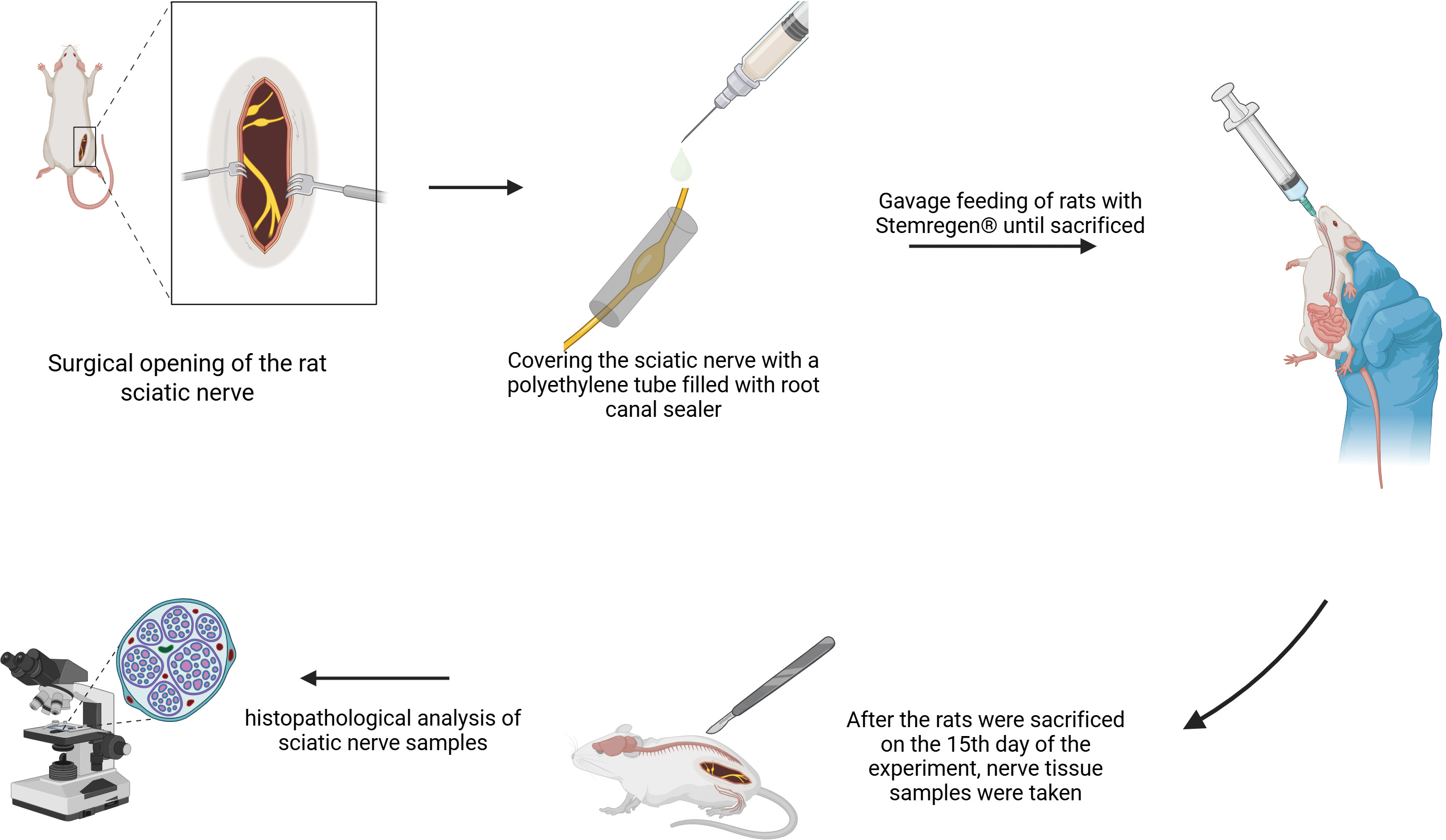
Materials and methods: Forty-eight male Sprague-Dawley rats were randomly assigned to four groups (n = 12): Control, Supplement only, Bioceramic only, and Bioceramic + Supplement. Sterile polyethylene tubes containing AH Plus® Bioceramic Sealer were applied in contact with the sciatic nerve in the relevant groups. Stemregen® was administered orally at 300 mg/kg/day for 15 days. At the end of the experiment, nerve tissues were harvested and examined histologically. Nerve tissues were evaluated histologically for axonal and myelin degeneration, inflammatory infiltration, fibrosis, and perineurial changes using semiquantitative scoring, and intergroup comparisons were performed using nonparametric statistical tests (Kruskal–Wallis test followed by Dunn’s post hoc analysis).
Results: Severe axonal degeneration, inflammation, and fibrosis were observed in the Bioceramic group compared to controls (p < 0.05). The Bioceramic + Supplement group showed lower histological damage scores than the Bioceramic-only group; however, these differences were not statistically significant (p > 0.05).
Conclusions: These findings indicate that direct exposure to a bioceramic root canal sealer is associated with histopathological alterations in peripheral nerve tissue, whereas systemic administration of Stemregen® was associated with lower injury scores without achieving statistical significance.
Downloads
References
Poveda R, Bagán JV, Fernández JM, Sanchis JM. Mental nerve paresthesia associated with endodontic paste within the mandibular canal: report of a case. Oral Surg Oral Med Oral Pathol Oral Radiol Endod. 2006;102(5):e46–9. DOI: https://doi.org/10.1016/j.tripleo.2006.03.022
Tamse A, Kaffe I, Littner MM, Kozlovsky A. Paresthesia following overextension of AH-26: report of two cases and review of the literature. J Endod. 1982;8(2):88–90. DOI: https://doi.org/10.1016/S0099-2399(82)80265-3
Morse DR. Infection-related mental and inferior alveolar nerve paresthesia: literature review and presentation of two cases. J Endod. 1997;23(7):457–60. DOI: https://doi.org/10.1016/S0099-2399(97)80303-2
Köseoğlu BG, Tanrikulu S, Sübay RK, Sencer S. Anesthesia following overfilling of a root canal sealer into the mandibular canal: a case report. Oral Surg Oral Med Oral Pathol Oral Radiol Endod. 2006;101(6):803–6. DOI: https://doi.org/10.1016/j.tripleo.2005.07.015
Gallas-Torreira MM, Reboiras-López MD, García-García A, Gándara-Rey J. Mandibular nerve paresthesia caused by endodontic treatment. Med Oral. 2003;8(4):299–303.
Tilotta-Yasukawa F, Millot S, El Haddioui A, Bravetti P, Gaudy JF. Labiomandibular paresthesia caused by endodontic treatment: an ana-tomic and clinical study. Oral Surg Oral Med Oral Pathol Oral Radiol Endod. 2006;102(4):e47–59. DOI: https://doi.org/10.1016/j.tripleo.2006.02.017
Pogrel MA. Damage to the inferior alveolar nerve as the result of root canal therapy. J Am Dent Assoc. 2007;138(1):65–9. DOI: https://doi.org/10.14219/jada.archive.2007.0022
Boiesen J, Brodin P. Neurotoxic effect of two root canal sealers with calcium hydroxide on rat phrenic nerve in vitro. Endod Dent Trau-matol. 1991;7(6):242–5. DOI: https://doi.org/10.1111/j.1600-9657.1991.tb00210.x
Ahlgren FK, Johannessen AC, Hellem S. Displaced calcium hydroxide paste causing inferior alveolar nerve paraesthesia: report of a case. Oral Surg Oral Med Oral Pathol Oral Radiol Endod. 2003;96(6):734–7. DOI: https://doi.org/10.1016/j.tripleo.2003.08.018
Plotino G, Venturi M, Grande NM. Complications due to root canal filling procedures. In: Jain PR, editor. Common complications in endodon-tics: prevention and management. Cham: Springer; 2018. p. 101–46. DOI: https://doi.org/10.1007/978-3-319-60997-3_6
Orstavik D, Brodin P, Aas E. Paraesthesia following endodontic treatment: survey of the literature and report of a case. Int Endod J. 1983;16(4):167–72. DOI: https://doi.org/10.1111/j.1365-2591.1983.tb01320.x
Yaltirik M, Ozbas H, Erisen R. Surgical management of overfilling of the root canal: a case report. Quintessence Int. 2002;33(9):670–2.
Grötz KA, Al-Nawas B, de Aguiar EG, Schulz A, Wagner W. Treatment of injuries to the inferior alveolar nerve after endodontic procedures. Clin Oral Investig. 1998;2(2):73–6. DOI: https://doi.org/10.1007/s007840050048
Awawdeh L, Al-Qudah A, Hamouri H, Chakra RJ. Outcomes of vital pulp therapy using mineral trioxide aggregate or biodentine: a prospec-tive randomized clinical trial. J Endod. 2018;44(11):1603–9. DOI: https://doi.org/10.1016/j.joen.2018.08.004
Parirokh M, Torabinejad M, Dummer PMH. Mineral trioxide aggregate and other bioactive endodontic cements: an updated overview – part I: vital pulp therapy. Int Endod J. 2018;51(2):177–205. DOI: https://doi.org/10.1111/iej.12841
Souza LC, Neves GST, Kirkpatrick T, Letra A, Silva R. Physicochemical and biological properties of AH plus bioceramic. J Endod. 2023;49(1):69–76. DOI: https://doi.org/10.1016/j.joen.2022.10.009
Sanz JL, López-García S, Rodríguez-Lozano FJ, Melo M, Lozano A, Llena C, et al. Cytocompatibility and bioactive potential of AH plus bioc-eramic sealer: an in vitro study. Int Endod J. 2022;55(10):1066–80. DOI: https://doi.org/10.1111/iej.13805
Chopra V. Bioceramics in endodontics. 1st ed. New Jersey: Wiley Blackwell; 2024. DOI: https://doi.org/10.1002/9781119898474
Fonseca DA, Paula AB, Marto CM, Coelho A, Paulo S, Martinho JP, et al. Biocompatibility of root canal sealers: a systematic review of in vitro and in vivo studies. Materials (Basel). 2019;12(24):4113. DOI: https://doi.org/10.3390/ma12244113
Tuğ Kılkış B, Er K, Taşdemir T, Yildirim M, Taskesen F, Tümkaya L, et al. Neurotoxicity of various root canal sealers on rat sciatic nerve: an electrophysiologic and histopathologic study. Clin Oral Investig. 2015;19(8):2091–100. DOI: https://doi.org/10.1007/s00784-015-1447-y
Theparambil SM, Hosford PS, Ruminot I, Kopach O, Reynolds JR, Sandoval PY, et al. Astrocytes regulate brain extracellular pH via a neu-ronal activity-dependent bicarbonate shuttle. Nat Commun. 2020;11(1):5073. DOI: https://doi.org/10.1038/s41467-020-18756-3
Ruffin VA, Salameh AI, Boron WF, Parker MD. Intracellular pH regulation by acid-base transporters in mammalian neurons. Front Physiol. 2014;5:43. DOI: https://doi.org/10.3389/fphys.2014.00043
Ataoğlu A. Lithium-induced neurotoxicity. Turkiye Klinikleri J Int Med Sci. 2006;2(29):52–9.
P
rettyman R. Lithium neurotoxicity at subtherapeutic serum levels. Br J Psychiatry. 1994;164(1):123. DOI: https://doi.org/10.1192/bjp.164.1.123a
Michot B, Yildirim G, Alzahrani L, Gibbs JL. Comparison of the neurocompatibility of 3 different classes of endodontic materials, resin-based (AH-plus), calcium silicate (BC-sealer), and hydrogel-based (OdneTMFill). J Endod. 2025;51(8):1094–101. DOI: https://doi.org/10.1016/j.joen.2025.05.010
Li X, Guan Y, Li C, Zhang T, Meng F, Zhang J, et al. Immunomodulatory effects of mesenchymal stem cells in peripheral nerve injury. Stem Cell Res Ther. 2022;13(1):18. DOI: https://doi.org/10.1186/s13287-021-02690-2
Sullivan R, Dailey T, Duncan K, Abel N, Borlongan CV. Peripheral nerve injury: stem cell therapy and peripheral nerve transfer. Int J Mol Sci. 2016;17(12):2101. DOI: https://doi.org/10.3390/ijms17122101
Hussain G, Wang J, Rasul A, Anwar H, Qasim M, Zafar S, et al. Current status of therapeutic approaches against peripheral nerve injuries: a detailed story from injury to recovery. Int J Biol Sci. 2020;16(1):116–34. DOI: https://doi.org/10.7150/ijbs.35653
Usubalieva A, Eyiz V, Totubaeva N, Tontul İ. Chemical composition and antioxidant activity of sea-buckthorn (Hippophae rhamnoides L.) grown in Issyk-Kul Region, Kyrgyz Republic. Akademik Gıda. 2022;20(4):321–8. DOI: https://doi.org/10.24323/akademik-gida.1224265
Drapeau C, Benson KF, Jensen GS. Rapid and selective mobilization of specific stem cell types after consumption of a polyphenol-rich extract from sea buckthorn berries (Hippophae) in healthy human subjects. Clin Interv Aging. 2019;14:253–63. DOI: https://doi.org/10.2147/CIA.S186893
Fatima T, Snyder CL, Schroeder WR, Cram D, Datla R, Wishart D, et al. Fatty acid composition of developing sea buckthorn (Hippophae rhamnoides L.) berry and the transcriptome of the mature seed. PLoS One. 2012;7(4):e34099. DOI: https://doi.org/10.1371/journal.pone.0034099
Jensen GS, Hart AN, Zaske LAM, Drapeau C, Gupta N, Schaeffer DJ, et al. Mobilization of human CD34+ CD133+ and CD34+ CD133(-) stem cells in vivo by consumption of an extract from Aphanizomenon flos-aquae – related to modulation of CXCR4 expression by an L-selectin ligand? Cardiovasc Revasc Med. 2007;8(3):189–202. DOI: https://doi.org/10.1016/j.carrev.2007.03.004
Ale MT, Mikkelsen JD, Meyer AS. Important determinants for fucoidan bioactivity: a critical review of structure-function relations and extraction methods for fucose-containing sulfated polysaccharides from brown seaweeds. Mar Drugs. 2011;9(10):2106–30. DOI: https://doi.org/10.3390/md9102106
Cabral EM, Mondala JRM, Oliveira M, Przyborska J, Fitzpatrick S, Rai DK, et al. Influence of molecular weight fractionation on the antimi-crobial and anticancer properties of a fucoidan rich-extract from the macroalgae Fucus vesiculosus. Int J Biol Macromol. 2021;186:994–1002. DOI: https://doi.org/10.1016/j.ijbiomac.2021.06.182
Obluchinskaya ED, Pozharitskaya ON, Zakharova LV, Daurtseva AV, Flisyuk EV, Shikov AN. Efficacy of natural deep eutectic solvents for extraction of hydrophilic and lipophilic compounds from Fucus vesiculosus. Molecules. 2021;26(14):4198. DOI: https://doi.org/10.3390/molecules26144198
Li Y, Suo L, Liu Y, Li H, Xue W. Protective effects of ginsenoside Rg1 against oxygen-glucose-deprivation-induced apoptosis in neural stem cells. J Neurol Sci. 2017;373:107–12. DOI: https://doi.org/10.1016/j.jns.2016.12.036
Zheng M, Xin Y, Li Y, Xu F, Xi X, Guo H, et al. Ginsenosides: a potential neuroprotective agent. Biomed Res Int. 2018;2018:8174345. DOI: https://doi.org/10.1155/2018/8174345
Drapeau C, Antarr D, Ma H, Yang Z, Tang L, Hoffman RM, et al. Mobilization of bone marrow stem cells with StemEnhance improves mus-cle regeneration in cardiotoxin-induced muscle injury. Cell Cycle. 2010;9(9):1819–23. DOI: https://doi.org/10.4161/cc.9.9.11540
Jacob SW, Herschler R. Pharmacology of DMSO. Cryobiology. 1986;23(1):14–27. DOI: https://doi.org/10.1016/0011-2240(86)90014-3
Gironi B, Kahveci Z, McGill B, Lechner BD, Pagliara S, Metz J, et al. Effect of DMSO on the mechanical and structural properties of model and biological membranes. Biophys J. 2020;119(2):274–86. DOI: https://doi.org/10.1016/j.bpj.2020.05.037
Lundborg G, Dahlin LB. The pathophysiology of nerve compression. Hand Clin. 1992;8(2):215–27. DOI: https://doi.org/10.1016/S0749-0712(21)00708-3
Hildenborg M. Systemic inflammation and neuroimmunity in surgical trauma [Internet]. Karolinska Institutet; 2024 [cited 2024 Dec 25]. Available from: https://openarchive.ki.se/articles/thesis/Systemic_inflammation_and_neuroimmunity_in_surgical_trauma/26018335?file=49081306 DOI: https://doi.org/10.69622/26018335
Dowdall T, Robinson I, Meert TF. Comparison of five different rat models of peripheral nerve injury. Pharmacol Biochem Behav. 2005;80(1):93–108. DOI: https://doi.org/10.1016/j.pbb.2004.10.016
Drapeau C, Benson KF, James J, Jensen GS. Aloe macroclada from Madagascar triggers transient bone marrow stem cell mobilization. J Stem Cell Res Ther. 2015;5(6):287. DOI: https://doi.org/10.4172/2157-7633.1000287
Cramer DE, Wagner S, Li B, Liu J, Hansen R, Reca R, et al. Mobilization of topoietic progenitor cells by yeast-derived beta-glucan requires activation of matrix metalloproteinase-9. Stem Cells. 2008;26(5):1231–40. DOI: https://doi.org/10.1634/stemcells.2007-0712
Published
How to Cite
Issue
Section
License
Copyright (c) 2026 Haydar İrfan Yücel, Mevlüt Sinan Ocak, Ebru Gökdere, Serkan Dundar, İbrahim Hanifi Özercan

This work is licensed under a Creative Commons Attribution 4.0 International License.




 Biomaterial Investigations in Dentistry is a Diamond Open Access peer-reviewed journal, publishing research in oral biomaterials science. The publishing of articles is free for authors, thanks to the support of Acta Odontologica Scandinavica Society (AOSS), a not-for-profit society.


