Sikri’s Magnetic Attachment Retained Two-Tray (SMART) impression technique for prosthodontic management of a knife-edge residual ridge: a case report
DOI:
https://doi.org/10.2340/biid.v13.46765Keywords:
Dual tray, knife-edge ridge, magnetically retained custom tray, modified custom tray, mucostatic impression, sharp residual ridge, Sikri’s technique, SMART tray impression technique, split tray, thin ridgeAbstract
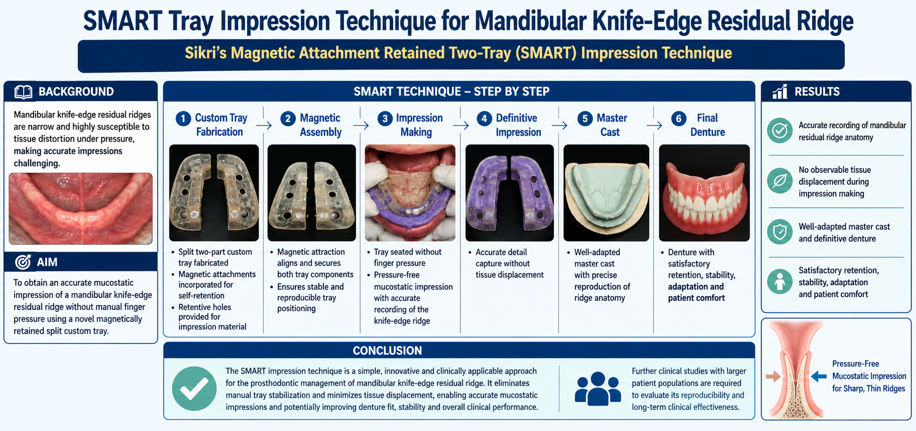
Background: Mandibular knife-edge residual ridges present a significant challenge during complete denture impression making because of their narrow morphology and susceptibility to tissue distortion under pressure. This report describes the clinical application of the novel Sikri’s Magnetic Attachment Retained Two-Tray (SMART) impression technique, conceptualized, developed, and patented by Dr. Arpit Sikri (Patent/ROC No. L-156844/2024), for obtaining an accurate mucostatic impression of a mandibular knife-edge residual ridge without the application of manual finger pressure. The SMART technique employs a magnetically retained split custom tray design that facilitates pressure-free tray stabilization and precise impression making in anatomically challenging mandibular residual ridges.
Materials and Methods: A completely edentulous patient with a pronounced knife-edge residual ridge in the anterior mandible was rehabilitated using the SMART impression technique. A custom-designed split two-part impression tray incorporating magnetic attachments for self-retention and retentive holes for impression material retention was fabricated. Magnetic attraction between the tray components ensured stable and reproducible tray positioning throughout the impression procedure, eliminating the need for operator-applied finger pressure and facilitating a pressure-free mucostatic impression of the residual ridge.
Results: The SMART technique enabled accurate recording of the mandibular residual ridge anatomy without observable tissue displacement during impression making. The definitive impression produced a well-adapted master cast, resulting in a complete denture with satisfactory retention, stability, adaptation, and patient comfort during clinical evaluation.
Conclusion: The SMART impression technique represents a simple, innovative, and clinically applicable approach for the prosthodontic management of mandibular knife-edge residual ridge. By eliminating manual tray stabilization and minimizing tissue displacement during impression making, it facilitates accurate mucostatic impressions and may improve denture fit, stability, and overall clinical performance. Further clinical studies involving larger patient populations are required to evaluate its reproducibility and long-term clinical effectiveness.
Downloads
References
Atwood DA. Reduction of residual ridges: a major oral disease entity. J Prosthet Dent. 1971;26:266–79. DOI: https://doi.org/10.1016/0022-3913(71)90069-2
Atwood DA, Coy WA. Clinical, cephalometric, and densitometric study of reduction of residual ridges. J Prosthet Dent. 1971;26:280–95. DOI: https://doi.org/10.1016/0022-3913(71)90070-9
The Glossary of Prosthodontic Terms 2023: Tenth Edition. J Prosthet Dent. 2023;130(4 Suppl 1):e3. DOI: https://doi.org/10.1016/j.prosdent.2023.03.003
Cawood JI, Howell RA. A classification of the edentulous jaws. Int J Oral Maxillofac Surg. 1988;17:232–6. DOI: https://doi.org/10.1016/S0901-5027(88)80047-X
Nishimura I, Atwood DA. Knife-edge residual ridges: a clinical report. J Prosthet Dent. 1994;71:231–4. DOI: https://doi.org/10.1016/0022-3913(94)90459-6
Meyer RA. Management of denture patients with sharp residual ridges. J Prosthet Dent. 1966;16:431–7. DOI: https://doi.org/10.1016/0022-3913(66)90046-1
Goodsell JO. Surgical aids to intraoral prosthesis. J Oral Surg (Chic). 1955;13:8–34.
Landa JS. Practical full denture prosthesis. Dent Items Interest. 1946;68:155 passim.
Nishimura I, Hosokawa R, Atwood DA. The knife-edge tendency in mandibular residual ridges in women. J Prosthet Dent. 1992;67:820–6. DOI: https://doi.org/10.1016/0022-3913(92)90592-X
Bear SE. Surgical preparation of the mouth for a prosthesis. J Oral Surg (Chic). 1958;16:3–19.
Bolender CL, Swenson RD. Cephalometric evaluation of a labial vestibular extension procedure. J Prosthet Dent. 1963;13:416–31. DOI: https://doi.org/10.1016/0022-3913(63)90096-9
Thoma KH. Oral surgery. St. Louis (MO): Mosby; 1948.
Boucher LJ. Injected silastic in ridge extension procedures. J Prosthet Dent. 1964;14:460–4. DOI: https://doi.org/10.1016/S0022-3913(64)80011-1
Devan MM. Basic principles in impression making. J Prosthet Dent. 1952;93:503–8. DOI: https://doi.org/10.1016/j.prosdent.2005.02.007
Winkler S. Essentials of complete denture prosthodontics. 2nd ed. Littleton, MA: PSG Publishing Co.; 1988.
Hyde TP. Case report: differential pressure impressions for complete dentures. Eur J Prosthodont Restor Dent. 2003;11:5–8.
Jamieson CH. A complete denture impression technique. J Prosthet Dent. 1954;4(1):17–29. DOI: https://doi.org/10.1016/0022-3913(54)90061-X
Weng BX, Khlevnoy V. Pressure control for complete denture impressions. Oral Health. 1995;85:21–3, 24, 27.
Addison PI. Mucostatic impressions. J Am Dent Assoc. 1944;31(13):941–6. DOI: https://doi.org/10.14219/jada.archive.1944.0211
Boucher CO. Complete denture impressions based upon the anatomy of the mouth. J Am Dent Assoc. 1944;31(17):1174–81. DOI: https://doi.org/10.14219/jada.archive.1944.0262
Boucher CO. A critical analysis of mid-century impression techniques for full dentures. J Prosthet Dent. 1951;1(4):472–91. DOI: https://doi.org/10.1016/0022-3913(51)90032-7
Sikri A, Sikri J, Singh N, Bali P, Mittal N. Prosthodontic management of knife-edge ridge using customized prefabricated metal mesh custom tray impression technique. Dent J Indira Gandhi Inst Med Sci. 2022;1(2):59–68. DOI: https://doi.org/10.25259/DJIGIMS_20220102_59
Sikri A, Sikri J. Prosthetic management of flabby ridge using customized prefabricated metal mesh custom tray impression technique: a case report. IP Ann Prosthodont Restor Dent. 2022;8(1):50–60. DOI: https://doi.org/10.18231/j.aprd.2022.011
Smith PW, Richmond R, McCord JF. The design and use of special trays in prosthodontics: guidelines to improve clinical effectiveness. Br Dent J 1999;187:423–6. DOI: https://doi.org/10.1038/sj.bdj.4800295a
Sikri A, Kalra V, Sikri J, Jassal P, Yamika. Precision prosthodontic rehabilitation of flabby ridge with a modified window technique: case report. Dental J Indira Gandhi Inst Med Sci. 2024;3(1):51–64. DOI: https://doi.org/10.25259/DJIGIMS_1_2024
Published
How to Cite
Issue
Section
License
Copyright (c) 2026 Arpit Sikri, Jyotsana Sikri, Rekha Thiruvengadam, Muthu Thiruvengadam

This work is licensed under a Creative Commons Attribution 4.0 International License.




 Biomaterial Investigations in Dentistry is a Diamond Open Access peer-reviewed journal, publishing research in oral biomaterials science. The publishing of articles is free for authors, thanks to the support of Acta Odontologica Scandinavica Society (AOSS), a not-for-profit society.