
Clinical factors associated with motor imagery ability in individuals with subacute stroke
DOI:
https://doi.org/10.2340/jrm.v58.46042Keywords:
motor imagery, stroke rehabilitation, working memory, proprioception, motor learningAbstract
Objective: To examine the associations of motor recovery stage, proprioceptive function, and working memory with motor imagery ability in patients with subacute stroke.
Design: Multicentre cross-sectional study.
Subjects/Patients: Forty patients with subacute stroke who had complete data.
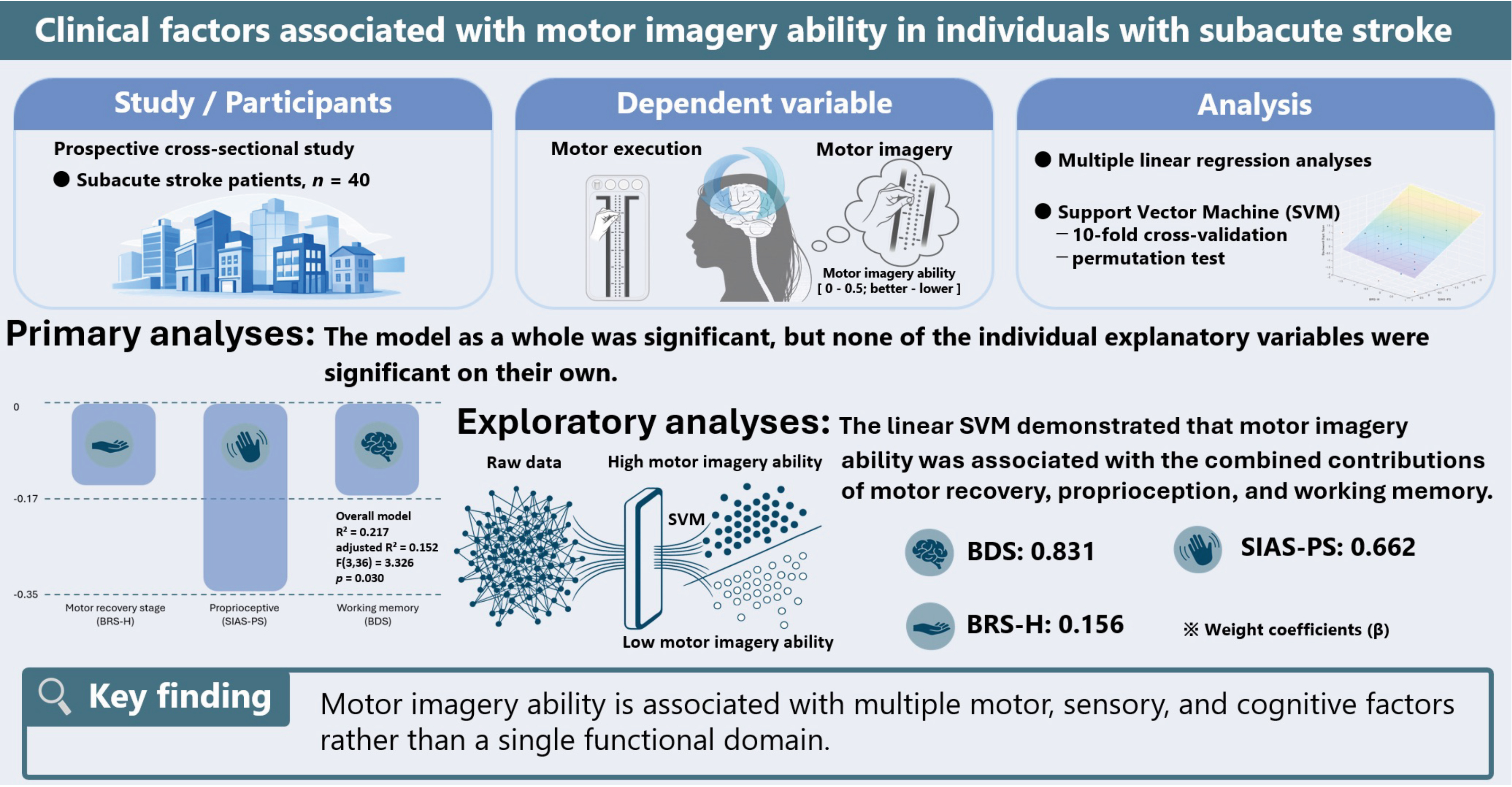
Methods: Motor imagery ability was assessed using mental chronometry during a Purdue Pegboard task. Associations with motor recovery stage, proprioceptive function, and working memory were examined using correlation and multiple regression analyses. An exploratory linear support vector machine analysis was performed to classify participants into groups with higher and lower motor imagery ability using these variables.
Results: Motor imagery ability was significantly associated with motor recovery stage and proprioceptive function in the correlation analyses. However, these associations were no longer independently significant after adjustment in the multiple regression analysis. A linear support vector machine achieved an area under the curve of 0.716, with an accuracy of 0.700, sensitivity of 0.800, and specificity of 0.600. Permutation testing confirmed that the classification performance was above chance.
Conclusion: Motor imagery ability may be related to a combination of motor, sensory, and cognitive functions rather than any single functional domain.
Downloads
References
Kolmos M, Munoz-Novoa M, Sunnerhagen K, Alt Murphy M, Kruuse C. Upper-extremity motor recovery after stroke: a systematic review and meta-analysis of usual care in trials and observational studies. J Neurol Sci 2025; 468: 123341. DOI: https://doi.org/10.1016/j.jns.2024.123341
Purton J, Sim J, Hunter SM. Stroke survivors’ views on their priorities for upper-limb recovery and the availability of therapy services after stroke: a longitudinal, phenomenological study. Disabil Rehabil 2023; 45: 3059–3069. DOI: https://doi.org/10.1080/09638288.2022.2120097
Wolpert DM, Flanagan JR. Motor prediction. Curr Biol 2001; 11: R729–R732. DOI: https://doi.org/10.1016/S0960-9822(01)00432-8
Wolpert DM, Diedrichsen J, Flanagan JR. Principles of sensorimotor learning. Nat Rev Neurosci 2011; 12: 739–751. DOI: https://doi.org/10.1038/nrn3112
Shmuelof L, Krakauer JW. Are we ready for a natural history of motor learning? Neuron 2011; 72: 469–476. DOI: https://doi.org/10.1016/j.neuron.2011.10.017
Debaere F, Wenderoth N, Sunaert S, Van Hecke P, Swinnen SP. Internal vs external generation of movements: differential neural pathways involved in bimanual coordination performed in the presence or absence of augmented visual feedback. Neuroimage 2003; 19: 764–776. DOI: https://doi.org/10.1016/S1053-8119(03)00148-4
Edwards LL, King EM, Buetefisch CM, Borich MR. Putting the “sensory” into sensorimotor control: the role of sensorimotor integration in goal-directed hand movements after stroke. Front Integr Neurosci 2019; 13: 16. DOI: https://doi.org/10.3389/fnint.2019.00016
Farah MJ. The neural basis of mental imagery. Trends Neurosci 1989; 12: 395–399. DOI: https://doi.org/10.1016/0166-2236(89)90079-9
Wolpert DM, Ghahramani Z, Jordan MI. An internal model for sensorimotor integration. Science 1995; 269: 1880–1882. DOI: https://doi.org/10.1126/science.7569931
Decety J, Jeannerod M, Prablanc C. The timing of mentally represented actions. Behav Brain Res 1989; 34: 35–42. DOI: https://doi.org/10.1016/S0166-4328(89)80088-9
Kawasaki T, Mikami K, Kamo T, Aoki R, Ishiguro R, Nakamura H, et al. Motor planning error in Parkinson’s disease and its clinical correlates. PLoS One 2018; 13: e0202228. DOI: https://doi.org/10.1371/journal.pone.0202228
Sakai K, Hosoi Y, Harada Y, Ikeda Y. Estimation error consisting of motor imagery and motor execution in patients with stroke. J Mot Behav 2023; 55: 435–442. DOI: https://doi.org/10.1080/00222895.2023.2219631
Guillot A, Collet C. Duration of mentally simulated movement: a review. J Mot Behav 2005; 37: 10–20. DOI: https://doi.org/10.3200/JMBR.37.1.10-20
Hardwick RM, Caspers S, Eickhoff SB, Swinnen SP. Neural correlates of action: comparing meta-analyses of imagery, observation, and execution. Neurosci Biobehav Rev 2018; 94: 31–44. DOI: https://doi.org/10.1016/j.neubiorev.2018.08.003
Fukumoto Y, Todo M, Bizen H, Kimura D, Suzuki T. Precision pinch force control via brain and spinal motor neuron excitability during motor imagery. Neurosci Lett 2021; 754: 135843. DOI: https://doi.org/10.1016/j.neulet.2021.135843
Fukumoto Y, Todo M, Bizen H, Kimura D, Suzuki T. Causal relationships between brain and spinal motor neuron excitability during motor imagery: using NIRS and evoked electromyogram study. Neuroimage Rep 2022; 2: 100083. DOI: https://doi.org/10.1016/j.ynirp.2022.100083
Krakauer JW, Carmichael ST, Corbett D, Wittenberg GF. Getting neurorehabilitation right: what can be learned from animal models? Neurorehabil Neural Repair 2012; 26: 923–931. DOI: https://doi.org/10.1177/1545968312440745
Suzuki T, Saitoh E, Tani M, Nabeta R, Daikuya S, Hirose H, et al. Characteristic appearances of the H-reflex and F-wave with increased stimulus intensity in patients with cerebrovascular disease. Electromyogr Clin Neurophysiol 2002; 42: 67–70.
Suzuki T, Bunno Y, Tani M, Onigata C, Fukumoto Y, Todo M, et al. F-wave waveform values based on the correlation coefficient of each waveform increased following improved voluntary movements in a patient with cerebrovascular disease: a case study with long-term follow-up. Int J Neurorehabilitation 2016; 3: 206. DOI: https://doi.org/10.4172/2376-0281.1000206
de Vries S, Tepper M, Feenstra W, Oosterveld H, Boonstra AM, Otten B. Motor imagery ability in stroke patients: the relationship between implicit and explicit motor imagery measures. Front Hum Neurosci 2013; 7: 790. DOI: https://doi.org/10.3389/fnhum.2013.00790
Kilteni K, Andersson BJ, Houborg C, Ehrsson HH. Motor imagery involves predicting the sensory consequences of the imagined movement. Nat Commun 2018; 9: 1617. DOI: https://doi.org/10.1038/s41467-018-03989-0
Liepert J, Greiner J, Nedelko V, Dettmers C. Reduced upper limb sensation impairs mental chronometry for motor imagery after stroke: clinical and electrophysiological findings. Neurorehabil Neural Repair 2012; 26: 470–478. DOI: https://doi.org/10.1177/1545968311425924
Lee H, Kim H, Ahn M, You Y. Effects of proprioception training with exercise imagery on balance ability of stroke patients. J Phys Ther Sci 2015; 27: 1–4. DOI: https://doi.org/10.1589/jpts.27.1
Malouin F, Richards CL, Durand A. Slowing of motor imagery after a right hemispheric stroke. Stroke Res Treat 2012; 2012: 297217. DOI: https://doi.org/10.1155/2012/297217
Fukumoto Y, Fujii K, Todo M, Suzuki T. Differences in working memory function are associated with motor imagery-induced changes in spinal motor nerve excitability and subsequent motor skill changes. Cogn Process 2025; 26: 15–27. DOI: https://doi.org/10.1007/s10339-024-01231-y
Fukumoto Y, Bizen H, Todo M, Yoshida N, Suzuki T. The combined effects of motor imagery and motor practice are influenced by differences in working memory function: examination of brain, spinal cord, and muscle performance data. IBRO Neurosci Rep 2025; 19: 210–222. DOI: https://doi.org/10.1016/j.ibneur.2025.06.016
Brunnstrom S. Motor testing procedures in hemiplegia: based on sequential recovery stages. Phys Ther 1966; 46: 357–375. DOI: https://doi.org/10.1093/ptj/46.4.357
Chino N, Sonoda S, Domen K, Saitoh E, Kimura A. Stroke Impairment Assessment Set (SIAS): a new evaluation instrument for stroke patients. Jpn J Rehabil Med 1994; 31: 119–125. DOI: https://doi.org/10.2490/jjrm1963.31.119
Fukumoto Y, Todo M, Bunno Y, Yoneda H, Tani M, Suzuki T. Differences in motor imagery strategy change behavioral outcome. Sci Rep 2022; 12: 13868. DOI: https://doi.org/10.1038/s41598-022-18164-1
Carlson SM, Moses LJ, Breton C. How specific is the relation between executive function and theory of mind? Contributions of inhibitory control and working memory. Infant Child Dev 2002; 11: 73–92. DOI: https://doi.org/10.1002/icd.298
Malouin F, Richards CL, Durand A, Doyon J. Reliability of mental chronometry for assessing motor imagery ability after stroke. Arch Phys Med Rehabil 2008; 89: 311–319. DOI: https://doi.org/10.1016/j.apmr.2007.11.006
Beauchet O, Annweiler C, Assal F, Bridenbaugh S, Herrmann FR, Kressig RW, et al. Imagined Timed Up & Go test: a new tool to assess higher-level gait and balance disorders in older adults? J Neurol Sci 2010; 294: 102–106. DOI: https://doi.org/10.1016/j.jns.2010.03.021
Allali G, van der Meulen M, Beauchet O, Rieger SW, Vuilleumier P, Assal F. The neural basis of age-related changes in motor imagery of gait: an fMRI study. J Gerontol A Biol Sci Med Sci 2014; 69: 1389–1398. https://doi.org/10.1093/gerona/glt207 DOI: https://doi.org/10.1093/gerona/glt207
de Vries S, Tepper M, Otten B, Mulder T. Recovery of motor imagery ability in stroke patients. Rehabil Res Pract 2011; 2011: 283840. DOI: https://doi.org/10.1155/2011/283840
Liepert J, Büsching I, Sehle A, Schoenfeld MA. Mental chronometry and mental rotation abilities in stroke patients with different degrees of sensory deficit. Restor Neurol Neurosci 2016; 34: 907–914. DOI: https://doi.org/10.3233/RNN-160640
Malouin F, Belleville S, Richards CL, Desrosiers J, Doyon J. Working memory and mental practice outcomes after stroke. Arch Phys Med Rehabil 2004; 85: 177–183. DOI: https://doi.org/10.1016/S0003-9993(03)00771-8
Sidarta A, van Vugt FT, Ostry DJ. Somatosensory working memory in human reinforcement-based motor learning. J Neurophysiol 2018; 120: 3275–3286. DOI: https://doi.org/10.1152/jn.00442.2018
Chiyohara S, Furukawa J-I, Noda T, Morimoto J, Imamizu H. Proprioceptive short-term memory in passive motor learning. Sci Rep 2023; 13: 20826. DOI: https://doi.org/10.1038/s41598-023-48101-9
Additional Files
Published
How to Cite
License
Copyright (c) 2026 Yuki Fukumoto, Tsubasa Kawasaki, Takanao Shirai, Tomomi Hase, Hinata Chiyo, Hiroyuki Hamada, Katsuya Sakai, Junpei Tanabe, Kota Sawa, Toshiaki Suzuki

This work is licensed under a Creative Commons Attribution 4.0 International License.
All digitalized JRM contents is available freely online. The Foundation for Rehabilitation Medicine owns the copyright for all material published until volume 40 (2008), as from volume 41 (2009) authors retain copyright to their work and as from volume 49 (2017) the journal has been published Open Access, under CC-BY-NC licences (unless otherwise specified). The CC-BY-NC licenses allow third parties to copy and redistribute the material in any medium or format and to remix, transform, and build upon the material for non-commercial purposes, provided proper attribution to the original work.
From 2024, articles are published under the CC-BY licence. This license permits sharing, adapting, and using the material for any purpose, including commercial use, with the condition of providing full attribution to the original publication.






